Product Info Summary
SKU: | A02260-1 |
---|---|
Size: | 100ul |
Reactive Species: | Human, Mouse, Rat |
Host: | Rabbit |
Application: | Flow Cytometry, IP, IF, IHC, ICC, WB |
Customers Who Bought This Also Bought
Product info
Product Name
Anti-Cytoplasmic protein NCK1 NCK1 Antibody
SKU/Catalog Number
A02260-1
Size
100ul
Form
Liquid
Description
Boster Bio Anti-Cytoplasmic protein NCK1 NCK1 Antibody catalog # A02260-1. Tested in WB,ICC,IF,IHC,IP,Flow Cytometry applications. This antibody reacts with Human,Mouse,Rat.
Storage & Handling
Store at -20°C for one year. For short term storage and frequent use, store at 4°C for up to one month. Avoid repeated freeze-thaw cycles.
Cite This Product
Anti-Cytoplasmic protein NCK1 NCK1 Antibody (Boster Biological Technology, Pleasanton CA, USA, Catalog # A02260-1)
Host
Rabbit
Contents
Rabbit IgG, 1mg/ml in PBS with 0.02% sodium azide, 50% glycerol, pH7.2
Clonality
Polyclonal
Isotype
IgG
Immunogen
Synthetic peptide, corresponding to amino acids 100-150 of Human UCP2.
*Blocking peptide can be purchased. Costs vary based on immunogen length. Contact us for pricing.
Reactive Species
A02260-1 is reactive to NCK1 in Human, Mouse, Rat
Applications
A02260-1 is guaranteed for Flow Cytometry, IP, IF, IHC, ICC, WB Boster Guarantee
Observed Molecular Weight
Calculated molecular weight
42.864kDa
Background of NCK1
Binds peptides derived from antigens that access the endocytic route of antigen presenting cells (APC) and presents them on the cell surface for recognition by the CD4 T-cells. The peptide binding cleft accommodates peptides of 10-30 residues. The peptides presented by MHC class II molecules are generated mostly by degradation of proteins that access the endocytic route, where they are processed by lysosomal proteases and other hydrolases. Exogenous antigens that have been endocytosed by the APC are thus readily available for presentation via MHC II molecules, and for this reason this antigen presentation pathway is usually referred to as exogenous. As membrane proteins on their way to degradation in lysosomes as part of their normal turn-over are also contained in the endosomal/lysosomal compartments, exogenous antigens must compete with those derived from endogenous components. Autophagy is also a source of endogenous peptides, autophagosomes constitutively fuse with MHC class II loading compartments. In addition to APCs, other cells of the gastrointestinal tract, such as epithelial cells, express MHC class II molecules and CD74 and act as APCs, which is an unusual trait of the GI tract. To produce a MHC class II molecule that presents an antigen, three MHC class II molecules (heterodimers of an alpha and a beta chain) associate with a CD74 trimer in the ER to form a heterononamer. Soon after the entry of this complex into the endosomal/lysosomal system where antigen processing occurs, CD74 undergoes a sequential degradation by various proteases, including CTSS and CTSL, leaving a small fragment termed CLIP (class-II-associated invariant chain peptide). The removal of CLIP is facilitated by HLA-DM via direct binding to the alpha-beta-CLIP complex so that CLIP is released. HLA-DM stabilizes MHC class II molecules until primary high affinity antigenic peptides are bound. The MHC II molecule bound to a peptide is then transported to the cell membrane surface. In B-cells, the interaction between HLA-DM and MHC class II molecules is regulated by HLA-DO. Primary dendritic cells (DCs) also to express HLA-DO. Lysosomal microenvironment has been implicated in the regulation of antigen loading into MHC II molecules, increased acidification produces increased proteolysis and efficient peptide loading.
Lawrance S.K., Nucleic Acids Res. 13:7515-7528(1985).
Gustafsson K., J. Biol. Chem. 262:8778-8786(1987).
Young J.A., Hum. Immunol. 23:37-44(1988).<
Antibody Validation
Boster validates all antibodies on WB, IHC, ICC, Immunofluorescence, and ELISA with known positive control and negative samples to ensure specificity and high affinity, including thorough antibody incubations.
Innovating Scientists Reward
If you are the first to review this product, or if you have results for a special sample, species or application this product is not validated in, share your results with us and receive product credits you can use towards any Boster products! Applicable to all scientists worldwide.
Submit A Review
Assay dilution & Images
Assay Dilutions Recommendation
The recommendations below provide a starting point for assay optimization. The actual working concentration varies and should be decided by the user.
WB: 1:500-1:2,000
ICC: 1:50-1:200
IHC: 1:50-1:200
FC: 1:50-1:100
IP: 1:10-1:50
Validation Images & Assay Conditions
Click image to see more details
Figure 2. Immunocytochemistry staining of NCK1 using Anti-Cytoplasmic protein NCK1 NCK1 Antibody (A02260-1).
ICC staining NCK1 in LOVO cells (green). The nuclear counter stain is DAPI (blue). Cells were fixed in paraformaldehydeClick image to see more details
Figure 1. Western blotting validation for Anti-Cytoplasmic protein NCK1 NCK1 Antibody A02260-1
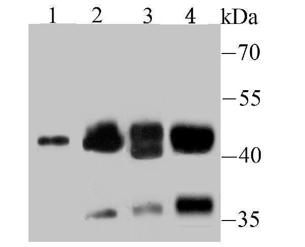
Western blot analysis of NCK1 on different lysates using anti-NCK1 antibody at 1/500 dilution.Positive control:
Lane 1: Hela
Lane 1: NIH-3T3
Lane 2: Rat kidney tissue
Lane 3: Mouse testis tissue
Electrophoresis was performed on a SDS-PAGE gel. To determine SDS-PAGE gel concentration
Protein Target Info & Infographic
Gene/Protein Information For NCK1 (Source: Uniprot.org, NCBI)
Gene Name
NCK1
Full Name
Cytoplasmic protein NCK1
Weight
42.864kDa
Alternative Names
cytoplasmic protein NCK1; MGC12668; NCK adaptor protein 1melanoma NCK protein; NCK1; nck-1; NCKalpha; NCKNCK tyrosine kinase; non-catalytic region of tyrosine kinase; SH2/SH3 adaptor protein NCK-alpha NCK1 NCK, NCKalpha, nck-1 NCK adaptor protein 1 cytoplasmic protein NCK1|NCK tyrosine kinase|SH2/SH3 adaptor protein NCK-alpha|melanoma NCK protein|non-catalytic region of tyrosine kinase
*If product is indicated to react with multiple species, protein info is based on the gene entry specified above in "Species".For more info on NCK1, check out the NCK1 Infographic
We have 30,000+ of these available, one for each gene! Check them out.
In this infographic, you will see the following information for NCK1: database IDs, superfamily, protein function, synonyms, molecular weight, chromosomal locations, tissues of expression, subcellular locations, post-translational modifications, and related diseases, research areas & pathways. If you want to see more information included, or would like to contribute to it and be acknowledged, please contact [email protected].
Specific Publications For Anti-Cytoplasmic protein NCK1 NCK1 Antibody (A02260-1)
Hello CJ!
No publications found for A02260-1
*Do you have publications using this product? Share with us and receive a reward. Ask us for more details.
Recommended Resources
Here are featured tools and databases that you might find useful.
- Boster's Pathways Library
- Protein Databases
- Bioscience Research Protocol Resources
- Data Processing & Analysis Software
- Photo Editing Software
- Scientific Literature Resources
- Research Paper Management Tools
- Molecular Biology Software
- Primer Design Tools
- Bioinformatics Tools
- Phylogenetic Tree Analysis
Customer Reviews
Have you used Anti-Cytoplasmic protein NCK1 NCK1 Antibody?
Submit a review and receive an Amazon gift card.
- $25/€18/£15/$25CAN/¥75 Yuan/¥1250 Yen for a review with an image
- $10/€7/£6/$10 CAD/¥70 Yuan/¥1110 Yen for a review without an image
Submit A Review
Be the first to review Anti-Cytoplasmic protein NCK1 NCK1 Antibody
*The first user to submit a review for a product is eligible for Boster's Innovating Scientists Reward, which gives product credits. This is in addition to the gift card reward.
Customer Q&As
Have a question?
Find answers in Q&As, reviews.
Can't find your answer?
Submit your question