Click image to see more details
-

-
-
-
-
+2
Product Info Summary
| SKU: | PA1970 |
|---|---|
| Size: | 100 μg/vial |
| Reactive Species: | Human, Mouse, Rat |
| Host: | Rabbit |
| Application: | IHC, WB |
Customers Who Bought This Also Bought

Product info
Product Name
Anti-DNA PKcs/PRKDC Antibody Picoband®
SKU/Catalog Number
PA1970
BA2908-2 is an alternative SKU for this antibody, used in previous lots.
Size
100 μg/vial
Form
Lyophilized
Description
Boster Bio Anti-DNA PKcs/PRKDC Antibody catalog # PA1970. Tested in WB, IHC applications. This antibody reacts with Human, Mouse, Rat. The brand Picoband indicates this is a premium antibody that guarantees superior quality, high affinity, and strong signals with minimal background in Western blot applications. Only our best-performing antibodies are designated as Picoband, ensuring unmatched performance.
Storage & Handling
Store at -20˚C for one year from date of receipt. After reconstitution, at 4˚C for one month. It can also be aliquotted and stored frozen at -20˚C for six months. Avoid repeated freeze-thaw cycles.
Cite This Product
Anti-DNA PKcs/PRKDC Antibody Picoband® (Boster Biological Technology, Pleasanton CA, USA, Catalog # PA1970)
Host
Rabbit
Contents
Each vial contains 4 mg Trehalose, 0.9 mg NaCl and 0.2 mg Na2HPO4.
Clonality
Polyclonal
Isotype
Rabbit IgG
Immunogen
A synthetic peptide corresponding to a sequence at the N-terminus of human DNA PKcs.
Cross-reactivity
No cross-reactivity with other proteins
Reactive Species
PA1970 is reactive to PRKDC in Human, Mouse, Rat
Observed Molecular Weight
469 kDa
Calculated molecular weight
469.1 kDa
Background of PRKDC
PRKDC (Protein Kinase DNA-Activated Catalytic Subunit), also called DNAPK, HYRC1, p350 or DNPK1, is an enzyme that in humans is encoded by the PRKDC gene. DNA-PKcs belongs to the phosphatidylinositol 3-kinase-related kinase protein family. Satoh et al. (1997) mapped the MCM4 gene to 8q11.2 by FISH. Based on the close proximity of the PRKDC and MCM4 genes, it was assumed that the PRKDC gene also maps to this location. Anderson and Lees-Miller (1992) noted that DNA-PK had been shown in vitro to phosphorylate several transcription factors, suggesting that it functions in cell homeostasis by modulating transcription. Daniel et al. (1999) demonstrated that the PRKDC protein participates in retroviral DNA integration, which is catalyzed by the viral protein integrase.
Antibody Validation
Boster validates all antibodies on WB, IHC, ICC, Immunofluorescence, and ELISA with known positive control and negative samples to ensure specificity and high affinity, including thorough antibody incubations.
Application & Images
Applications
PA1970 is guaranteed for IHC, WB Boster Guarantee
Recommend Dilution
| Application | Dilution | Species |
|---|---|---|
| Western blot | 0.1-0.5μg/ml | Human |
| Immunohistochemistry (Paraffin-embedded Section) | 2-5μg/ml | Human, Mouse, Rat |
Tested application
Suggested blocking solution with 5% non-fat milk or BSA; (*)Recommended protein loading: 20-40 µg per lane
Use TE buffer pH 9.0 for antigen retrieval; (*) citrate buffer pH 6.0 is an alternative.
Validation Images & Assay Conditions
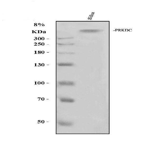
Click image to see more details
Western blot analysis of PRKDC using anti-PRKDC antibody (PA1970).
Electrophoresis was performed on a 8% SDS-PAGE gel at 80V (Stacking gel) / 120V (Resolving gel) for 2 hours. The sample well of each lane was loaded with 30 ug of sample under reducing conditions.
Lane 1: human SiHa whole cell lysates.
After electrophoresis, proteins were transferred to a nitrocellulose membrane at 150 mA for 50-90 minutes. Blocked the membrane with 5% non-fat milk/TBS for 1.5 hour at RT. The membrane was incubated with rabbit anti-PRKDC antigen affinity purified polyclonal antibody (PA1970) at 0.5 μg/mL overnight at 4°C, then washed with TBS-0.1%Tween 3 times with 5 minutes each and probed with a goat anti-rabbit IgG-HRP secondary antibody (Catalog # BA1054) at a dilution of 1:5000 for 1.5 hour at RT. The signal is developed using an ECL Plus Western Blotting Substrate (Catalog # AR1196-200) with Tanon 5200 system. A specific band was detected for PRKDC at approximately 469 kDa. The expected band size for PRKDC is at 469 kDa.

Click image to see more details
IHC analysis of PRKDC using anti-PRKDC antibody (PA1970).
PRKDC was detected in a paraffin-embedded section of huma colon cancer tissue. Heat mediated antigen retrieval was performed in EDTA buffer (pH 8.0, epitope retrieval solution). The tissue section was blocked with 10% goat serum. The tissue section was then incubated with 2 μg/ml rabbit anti-PRKDC Antibody (PA1970) overnight at 4°C. Peroxidase Conjugated Goat Anti-rabbit IgG was used as secondary antibody and incubated for 30 minutes at 37°C. The tissue section was developed using HRP Conjugated Rabbit IgG Super Vision Assay Kit (Catalog # SV0002) with DAB as the chromogen.

Click image to see more details
IHC analysis of PRKDC using anti-PRKDC antibody (PA1970).
PRKDC was detected in a paraffin-embedded section of human gastric carcinoma tissue. Heat mediated antigen retrieval was performed in EDTA buffer (pH 8.0, epitope retrieval solution). The tissue section was blocked with 10% goat serum. The tissue section was then incubated with 2 μg/ml rabbit anti-PRKDC Antibody (PA1970) overnight at 4°C. Peroxidase Conjugated Goat Anti-rabbit IgG was used as secondary antibody and incubated for 30 minutes at 37°C. The tissue section was developed using HRP Conjugated Rabbit IgG Super Vision Assay Kit (Catalog # SV0002) with DAB as the chromogen.

Click image to see more details
IHC analysis of PRKDC using anti-PRKDC antibody (PA1970).
PRKDC was detected in a paraffin-embedded section of human ovarian cancer tissue. Heat mediated antigen retrieval was performed in EDTA buffer (pH 8.0, epitope retrieval solution). The tissue section was blocked with 10% goat serum. The tissue section was then incubated with 2 μg/ml rabbit anti-PRKDC Antibody (PA1970) overnight at 4°C. Peroxidase Conjugated Goat Anti-rabbit IgG was used as secondary antibody and incubated for 30 minutes at 37°C. The tissue section was developed using HRP Conjugated Rabbit IgG Super Vision Assay Kit (Catalog # SV0002) with DAB as the chromogen.

Click image to see more details
IHC analysis of PRKDC using anti-PRKDC antibody (PA1970).
PRKDC was detected in a paraffin-embedded section of mouse brain tissue. Heat mediated antigen retrieval was performed in EDTA buffer (pH 8.0, epitope retrieval solution). The tissue section was blocked with 10% goat serum. The tissue section was then incubated with 2 μg/ml rabbit anti-PRKDC Antibody (PA1970) overnight at 4°C. Peroxidase Conjugated Goat Anti-rabbit IgG was used as secondary antibody and incubated for 30 minutes at 37°C. The tissue section was developed using HRP Conjugated Rabbit IgG Super Vision Assay Kit (Catalog # SV0002) with DAB as the chromogen.

Click image to see more details
IHC analysis of PRKDC using anti-PRKDC antibody (PA1970).
PRKDC was detected in a paraffin-embedded section of rat brain tissue. Heat mediated antigen retrieval was performed in EDTA buffer (pH 8.0, epitope retrieval solution). The tissue section was blocked with 10% goat serum. The tissue section was then incubated with 2 μg/ml rabbit anti-PRKDC Antibody (PA1970) overnight at 4°C. Peroxidase Conjugated Goat Anti-rabbit IgG was used as secondary antibody and incubated for 30 minutes at 37°C. The tissue section was developed using HRP Conjugated Rabbit IgG Super Vision Assay Kit (Catalog # SV0002) with DAB as the chromogen.
Specific Publications For Anti-DNA PKcs/PRKDC Antibody Picoband® (PA1970)
Loading publications
Recommended Resources
Here are featured tools and databases that you might find useful.
- Boster's Pathways Library
- Protein Databases
- Bioscience Research Protocol Resources
- Data Processing & Analysis Software
- Photo Editing Software
- Scientific Literature Resources
- Research Paper Management Tools
- Molecular Biology Software
- Primer Design Tools
- Bioinformatics Tools
- Phylogenetic Tree Analysis
Customer Reviews
Have you used Anti-DNA PKcs/PRKDC Antibody Picoband®?
Share your experimental results or join a short interview to earn up to $1,000 in product credits or other rewards.
0 Reviews For Anti-DNA PKcs/PRKDC Antibody Picoband®
Customer Q&As
Have a question?
Find answers in Q&As, reviews.
Can't find your answer?
Submit your question
4 Customer Q&As for Anti-DNA PKcs/PRKDC Antibody Picoband®
Question
We are currently using anti-DNA PKcs/PRKDC antibody PA1970 for human tissue, and we are happy with the WB results. The species of reactivity given in the datasheet says human. Is it possible that the antibody can work on pig tissues as well?
Verified Customer
Verified customer
Asked: 2020-02-27
Answer
The anti-DNA PKcs/PRKDC antibody (PA1970) has not been validated for cross reactivity specifically with pig tissues, though there is a good chance of cross reactivity. We have an innovator award program that if you test this antibody and show it works in pig you can get your next antibody for free. Please contact me if I can help you with anything.
Boster Scientific Support
Answered: 2020-02-27
Question
Our team were well pleased with the WB result of your anti-DNA PKcs/PRKDC antibody. However we have seen positive staining in placenta nucleus using this antibody. Is that expected? Could you tell me where is PRKDC supposed to be expressed?
C. Krishna
Verified customer
Asked: 2019-12-24
Answer
Based on literature, placenta does express PRKDC. Generally PRKDC expresses in nucleus. Regarding which tissues have PRKDC expression, here are a few articles citing expression in various tissues:
Brain, Pubmed ID: 2507541, 2247066
Cervix carcinoma, Pubmed ID: 7671312, 17081983, 18669648, 18691976, 20068231
Cervix carcinoma, and Erythroleukemia, Pubmed ID: 23186163
Embryonic kidney, Pubmed ID: 17525332
Fetal lung, Pubmed ID: 7594449
Leukemic T-cell, Pubmed ID: 19690332
Liver, Pubmed ID: 24275569
Placenta, Pubmed ID: 7638222
Boster Scientific Support
Answered: 2019-12-24
Question
I am looking for using your anti-DNA PKcs/PRKDC antibody for regulation of smooth muscle cell proliferation studies. Has this antibody been tested with western blotting on hela cell lysate? We would like to see some validation images before ordering.
Verified Customer
Verified customer
Asked: 2018-08-24
Answer
We appreciate your inquiry. This PA1970 anti-DNA PKcs/PRKDC antibody is tested on hela cell lysate. It is guaranteed to work for WB in human. Our Boster guarantee will cover your intended experiment even if the sample type has not been be directly tested.
Boster Scientific Support
Answered: 2018-08-24
Question
We have observed staining in human cervix carcinoma. What should we do? Is anti-DNA PKcs/PRKDC antibody supposed to stain cervix carcinoma positively?
Verified Customer
Verified customer
Asked: 2017-10-23
Answer
From literature cervix carcinoma does express PRKDC. From Uniprot.org, PRKDC is expressed in kidney, cervix carcinoma, placenta, fetal lung, brain, embryonic kidney, leukemic t-cell, cervix carcinoma erythroleukemia, liver, among other tissues. Regarding which tissues have PRKDC expression, here are a few articles citing expression in various tissues:
Brain, Pubmed ID: 2507541, 2247066
Cervix carcinoma, Pubmed ID: 7671312, 17081983, 18669648, 18691976, 20068231
Cervix carcinoma, and Erythroleukemia, Pubmed ID: 23186163
Embryonic kidney, Pubmed ID: 17525332
Fetal lung, Pubmed ID: 7594449
Leukemic T-cell, Pubmed ID: 19690332
Liver, Pubmed ID: 24275569
Placenta, Pubmed ID: 7638222
Boster Scientific Support
Answered: 2017-10-23
