Click image to see more details
Product Info Summary
| SKU: | A00023-1 |
|---|---|
| Size: | 250uL |
| Reactive Species: | Human, Rat |
| Host: | Rabbit |
| Application: | ELISA, IP, IHC, WB |
Customers Who Bought This Also Bought

Product info
Product Name
Anti-EGFR Antibody
SKU/Catalog Number
A00023-1
Size
250uL
Form
Liquid (sterile filtered)
Description
Boster Bio Anti-EGFR Antibody (Catalog # A00023-1). Tested in IHC, IP, WB applications. This antibody reacts with Human, Rat.
Storage & Handling
Store vial at -20°C prior to opening. Aliquot contents and freeze at -20°C or below for extended storage. Avoid cycles of freezing and thawing. Centrifuge product if not completely clear after standing at room temperature. This product is stable for several weeks at 4°C as an undiluted liquid. Dilute only prior to immediate use. Expiration date is one (1) year from date of opening. (Ship on dry ice.)
Cite This Product
Anti-EGFR Antibody (Boster Biological Technology, Pleasanton CA, USA, Catalog # A00023-1)
Host
Rabbit
Contents
0.01% (w/v) Sodium Azide
Clonality
Polyclonal
Isotype
Antiserum
Immunogen
This whole rabbit serum was prepared by repeated immunizations with a peptide synthesized using conventional technology. The sequence of the epitope maps to a region near the carboxy terminus which is identical in human, mouse and rat EGFR.
Reactive Species
A00023-1 is reactive to EGFR in Human, Rat
Observed Molecular Weight
68 kDa
Calculated molecular weight
134.3 kDa
Background of EGFR
EGFR is a transmembrane glycoprotein that is a member of a family of protein tyrosine kinases crucial to maintaining a normal balance in cell growth and development. Growth factor receptors are involved not only in promoting the proliferation of normal cells but also in the aberrant growth of many types of human tumors. For example, the epidermal growth factor receptor (EGFR) is mutated and/or over-expressed in many common solid human squamous cell carcinomas including breast, brain, bladder, lung, gastric, head & neck, esophagus, cervix, vulva, ovary, and endometrium. Over-expression of the EGFR gene occurs in carcinomas with and without gene amplification. EGFR and ErbB-2 are particularly important in breast cancer because increased production or activation has been associated with poor prognosis. EGFR belongs to a family of growth factor receptors, which also includes ErbB-2/HER-2/neu, ErbB-3/HER-3/neu and ErbB-4/HER-4/neu. EGFR can heterodimerize with each of the members of this family.
Antibody Validation
Boster validates all antibodies on WB, IHC, ICC, Immunofluorescence, and ELISA with known positive control and negative samples to ensure specificity and high affinity, including thorough antibody incubations.
Application & Images
Applications
A00023-1 is guaranteed for ELISA, IP, IHC, WB Boster Guarantee
Recommend Dilution
| Application | Dilution | Species |
|---|---|---|
| ELISA: 1:10 | 000 - 1:50 | 000 |
| WB: 1:1 | 000 - 1:10 | 000 |
| Anti-EGFR antibody has been tested by and is specifically designed for ELISA | immunoblotting | immunoprecipitation, and immunohistochemistry. Reactivity in other assays is likely, but has not been determined. Recognition of EGFR is independent of the phosphorylation status at tyrosine 1173. A431 cells, keratinocytes in normal epidermis, or placenta are typically used as positive control sources. The antigen is typically localized in the cell membrane. For western blotting, good results are also achieved on PVDF membranes blocked with 5% lowfat milk diluted in TTBS for 1 hour at room temperature. Also, dilute the primary antibody and secondary in 5% lowfat milk in TTBS. Anti-EGFR can be diluted up to 1:10,000 for immunoblot depending on the cell line and the amount of EGFR in a particular lysate. For immunoprecipitation, use approximately 10 µl of the antibody. The immunoprecipitation mix should contain the antibody, 25 µl of Protein A-agarose beads and 1.0 ml of lysate (lysate contains approximately 1.0 mg of total protein). This mixture should be rotated overnight at 4°C and then washed 3 times with lysis buffer (used to prepare the lysate). The resulting bead complex is dissolved in 20-30 µl of 3X SDS-PAGE sample buffer and approximately 15 µl is loaded per lane on an 8% polyacrylamide gel. |
Validation Images & Assay Conditions
Click image to see more details
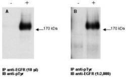
Combined immunoprecipitation and western blot using anti-EGFR antibody. Lysates were prepared from GN4 rat liver epithelial cells both with (+) EGF treatment for 15' at 100 ng/ml and without (-) the addition of EGF. The combination of immunoprecipitation and western blotting was performed using the anti-EGFR antibody for immunoprecipitation (10 µL) followed by western blot detection using an anti-phosphotyrosine antibody (Panel A). This was repeated in reverse order using a 1:2000 dilution of anti-EGFR for western blot (Panel B). Visualization occurred using an ECL system. Film exposure was approximately 1’. Other detection systems will yield similar results.

Click image to see more details
Immunohistochemistry of Anti-EGFR Antibody with positive staining.

Click image to see more details
Western blot using Boster's anti-EGFR antibody. Lane 1: unstimulated A431 whole cell lysates . Lane 2: EGF stimulated A431 whole cell lysates . Shows detection of a band at ~170 kDa corresponding to human EGFR present in unstimulated and stimulated (50 ng/ml for 15 min) lysates (arrowhead). Loaded: 30µg lysate was resolved on a 4-20% Tris-Glycine gel by SDS-PAGE and transferred onto nitrocellulose. Primary Antibody: Anti-EGFR at 1:1,000 overnight at 4° C. Secondary Antibody: IRDye® 800 conjugated Gt-a-Rabbit IgG (H&L) MX10 at 1:10,000 dilution of for 45 min at room temperature (800 nm channel, green). Molecular weight estimation was made by comparison to prestained MW markers in lane M (700 nm channel, red). IRDye® 800 fluorescence image was captured using the Odyssey® Infrared Imaging System developed by LI-COR. IRDye is a trademark of LI-COR, Inc. Other detection systems will yield similar results.
Specific Publications For Anti-EGFR Antibody (A00023-1)
Loading publications
Recommended Resources
Here are featured tools and databases that you might find useful.
- Boster's Pathways Library
- Protein Databases
- Bioscience Research Protocol Resources
- Data Processing & Analysis Software
- Photo Editing Software
- Scientific Literature Resources
- Research Paper Management Tools
- Molecular Biology Software
- Primer Design Tools
- Bioinformatics Tools
- Phylogenetic Tree Analysis
Customer Reviews
Have you used Anti-EGFR Antibody?
Share your experimental results or join a short interview to earn up to $1,000 in product credits or other rewards.
0 Reviews For Anti-EGFR Antibody
Customer Q&As
Have a question?
Find answers in Q&As, reviews.
Can't find your answer?
Submit your question
