Click image to see more details
-

-
-
-
-
+5
Product Info Summary
| SKU: | M00057-2 |
|---|---|
| Size: | 100 μl |
| Reactive Species: | Human, Mouse, Rat |
| Host: | Rabbit |
| Application: | Flow Cytometry, IF, IHC, ICC, WB |
Customers Who Bought This Also Bought

Product info
Product Name
Anti-ER alpha ESR1 Rabbit Monoclonal Antibody
SKU/Catalog Number
M00057-2
BM3947 is an alternative SKU for this antibody, used in previous lots.
Size
100 μl
Form
Liquid
Description
Boster Bio Anti-ER alpha ESR1 Rabbit Monoclonal Antibody catalog # M00057-2. Tested in WB, IHC, ICC/IF, Flow Cytometry applications. This antibody reacts with Human, Mouse, Rat.
Storage & Handling
Store at -20°C for one year. For short term storage and frequent use, store at 4°C for up to one month. Avoid repeated freeze-thaw cycles.
Cite This Product
Anti-ER alpha ESR1 Rabbit Monoclonal Antibody (Boster Biological Technology, Pleasanton CA, USA, Catalog # M00057-2)
Host
Rabbit
Contents
Rabbit IgG in stabilizing components, phosphate buffered saline, pH 7.4, 150mM NaCl, 0.02% sodium azide and 50% glycerol.
*This antibody is supplied in a stabilized formulation.
Compatibility with conjugation reactions depends on the chemistry of the conjugation method used.
For conjugation methods that are not compatible with the stabilizing components present in this formulation, a carrier-free antibody format is required.
Clonality
Monoclonal
Clone Number
IE-5
Isotype
Rabbit IgG
Immunogen
A synthesized peptide derived from human ER alpha
Reactive Species
M00057-2 is reactive to ESR1 in Human, Mouse, Rat
Observed Molecular Weight
60 kDa
Calculated molecular weight
66.2 kDa
Antibody Validation
Boster validates all antibodies on WB, IHC, ICC, Immunofluorescence, and ELISA with known positive control and negative samples to ensure specificity and high affinity, including thorough antibody incubations.
Application & Images
Applications
M00057-2 is guaranteed for Flow Cytometry, IF, IHC, ICC, WB Boster Guarantee
Recommend Dilution
WB 1:500-1:2000
IHC 1:50-1:100
ICC/IF 1:50-1:100
FC 1:30
Tested application
Use TE buffer pH 9.0 for antigen retrieval; (*) citrate buffer pH 6.0 is an alternative.
Validation Images & Assay Conditions
Click image to see more details
All lanes use the Antibody at 1:1K dilution for 1 hour at room temperature.

Click image to see more details
All lanes use the Antibody at 1:500 dilution for 1 hour at room temperature.

Click image to see more details
All lanes use the Antibody at 1:1K dilution for 1 hour at room temperature.

Click image to see more details
All lanes use the Antibody at 1:500 dilution for 1 hour at room temperature.

Click image to see more details
All lanes use the Antibody at 1:1K dilution for 1 hour at room temperature.

Click image to see more details
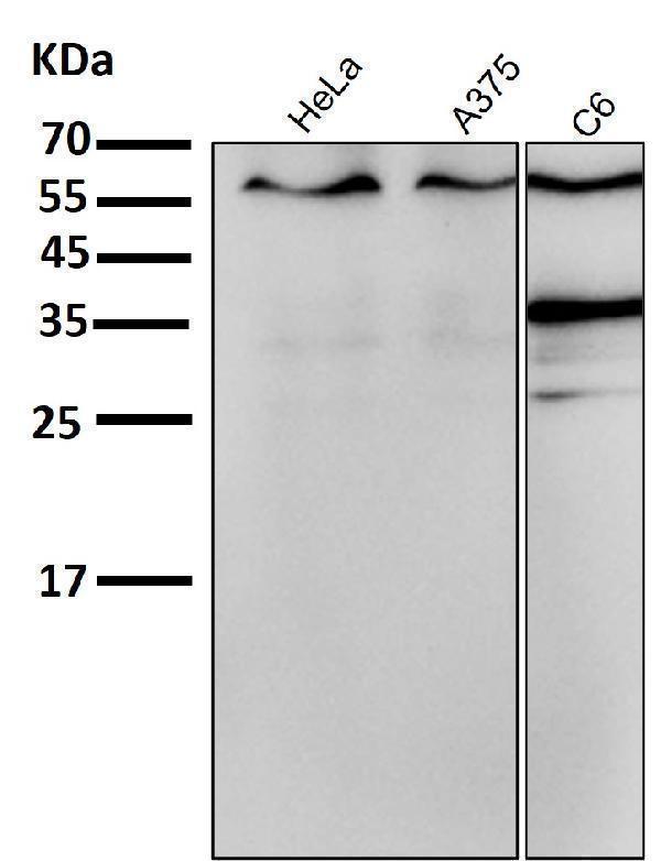
Western blot analysis of ER alpha expression in (1) MCF7 cell lysate; (2)T47-D cell lysate.

Click image to see more details
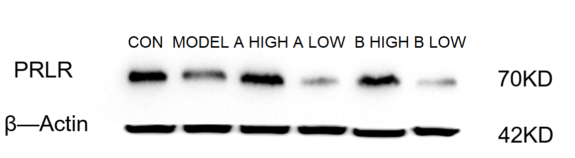
Western blot analysis of ER alpha using anti-ER alpha antibody (M00057-2).
Electrophoresis was performed on a 10% SDS-PAGE gel at 80V (Stacking gel) / 120V (Resolving gel) for 2 hours. The sample well of each lane was loaded with 30 ug of sample under reducing conditions.
Lane 1: control group-mouse brain tissue lysates,
Lane 2: model group-model mouse brain tissue lysates,
Lane 3: high dose A medicine treated group-model mouse brain tissue lysates,
Lane 4: low dose A medicine treated group-model mouse brain tissue lysates,
Lane 5: high dose B medicine treated group-model mouse brain tissue lysates,
Lane 6: low dose B medicine treated group-model mouse brain tissue lysates.
After electrophoresis, proteins were transferred to a nitrocellulose membrane at 150 mA for 50-90 minutes. Blocked the membrane with 5% non-fat milk/TBS for 1.5 hour at RT. The membrane was incubated with rabbit anti-ER alpha antigen affinity purified monoclonal antibody (M00057-2) at 1:1000 overnight at 4°C, then washed with TBS-0.1%Tween 3 times with 5 minutes each and probed with a goat anti-rabbit IgG-HRP secondary antibody at a dilution of 1:2000 for 1 hour at RT. The signal is developed using an ECL Plus Western Blotting Substrate (Catalog # AR1196-200) with ChemiDoc MP system. A specific band was detected for ER alpha at approximately 70 kDa. The expected band size for ER alpha is at 66 kDa.

Click image to see more details
Immunohistochemical analysis of paraffin-embedded human cervix carcinoma, using ER alpha Antibody.

Click image to see more details
Immunofluorescent analysis of MCF7 cells, using ER alpha Antibody .
Specific Publications For Anti-ER alpha ESR1 Rabbit Monoclonal Antibody (M00057-2)
Loading publications
Recommended Resources
Here are featured tools and databases that you might find useful.
- Boster's Pathways Library
- Protein Databases
- Bioscience Research Protocol Resources
- Data Processing & Analysis Software
- Photo Editing Software
- Scientific Literature Resources
- Research Paper Management Tools
- Molecular Biology Software
- Primer Design Tools
- Bioinformatics Tools
- Phylogenetic Tree Analysis
Customer Reviews
Have you used Anti-ER alpha ESR1 Rabbit Monoclonal Antibody?
Share your experimental results or join a short interview to earn up to $1,000 in product credits or other rewards.
1 Reviews For Anti-ER alpha ESR1 Rabbit Monoclonal Antibody
Western blot using Anti-ER/ESR1 Antibody (M00057-2) in mouse brain tissues showed clear, specific ESR1 bands across normal, disease model, and AB drug-treated groups, with β‑actin as a consistent loading control.
Excellent
| SKU | M00057-2 |
|---|---|
| Application | Western Blot |
| Sample | mouse brain tissue |
| Sample Processing Description | Mouse brain tissues were lysed in RIPA buffer containing a protease inhibitor cocktail at 4°C for 2 hours, centrifuged to collect the supernatant, and protein concentration was determined. After adjusting the concentration, samples were mixed with 5× protein loading buffer, denatured by heating at 95–100°C for 10 minutes, and 15 μL of protein was loaded per lane for SDS-PAGE. |
| Other Reagents | 5% non-fat milk |
| Primary Antibody | GAPDH Antibody Picoband® |
| Primary Incubation | 1:1000, overnight at 4 ℃ |
| Secondary Antibody | goat anti rabbit secondary antibodies |
| Secondary Incubation | 1:5000, 1 h in RT |
| Detection | Substrate: ECL substrate, Image system:ChemiDoc MP |
| Results Summary | This antibody is highly sensitive, produces clear WB bands, is reusable, offers excellent cost-effectiveness, and demonstrates a clear advantage over similar international products, making it highly recommended for use. |
Yetao Ju, Liaoning University of Traditional Chinese Medicine
Verified customer
Submitted 2026-03-05
Customer Q&As
Have a question?
Find answers in Q&As, reviews.
Can't find your answer?
Submit your question
2 Customer Q&As for Anti-ER alpha ESR1 Rabbit Monoclonal Antibody
Question
What is a secondary antibody for the M00057-2 antibody that is HRP or biotin conjugate?
Verified customer
Asked: 2019-11-04
Answer
For the Anti-ER alpha Rabbit Monoclonal Antibody M00057-2, we suggest Goat Anti-Rabbit IgG (H+L) Secondary Antibody, HRP Conjugate BA1054 and Goat Anti-Rabbit IgG (H+L) Secondary Antibody, Biotin Conjugate BA1003 as secondary antibodies that is HRP (Horseradish Peroxidase) or biotin conjugate.
Boster Scientific Support
Answered: 2019-11-04
Question
For M00057-2 (Abways CY5041) , what is the immunogen sequence?
Verified Customer
Verified customer
Asked: 2017-11-13
Answer
The immunogen sequence for M00057-2 (Abways CY5041) is CPPLNSVSPSPLMLLHPPPQ.
Boster Scientific Support
Answered: 2017-11-13
