Click image to see more details
-

-
-
-
-
+13
Product Info Summary
| SKU: | M00220-1 |
|---|---|
| Size: | 100 μl |
| Reactive Species: | Human, Mouse, Rat |
| Host: | Rabbit |
| Application: | Flow Cytometry, IP, IF, IHC, ICC, WB |
Customers Who Bought This Also Bought

Product info
Product Name
Anti-CD86/B7 2 Rabbit Monoclonal Antibody
SKU/Catalog Number
M00220-1
BM4121 is an alternative SKU for this antibody, used in previous lots.
Size
100 μl
Form
Liquid
Description
Boster Bio Anti-CD86/B7 2 Rabbit Monoclonal Antibody catalog # M00220-1. Tested in WB, IHC, ICC/IF, IP, Flow Cytometry applications. This antibody reacts with Human, Mouse, Rat.
Storage & Handling
Store at -20°C for one year. For short term storage and frequent use, store at 4°C for up to one month. Avoid repeated freeze-thaw cycles.
Cite This Product
Anti-CD86/B7 2 Rabbit Monoclonal Antibody (Boster Biological Technology, Pleasanton CA, USA, Catalog # M00220-1)
Host
Rabbit
Contents
Rabbit IgG in stabilizing components, phosphate buffered saline, pH 7.4, 150mM NaCl, 0.02% sodium azide and 50% glycerol.
*This antibody is supplied in a stabilized formulation.
Compatibility with conjugation reactions depends on the chemistry of the conjugation method used.
For conjugation methods that are not compatible with the stabilizing components present in this formulation, a carrier-free antibody format is required.
Clonality
Monoclonal
Clone Number
BFF-3
Isotype
Rabbit IgG
Immunogen
A synthesized peptide derived from human CD86
Reactive Species
M00220-1 is reactive to CD86 in Human, Mouse, Rat
Observed Molecular Weight
38 kDa
Calculated molecular weight
37.7 kDa
Antibody Validation
Boster validates all antibodies on WB, IHC, ICC, Immunofluorescence, and ELISA with known positive control and negative samples to ensure specificity and high affinity, including thorough antibody incubations.
Application & Images
Applications
M00220-1 is guaranteed for Flow Cytometry, IP, IF, IHC, ICC, WB Boster Guarantee
Recommend Dilution
WB 1:500-2000
IHC 1:50-200
ICC/IF 1:50-200
IP 1:20
FC 1:20
Tested application
Suggested blocking solution with 5% non-fat milk or BSA; (*)Recommended protein loading: 20-40 µg per lane
Use TE buffer pH 9.0 for antigen retrieval; (*) citrate buffer pH 6.0 is an alternative.
Validation Images & Assay Conditions
Click image to see more details
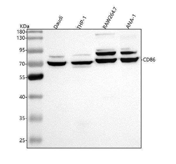
Western blot analysis of CD86 using anti-CD86 antibody (M00220-1).
Electrophoresis was performed on a 5-20% SDS-PAGE gel at 70V (Stacking gel) / 90V (Resolving gel) for 2-3 hours. The sample well of each lane was loaded with 30 ug of sample under reducing conditions.
Lane 1: human Daudi whole cell lysates,
Lane 2: human THP-1 whole cell lysates,
Lane 3: mouse RAW264.7 whole cell lysates,
Lane 4: mouse ANA-1 whole cell lysates.
After electrophoresis, proteins were transferred to a nitrocellulose membrane at 150 mA for 50-90 minutes. Blocked the membrane with 5% non-fat milk/TBS for 1.5 hour at RT. The membrane was incubated with rabbit anti-CD86 antigen affinity purified monoclonal antibody (Catalog # M00220-1) at 1:1000 overnight at 4°C, then washed with TBS-0.1%Tween 3 times with 5 minutes each and probed with a goat anti-rabbit IgG-HRP secondary antibody at a dilution of 1:5000 for 1.5 hour at RT. The signal is developed using an Enhanced Chemiluminescent detection (ECL) kit (Catalog # EK1002) with Tanon 5200 system. A specific band was detected for CD86 at approximately 70 kDa. The expected band size for CD86 is at 38 kDa.

Click image to see more details
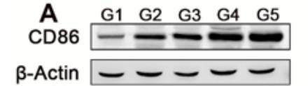
Western blot analysis of CD86 using anti-CD86 antibody (A04887-1).
Electrophoresis was performed on a 5-20% SDS-PAGE gel at 80V (Stacking gel) / 120V (Resolving gel) for 2 hours. The sample well of each lane was loaded with 30 ug of sample under reducing conditions.
Lane 1-5: mouse lung cancer tissue.
After electrophoresis, proteins were transferred to a nitrocellulose membrane at 150 mA for 50-90 minutes. Blocked the membrane with 5% non-fat milk/TBS for 1.5 hour at RT. The membrane was incubated with rabbit anti-CD86 antigen affinity purified polyclonal antibody (A04887-1) at 1:2000 overnight at 4°C, then washed with TBS-0.1%Tween 3 times with 5 minutes each and probed with a (HRP)-conjugated Anti-Rabbit IgG Secondary Antibody for 1 hour at RT. The signal is developed using an ECL Plus Western Blotting Substrate (Catalog # AR1196-200) with Tanon 5200 system. The expected band size for CD86 is at 38 kDa.

Click image to see more details
Western blot analysis of CD86 using anti-CD86 antibody (M00220-1).
Electrophoresis was performed on a 10% SDS-PAGE gel at 80V (Stacking gel) / 120V (Resolving gel) for 2 hours. The sample well of each lane was loaded with 30 ug of sample under reducing conditions.
Lane 1: human Daudi whole cell lysates,
Lane 2: human Raji whole cell lysates,
Lane 3: human HEL whole cell lysates,
Lane 4: rat PC-12 whole cell lysates,
Lane 5: rat C6 whole cell lysates,
Lane 6: mouse RAW264.7 whole cell lysates,
Lane 7: mouse A20 whole cell lysates.
After electrophoresis, proteins were transferred to a nitrocellulose membrane at 150 mA for 50-90 minutes. Blocked the membrane with 5% non-fat milk/TBS for 1.5 hour at RT. The membrane was incubated with rabbit anti-CD86 antigen affinity purified monoclonal antibody (M00220-1) at 1:500 overnight at 4°C, then washed with TBS-0.1%Tween 3 times with 5 minutes each and probed with a goat anti-rabbit IgG-HRP secondary antibody at a dilution of 1:5000 for 1.5 hour at RT. The signal is developed using an ECL Plus Western Blotting Substrate (Catalog # AR1196-200) with Tanon 5200 system. A specific band was detected for CD86 at approximately 72 kDa. The expected band size for CD86 is at 38 kDa.

Click image to see more details
Immunohistochemical analysis of paraffin-embedded human tonsil, using CD86 Antibody .

Click image to see more details
Immunofluorescent analysis using the Antibody at 1:50 dilution.

Click image to see more details
Immunofluorescent analysis using the Antibody at 1:150 dilution.

Click image to see more details
Immunofluorescent analysis using the Antibody at 1:50 dilution.

Click image to see more details
Immunofluorescent analysis of K562 cells, using CD86 Antibody .

Click image to see more details
WJMSCs inhibited pro-inflammatory macrophages and promoted anti-inflammatory macrophages in RV. (A) Representative images showed the CD86 + pro-inflammatory macrophages (green) and CD206 + anti-inflammatory macrophages (green) in each group. The nuclei were stained with DAPI (blue), and macrophages were stained with F4/80 (red). Scale bar, 20μm. (B–C) The percentage of CD86 + or CD206 + cells were calculated by Image-Pro Plus. (D–H) The protein level of CD86, CD206, TNF-α and IL-10 were measured by western blot, and quantification of the relative expression in the bands in different groups was calculated by Image-Pro Plus. All data were analyzed by two-way analysis of variance (ANOVA). Values are expressed as mean ± SD (n = 3-5). * p < 0.05, ** p < 0.01, *** p < 0.001. ns, not significant; HH, hypobaric hypoxia; NN, normobaric normoxia; IL-10, interleukin 10; TNF-α, tumor necrosis factor α. NS, neutral saline; WJMSCs, Wharton’s jelly-derived mesenchymal stem cells.
Index in PubMed under a CC BY license. PMID: 40547034

Click image to see more details
CPEB3 attenuates tumorigenesis and TAM polarization in vivo (a) Schematic of the procedure for separating tumor cells and TAMs. (b) HCT116 cells were stably infected with Ctrl and CPEB3 lentivirus, and LoVo cells were stably infected with shCtrl and shCPEB3 sequences. Tumorigenesis assay of Balb/c nude mice subcutaneously injected with HCT116-Ctrl/CPEB3 cells and LoVo-shCtrl/shCPEB3 cells ( n = 20). Representative photos of tumors from mice in various groups. ( c) IHC staining of Ki67 positive cells was counted per high-power field (PHF), while E-cadherin and vimentin expression scores were counted in tumor tissues in a xenograft model; error bars, SEM. (d) The mice with intra-spleen injection of LoVo-shCtrl/shCPEB3 cells were treated with tocilizumab (5 mg/kg) weekly via intraperitoneally injection. The number of liver metastatic sites (indicated by arrows) was counted under the microscope; error bars, SEM. (e) Macrophages were separated from murine tumor tissues using Percoll-layered liquid. Surface expression of CD86 and CD163 was detected in macrophages using flow cytometry. The percentage of CD86 + or CD163 + cells in macrophages was reported using error bars and SEM. (f) Expression of JAK1, pJAK1, STAT3, and pSTAT3 in the tumor tissues of the two groups were analyzed by western blot analysis. (g) Schematic overview of the mechanisms by which CPEB3 modulate TAM polarization and inhibit colorectal cancer EMT. ** P < 0.01; *** P < 0.001; **** P < 0.0001
Index in PubMed under a CC BY license. PMID: 32653013

Click image to see more details
Decreased CPEB3 in human CRC correlates with low CD86 + TAM content and high CD163 + TAM content (a) The expression of CPEB3 and CD86 in 82 pairs of CRC tissues and adjacent non-tumor tissues was detected using qRT-PCR. Correlation between CPEB3 and CD86 or CD163 expression levels in 82 colorectal cancer tissues; error bars, SEM. (b) The protein expression of CPEB3, CD68, CD86, and CD163 in a human colorectal cancer tissue array was detected by IHC staining. Representative photos are shown (400× magnification). The number of CD68 + , CD86 + and CD163 + cells per high-power field in tissues from colorectal cancer patients with different levels of CPEB3 expression; error bars, SEM. (c) Schema for an in vitro model of stably transfected CRC cells co-cultured with TAMs. (d) Flow cytometry was used to explore the surface expression of CD86 and CD163 in SW480-Ctrl/CPEB3 and HCT116-Ctrl/CPEB3 cells; error bars, SEM. (e) Flow cytometry was used to explore the surface expression of CD86 and CD163 in LoVo-shCtrl/shCPEB3 and RKO-shCtrl/shCPEB3 cells; error bars, SEM. (f) We measured the expression of the respective inflammatory cytokines in cell culture supernatants of TAMs co-cultured HCT116-Ctrl/CPEB3 cells using ProcartaPlex combinable panels; error bars, SEM. (g) We measured the expression of the respective inflammatory cytokines in cell culture supernatants of TAM-co-cultured LoVo-shCtrl/shCPEB3 cells using ProcartaPlex combinable panels; error bars, SEM; ns, not significant; * P < 0.05; ** P < 0.01; *** P < 0.001; **** P < 0.0001
Index in PubMed under a CC BY license. PMID: 32653013

Click image to see more details
CPEB3 modulates CCL2 secretion in CRC cell supernatants to regulate TAM polarization (a) We measured the expression of the respective inflammatory cytokines in cell culture supernatants of HCT116-Ctrl/CPEB3 cells by ProcartaPlex combinable panels; error bars, SEM. (b) We measured the expression of the respective inflammatory cytokines in the supernatants of LoVo-shCtrl/shCPEB3 cells by ProcartaPlex combinable panels; error bars, SEM. (c) THP-1 macrophages were co-cultured with LoVo-shCtrl/shCPEB3 with or without CCL2-neutralizing antibody (1 μg/mL) for 24 h. Flow cytometry was used to explore the surface expression of CD86 and CD163 in the differentiated macrophages; error bars, SEM. (d) THP-1 macrophages were co-cultured with RKO-shCtrl/shCPEB3 cells with or without a CCL2-neutralizing antibody (1 μg/mL) for 24 h. Flow cytometry was used to explore the surface expression of CD86 and CD163 in the differentiated macrophages. Error bars, SEM. * P < 0.05; ** P < 0.01; *** P < 0.001; **** P < 0.0001
Index in PubMed under a CC BY license. PMID: 32653013

Click image to see more details
Sanguinarine increased cellular levels of H3K4me2 and H3K9me2 and up-regulated the expression of CD86 in NSCLC cell lines. (A) and (B) Expression of H3K4me2 and H3K9me2 in H1975 and H1299 cells treated with sanguinarine for 48 hr, respectively, with H3 as loading control; (C) and (D) The expression of CD86 was detected, GADPH was used as a loading control. Data are the mean ± SEM of three independent experiments. * P <0.05, ** P <0.01, *** P <0.005 NSCLC: Non-small cell lung cancer
Index in PubMed under a CC BY license. PMID: 35949313

Click image to see more details
The expression levels of p-mTOR/mTOR ratio, HIF-1α, and IL-1β in BV2 cells tested by western blot, the levels of GLU, LD, ATP, LDH and PDH quantified by biochemical test kit and the levels of polarization biomarkers of CD86 and CD206 detected by flow cytometry, the expression levels of IL-6 and TNF-α in BV2 cells tested by ELISA. (A) Blotting of p-mTOR, mTOR and HIF-1α protein in BV2 cells in each group; (B, C) Expression levels of p-mTOR/mTOR ratio and HIF-1α in BV2 cells in each group. (D) GLU content analysis chart; (E) LD content analysis chart; (F) ATP content analysis chart; (G) . LDH activity analysis graph; (H) PDH activity analysis graph. (I) Flow cytometry analysis charts; (J) Flow cytograms of BV2 cells in each group; (K) Blotting of IL-1β protein in BV2 cells in each group; (L) Expression level of IL-1β in BV2 cells in each group. (M, N) Expression level of IL-6 and TNF-α in BV2 cells in each group. Model group compared with control group * p < 0.05, ** p < 0.01, *** p < 0.001, HSD-containing serum-treated group compared with the model group $ p < 0.05, $$ p < 0.01, $$$ p < 0.001, Reverse validation group compared with the HSD-containing serum-treated group & p < 0.05, && p < 0.01, &&& p < 0.001.
Index in PubMed under a CC BY license. PMID: 39130642

Click image to see more details
Expression levels of CD86, Arg1 and IL-1β protein in hippocampus and cortex of SAMP8 mice detected by Western-blot, the expression levels of IL-6 and TNF-α in hippocampus and cortex of SAMP8 mice by ELISA. (A,E) Blotting of CD86, Arg1 and IL-1β protein in hippocampus and cortex of mice in each group; (B–D) Expression of CD86, Arg1 and IL-1β protein in hippocampus of mice in each group; (F–H) Expression of CD86, Arg1 and IL-1β protein in cortex of mice in each group; (I–L) Expression of IL-6 and TNF-α protein in hippocampus and cortex of mice in each group. Model group compared with the control group * p < 0.05, ** p < 0.01, *** p < 0.001, Donepezil group compared with the model group # p < 0.05, ## p < 0.01, ### p < 0.001, HSD group compared with the model group $ p < 0.05, $$ p < 0.01, $$$ p < 0.001.
Index in PubMed under a CC BY license. PMID: 39130642

Click image to see more details
Mice reconstituted with FGF2 KO macrophages and subjected to CLP demonstrate increased M1 polarization in lung tissue. a - f The presence and levels of CD206, CD86, and F4/80 markers on macrophages within lung tissue were identified and quantitatively assessed using immunofluorescence staining. Bar is 20 μm. * p < 0.05, vs. WT; Δ p < 0.05 vs. WT + LPS; # p < 0.05 vs. FGF2 KO
Index in PubMed under a CC BY license. PMID: 39436561

Click image to see more details
Effect of FGF2 deficiency on BMDM apoptosis and polarization. a – c FGF2 deletion increased BMDM apoptosis. a Apoptosis in BMDM deprived of FBS for 24 h was assessed by flow cytometry ( n = 4). b - c Percentage of PI + Annexin V + and PI- Annexin V + BMDM after starvation. d - k FGF2 deletion in BMDM promoted M1 polarization. d - g Flow cytometric analysis of macrophage markers in BMDM treated with LPS or IL4, including CD86, iNOS, CD206, and Arg1 ( n = 3). h - k The levels of CD86, iNOS, CD206 and Arg1 in BMDM after treatment with LPS or IL4. N represents no treatment; * p < 0.05, vs. WT; Ψ p < 0.05, vs. N + WT; Ω p < 0.05, vs. N + FGF2 KO
Index in PubMed under a CC BY license. PMID: 39436561
Specific Publications For Anti-CD86/B7 2 Rabbit Monoclonal Antibody (M00220-1)
Loading publications
Recommended Resources
Here are featured tools and databases that you might find useful.
- Boster's Pathways Library
- Protein Databases
- Bioscience Research Protocol Resources
- Data Processing & Analysis Software
- Photo Editing Software
- Scientific Literature Resources
- Research Paper Management Tools
- Molecular Biology Software
- Primer Design Tools
- Bioinformatics Tools
- Phylogenetic Tree Analysis
Customer Reviews
Have you used Anti-CD86/B7 2 Rabbit Monoclonal Antibody?
Share your experimental results or join a short interview to earn up to $1,000 in product credits or other rewards.
2 Reviews For Anti-CD86/B7 2 Rabbit Monoclonal Antibody
This antibody is highly specific and efficient, with a clear target band and no nonspecific bands.
Excellent

| SKU | M00220-1 |
|---|---|
| Application | Western Blot |
| Sample | mouse lung cancer tissue |
| Sample Processing Description | Tissue samples were directly lysed in RIPA buffer, mixed with loading buffer at the appropriate ratio, and denatured by heating at 98°C. Twenty microliters of each protein sample were loaded per lane onto an SDS-PAGE gel. |
| Other Reagents | 5% Non-fat milk |
| Primary Antibody | CD86/B7 2 Rabbit Monoclonal Antibody |
| Primary Incubation | 1:2000, overnight at 4 ℃ |
| Secondary Antibody | HRP-conjugated Anti-Rabbit IgG Secondary Antibody |
| Secondary Incubation | 1 hour in room temperature |
| Detection | Substrate: ECL reagent, Imaging system:Tanon |
| Results Summary | This antibody is highly specific and efficient, with a clear target band and no nonspecific bands. |
Jiahui Yan, Ocean University of China
Verified customer
Submitted 2025-11-11
The antibody is highly efficient and specific, showing a clear target band with no nonspecific bands.
Excellent
| SKU | M00220-1 |
|---|---|
| Application | Western Blot |
| Sample | Mouse lung cancer tissue |
| Sample Processing Description | Tissue samples were directly lysed in RIPA buffer, mixed with loading buffer at the appropriate ratio, and denatured by heating at 98 °C. Load 20 µL of protein sample per lane onto SDS-PAGE. |
| Primary Incubation | The membrane was incubated with the CD86 primary antibodies (1:2000) overnight at 4 °C. |
| Secondary Antibody | HRP-conjugated Goat Anti-Rabbit IgG Secondary Antibody |
| Secondary Incubation | Incubate at room temperature for 1 hour |
| Other Reagents used | 5% non-fat milk |
| Detection | Signal was developed using ECL substrate on an Tanon system. |
| Results Summary | The antibody is highly efficient and specific, showing a clear target band with no nonspecific bands. I will definitely continue using BosterBio products and will recommend them to my classmates and colleagues. |
Jiahui Yan, College of Chemistry and Chemical Engineering, Ocean University of China
Verified customer
Submitted 2025-09-25
Customer Q&As
Have a question?
Find answers in Q&As, reviews.
Can't find your answer?
Submit your question
17 Customer Q&As for Anti-CD86/B7 2 Rabbit Monoclonal Antibody
Question
We have seen staining in rat foreskin. Any tips? Is anti-CD86/B7 2 Rabbit Monoclonal antibody supposed to stain foreskin positively?
Verified Customer
Verified customer
Asked: 2020-05-07
Answer
From what I have seen in literature foreskin does express CD86. From what I have seen in Uniprot.org, CD86 is expressed in leukocyte, brain, foreskin, among other tissues. Regarding which tissues have CD86 expression, here are a few articles citing expression in various tissues:
Brain, Pubmed ID: 14702039, 15489334
Foreskin, Pubmed ID: 7541777
Boster Scientific Support
Answered: 2020-05-07
Question
Is a blocking peptide available for product anti-CD86/B7 2 Rabbit Monoclonal antibody (M00220-1)?
Verified Customer
Verified customer
Asked: 2020-03-05
Answer
We do provide the blocking peptide for product anti-CD86/B7 2 Rabbit Monoclonal antibody (M00220-1). If you would like to place an order for it please contact support@bosterbio.com and make a special request.
Boster Scientific Support
Answered: 2020-03-05
Question
We bought anti-CD86/B7 2 Rabbit Monoclonal antibody for IHC on leukocyte in a previous experiment. I am using human, and We intend to use the antibody for ICC next. Our lab want to know about examining leukocyte as well as brain in our next experiment. Could give a recommendation on which antibody would work the best for ICC?
Verified Customer
Verified customer
Asked: 2020-02-24
Answer
I looked at the website and datasheets of our anti-CD86/B7 2 Rabbit Monoclonal antibody and it seems that M00220-1 has been validated on human in both IHC and ICC. Thus M00220-1 should work for your application. Our Boster satisfaction guarantee will cover this product for ICC in human even if the specific tissue type has not been validated. We do have a comprehensive range of products for ICC detection and you can check out our website bosterbio.com to find out more information about them.
Boster Scientific Support
Answered: 2020-02-24
Question
I was wanting to use your anti-CD86/B7 2 Rabbit Monoclonal antibody for Flow Cytometry for human leukocyte on frozen tissues, but I want to know if it has been validated for this particular application. Has this antibody been validated and is this antibody a good choice for human leukocyte identification?
Verified Customer
Verified customer
Asked: 2019-12-12
Answer
You can see on the product datasheet, M00220-1 anti-CD86/B7 2 Rabbit Monoclonal antibody has been validated for Flow Cytometry, IP, IF, IHC, ICC, WB on human, rat tissues. We have an innovator award program that if you test this antibody and show it works in human leukocyte in IHC-frozen, you can get your next antibody for free.
Boster Scientific Support
Answered: 2019-12-12
Question
I have attached the WB image, lot number and protocol we used for leukocyte using anti-CD86/B7 2 Rabbit Monoclonal antibody M00220-1. Please let me know if you require anything else.
Verified Customer
Verified customer
Asked: 2019-09-23
Answer
Thank you very much for the data. Our lab team are working to resolve this as quickly as possible, and we appreciate your patience and understanding! You have provided everything we needed. Please let me know if there is anything you need in the meantime.
Boster Scientific Support
Answered: 2019-09-23
Question
Will anti-CD86/B7 2 Rabbit Monoclonal antibody M00220-1 work for Flow Cytometry with leukocyte?
E. Roberts
Verified customer
Asked: 2019-04-26
Answer
According to the expression profile of leukocyte, CD86 is highly expressed in leukocyte. So, it is likely that anti-CD86/B7 2 Rabbit Monoclonal antibody M00220-1 will work for Flow Cytometry with leukocyte.
Boster Scientific Support
Answered: 2019-04-26
Question
We are currently using anti-CD86/B7 2 Rabbit Monoclonal antibody M00220-1 for human tissue, and we are well pleased with the IP results. The species of reactivity given in the datasheet says human, rat. Is it possible that the antibody can work on canine tissues as well?
Verified Customer
Verified customer
Asked: 2019-01-28
Answer
The anti-CD86/B7 2 Rabbit Monoclonal antibody (M00220-1) has not been tested for cross reactivity specifically with canine tissues, though there is a good chance of cross reactivity. We have an innovator award program that if you test this antibody and show it works in canine you can get your next antibody for free. Please contact me if I can help you with anything.
Boster Scientific Support
Answered: 2019-01-28
Question
Will M00220-1 anti-CD86/B7 2 Rabbit Monoclonal antibody work on parafin embedded sections? If so, which fixation method do you recommend we use (PFA, paraformaldehyde, other)?
T. Zhao
Verified customer
Asked: 2019-01-08
Answer
You can see on the product datasheet, M00220-1 anti-CD86/B7 2 Rabbit Monoclonal antibody as been validated on Flow Cytometry. It is best to use PFA for fixation because it has better tissue penetration ability. PFA needs to be prepared fresh before use. Long term stored PFA turns into formalin, as the PFA molecules congregate and become formalin.
Boster Scientific Support
Answered: 2019-01-08
Question
I see that the anti-CD86/B7 2 Rabbit Monoclonal antibody M00220-1 works with Flow Cytometry, what is the protocol used to produce the result images on the product page?
T. Banerjee
Verified customer
Asked: 2018-03-12
Answer
You can find protocols for Flow Cytometry on the "support/technical resources" section of our navigation menu. If you have any further questions, please send an email to support@bosterbio.com
Boster Scientific Support
Answered: 2018-03-12
Question
I am interested in using your anti-CD86/B7 2 Rabbit Monoclonal antibody for cd28 co-stimulation studies. Has this antibody been tested with western blotting on raji cell lysate? We would like to see some validation images before ordering.
C. Wu
Verified customer
Asked: 2018-01-12
Answer
Thank you for your inquiry. This M00220-1 anti-CD86/B7 2 Rabbit Monoclonal antibody is tested on raji cell lysate. It is guaranteed to work for Flow Cytometry, IP, IF, IHC, ICC, WB in human, rat. Our Boster guarantee will cover your intended experiment even if the sample type has not been be directly tested.
Boster Scientific Support
Answered: 2018-01-12
Question
Thank you for helping with my inquiry over the phone. Here are the WB image, lot number and protocol we used for leukocyte using anti-CD86/B7 2 Rabbit Monoclonal antibody M00220-1. Let me know if you need anything else.
Verified Customer
Verified customer
Asked: 2017-06-19
Answer
Thanks for the data. You have provided everything we needed. Our lab team are working to resolve your inquiry as quickly as possible, and we appreciate your patience and understanding! Please let me know if there is anything you need in the meantime.
Boster Scientific Support
Answered: 2017-06-19
Question
Would anti-CD86/B7 2 Rabbit Monoclonal antibody M00220-1 work on horse ICC with brain?
Verified Customer
Verified customer
Asked: 2017-05-11
Answer
Our lab technicians have not tested anti-CD86/B7 2 Rabbit Monoclonal antibody M00220-1 on horse. You can run a BLAST between horse and the immunogen sequence of anti-CD86/B7 2 Rabbit Monoclonal antibody M00220-1 to see if they may cross-react. If the sequence homology is close, then you can perform a pilot test. Keep in mind that since we have not validated horse samples, this use of the antibody is not covered by our guarantee. However we have an innovator award program that if you test this antibody and show it works in horse brain in ICC, you can get your next antibody for free.
Boster Scientific Support
Answered: 2017-05-11
Question
My question regarding product M00220-1, anti-CD86/B7 2 Rabbit Monoclonal antibody. I was wondering if it would be possible to conjugate this antibody with biotin. I would need it to be without BSA or sodium azide. I am planning on using a buffer exchange of sodium azide with PBS only. Would there be problems for me to conjugate the antibody and store it in -20 degrees in small aliquots?
L. Zhang
Verified customer
Asked: 2017-03-27
Answer
We suggest not storing this antibody with PBS buffer only in -20 degrees. If you want to store it in -20 degrees it is best to add some cryoprotectant like glycerol. If you want carrier free M00220-1 anti-CD86/B7 2 Rabbit Monoclonal antibody, we can provide it to you in a special formula with trehalose and/or glycerol. These molecules will not interfere with conjugation chemistry and provide a good level of protection for the antibody from degradation. Please be sure to specify this in your purchase order.
Boster Scientific Support
Answered: 2017-03-27
Question
We want to test anti-CD86/B7 2 Rabbit Monoclonal antibody M00220-1 on human leukocyte for research purposes, then I may be interested in using anti-CD86/B7 2 Rabbit Monoclonal antibody M00220-1 for diagnostic purposes as well. Is the antibody suitable for diagnostic purposes?
A. Jones
Verified customer
Asked: 2015-03-02
Answer
The products we sell, including anti-CD86/B7 2 Rabbit Monoclonal antibody M00220-1, are only intended for research use. They would not be suitable for use in diagnostic work. If you have the means to develop a product into diagnostic use, and are interested in collaborating with us and develop our product into an IVD product, please contact us for more discussions.
Boster Scientific Support
Answered: 2015-03-02
Question
Is this M00220-1 anti-CD86/B7 2 Rabbit Monoclonal antibody reactive to the isotypes of CD86?
N. Roberts
Verified customer
Asked: 2014-11-05
Answer
The immunogen of M00220-1 anti-CD86/B7 2 Rabbit Monoclonal antibody is A synthesized peptide derived from human CD86 . Could you tell me which isotype you are interested in so I can help see if the immunogen is part of this isotype?
Boster Scientific Support
Answered: 2014-11-05
Question
Do you have a BSA free version of anti-CD86/B7 2 Rabbit Monoclonal antibody M00220-1 available?
C. Zhang
Verified customer
Asked: 2014-04-29
Answer
I appreciate your recent telephone inquiry. I can confirm that some lots of this anti-CD86/B7 2 Rabbit Monoclonal antibody M00220-1 are BSA free. For now, these lots are available and we can make a BSA free formula for you free of charge. It will take 3 extra days to prepare. If you require this antibody BSA free again in future, please do not hesitate to contact me and I will be pleased to check which lots we have in stock that are BSA free.
Boster Scientific Support
Answered: 2014-04-29
Question
Our team were happy with the WB result of your anti-CD86/B7 2 Rabbit Monoclonal antibody. However we have observed positive staining in foreskin cell membrane using this antibody. Is that expected? Could you tell me where is CD86 supposed to be expressed?
C. Gonzalez
Verified customer
Asked: 2014-02-14
Answer
From what I have seen in literature, foreskin does express CD86. Generally CD86 expresses in cell membrane. Regarding which tissues have CD86 expression, here are a few articles citing expression in various tissues:
Brain, Pubmed ID: 14702039, 15489334
Foreskin, Pubmed ID: 7541777
Boster Scientific Support
Answered: 2014-02-14
