This website uses cookies to ensure you get the best experience on our website.
- Table of Contents
12 Citations 1 Q&As
1 Citations 2 Q&As


Facts about Estrogen receptor.

Ligand binding induces a conformational change allowing subsequent or combinatorial association with multiprotein coactivator complexes through LXXLL motifs of their various components. Mutual transrepression occurs between the estrogen receptor (ER) and NF- kappa-B in a cell-type particular manner.
| Human | |
|---|---|
| Gene Name: | ESR1 |
| Uniprot: | P03372 |
| Entrez: | 2099 |

| Belongs to: |
|---|
| nuclear hormone receptor family |

ER alpha; ER; Era; ER-alpha; ESR1; ESRESRA; Estradiol receptor; estrogen receptor 1; estrogen receptor alpha delta 3*4,56,7*/819-2 isoform; estrogen receptor alpha delta 4 +49 isoform; estrogen receptor alpha delta 4*5,6,7*/654 isoform; estrogen receptor alpha; estrogen receptor; NR3A1; NR3A1DKFZp686N23123; Nuclear receptor subfamily 3 group A member 1
Mass (kDA):
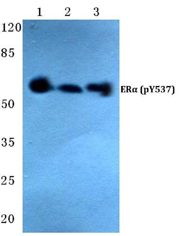
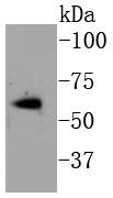
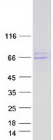
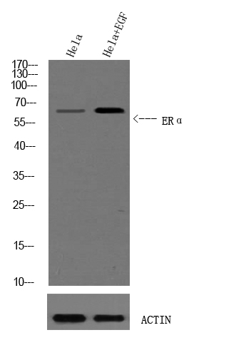
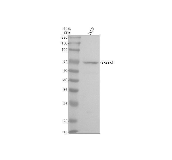
66.216 kDA

| Human | |
|---|---|
| Location: | 6q25.1-q25.2 |
| Sequence: | 6; NC_000006.12 (151654148..152129619) |
Widely expressed. Isoform 3 is not expressed in the pituitary gland.
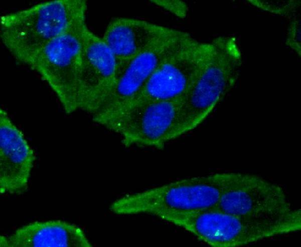
[Isoform 1]: Nucleus. Cytoplasm. Cell membrane; Peripheral membrane protein; Cytoplasmic side. A minor fraction is associated with the inner membrane.; [Isoform 3]: Nucleus. Cytoplasm. Cell membrane; Peripheral membrane protein; Cytoplasmic side. Cell membrane; Single-pass type I membrane protein. Associated with the inner membrane via palmitoylation (Probable). At least a subset exists as a transmembrane protein with a N-terminal extracellular domain.; Nucleus. Golgi apparatus. Cell membrane. Colocalizes with ZDHHC7 and ZDHHC21 in the Golgi apparatus where most probably palmitoylation occurs.


PMID: 3754034 by Green S., et al. Human oestrogen receptor cDNA: sequence, expression and homology to v-erb-A.
PMID: 3753802 by Greene G.L., et al. Sequence and expression of human estrogen receptor complementary DNA.
*More publications can be found for each product on its corresponding product page