Click image to see more details
-

-
-
-
-
+7
Product Info Summary
| SKU: | M00206-1 |
|---|---|
| Size: | 100 μl/vial |
| Reactive Species: | Human, Mouse, Rat |
| Host: | Rabbit |
| Application: | IHC, WB |
Customers Who Bought This Also Bought

Product info
Product Name
Anti-FGF1 Rabbit Monoclonal Antibody
SKU/Catalog Number
M00206-1
BM5544 is an alternative SKU for this antibody, used in previous lots.
Size
100 μl/vial
Form
Liquid
Description
Boster Bio Anti-FGF1 Rabbit Monoclonal Antibody catalog # M00206-1. Tested in WB, IHC applications. This antibody reacts with Human, Mouse, Rat.
Storage & Handling
Store at -20°C for one year. For short term storage and frequent use, store at 4°C for up to one month. Avoid repeated freeze-thaw cycles.
Cite This Product
Anti-FGF1 Rabbit Monoclonal Antibody (Boster Biological Technology, Pleasanton CA, USA, Catalog # M00206-1)
Host
Rabbit
Contents
Rabbit IgG in stabilizing components, phosphate buffered saline, pH 7.4, 150mM NaCl, 0.02% sodium azide and 50% glycerol.
*This antibody is supplied in a stabilized formulation.
Compatibility with conjugation reactions depends on the chemistry of the conjugation method used.
For conjugation methods that are not compatible with the stabilizing components present in this formulation, a carrier-free antibody format is required.
Clonality
Monoclonal
Clone Number
18F58
Isotype
IgG
Immunogen
A synthesized peptide derived from human FGF1
Reactive Species
M00206-1 is reactive to FGF1 in Human, Mouse, Rat
Observed Molecular Weight
17 kDa
Calculated molecular weight
17.5 kDa
Antibody Validation
Boster validates all antibodies on WB, IHC, ICC, Immunofluorescence, and ELISA with known positive control and negative samples to ensure specificity and high affinity, including thorough antibody incubations.
Application & Images
Applications
M00206-1 is guaranteed for IHC, WB Boster Guarantee
Recommend Dilution
WB 1:500-2000
IHC 1:50-200
Validation Images & Assay Conditions
Click image to see more details
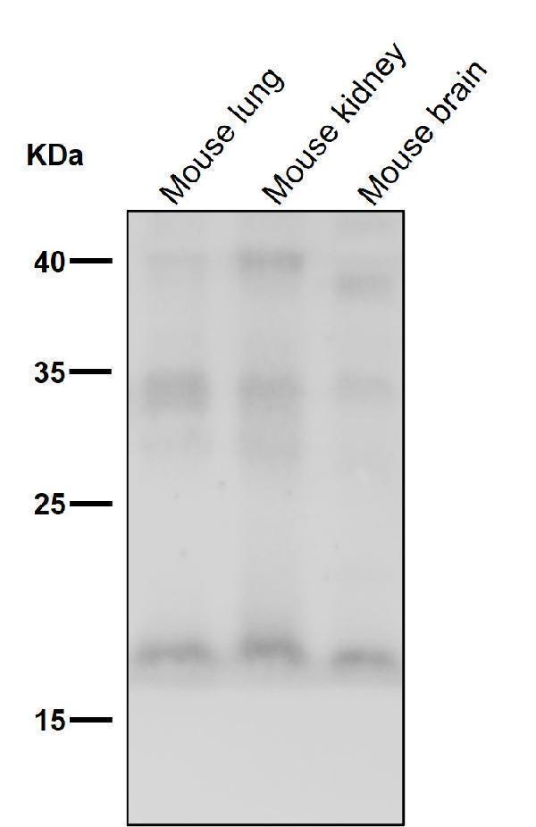
All lanes use the Antibody at 1:500 dilution for 1 hour at room temperature.

Click image to see more details
Illustration of experimental procedure. (A) Schematic illustration of animal experimental timeline showing the duration exogenous non-mitogenic fibroblast growth factor 1 (nmFGF1) administration after middle cerebral artery occlusion (MCAO) in adult mice and analysis. (B) Schematic illustration of cell experimental procedure after oxygen-glucose deprivation (OGD)/R and analysis. (C) Representative bands of endogenous FGF1 expression detected by western blot. (D) Densitometric analysis for the protein expression of FGF1. Data are expressed as means ± SEM (n = 5). **p < 0.01.
Index in PubMed under a CC BY license. PMID: 32194396

Click image to see more details
Non-mitogenic fibroblast growth factor 1 (nmFGF1) promoted angiogenesis in ischemic boundary following ischemia stroke. (A) A representative image of hematoxylin-eosin (HE) staining in a coronal brain section at 10 days after middle cerebral artery occlusion (MCAO). Box illustrates the peri-infarct areas from the corpus callosum where images in B were taken. (B) Representative immunofluorescence images of DAPI and EdU staining along with the staining for the vascular endothelium marker CD31 at 10 days after stroke. (C) Quantitative analysis of EdU- labeled cells in ischemic boundary following stroke. (D) Quantitative analysis of EdU + /CD31 + co-labeled cells in ischemic boundary following stroke. (E) Representative fluorescence image for vascular density analysis following staining with CD31 at 10 days after stroke. (F) Quantitative analysis of vascular density by CD31 labeling. Data are expressed as means ± SEM (n = 5). * p < 0.05 and **p < 0.01 as compared with the MCAO + vehicle group.
Index in PubMed under a CC BY license. PMID: 32194396

Click image to see more details
Non-mitogenic fibroblast growth factor 1 (nmFGF1) enhanced angiogenesis in ischemic boundary following ischemia stroke by S1P1 signaling pathway. (A) A representative image of HE staining in a coronal brain section at 10 days after middle cerebral artery occlusion (MCAO). Box illustrates the peri-infarct areas from the corpus callosum where images in B were taken. (B) Representative immunofluorescence images showing the co-localization of S1P1 and CD31 at 10 days after stroke. (C) Quantitative analysis of S1P1-positive cells in ischemic boundary following stroke. (D) Quantitative analysis of co-localization of S1P1 and CD31 at 10 days after stroke. Date are means ± SEM (n = 5). * p < 0.05, and **p < 0.01 as compared to the middle cerebral artery occlusion (MCAO) + vehicle group.
Index in PubMed under a CC BY license. PMID: 32194396

Click image to see more details
Non-mitogenic fibroblast growth factor 1 (nmFGF1) enhanced angiogenesis and wound healing in oxygen-glucose deprivation (OGD)-exposed HBMECs without affecting viability. (A) OGD and three concentrations of nmFGF1 (50, 100, and 200 nm) had no effect on cell viability. (B) Representative images to study the effect of OGD exposure and nmFGF1 treatment on tube formation ability of cells. (C) Quantification of the effects of nmFGF1 on the number of capillary-like tubes in OGD-treated cells. (D) Representative images to study the effect of nmFGF1 on wound healing in OGD-stimulated cells. (E) Quantification of the effects of nmFGF1 on wound closure in OGD-treated cells. Date are expressed as means ± SEM (n = 6). * p < 0.05 as compared to the control group, # p < 0.05 as compared to the OGD-treated group.
Index in PubMed under a CC BY license. PMID: 32194396

Click image to see more details
Non-mitogenic fibroblast growth factor 1 (nmFGF1) promoted angiogenesis through FGFR1 activation. (A) Representative image of western blot analysis for FGFR1 and p-FGFR1 expression. (B) Quantification of relative protein levels of p-FGFR1 and FGFR1. (C) Representative images for the analysis of the effects of cotreatment with the FGFR1 inhibitor PD173074 and nmFGF1 (100 nM) on tube formation ability of oxygen-glucose deprivation (OGD)-treated HBMECs. (D) Quantification of the effects of nmFGF1 and PD173074 cotreatment on the number of capillary-like tubes in OGD-treated cells. (E) Representative images showing the effects of PD173074 and nmFGF1 (100 nM) co-administration on wounding healing in OGD-stimulated cells. (F) Quantification of the effects of PD173074 and nmFGF1 (100 nM) co-administration on gap closure in OGD-treated cells. Date are means ± SEM (n = 6). * p < 0.05 compared to control group, # p < 0.05 as compared to the OGD-treated group, & p < 0.05 as compared to the nmFGF1 treated group.
Index in PubMed under a CC BY license. PMID: 32194396

Click image to see more details
Non-mitogenic fibroblast growth factor 1 (nmFGF1) promoted angiogenesis of oxygen-glucose deprivation (OGD)-exposed HBMECs via S1P signaling pathway. (A) Representative images showing the tube formation ability of OGD-exposed cells treated with the S1PR1 inhibitor VPC23019 (40 nM) and agonist FTY720 (50 nM) in the presence or absence of nmFGF1. (B) Quantification of the effects of the S1P1 inhibitor VPC23019 (40 nM) and agonist FTY720 (50 nM) in the presence or absence of nmFGF1 on tube formation ability. Date are means ± SEM (n = 6). * p < 0.05 as compared to the control group, # p < 0.05 as compared to the OGD-treated group.
Index in PubMed under a CC BY license. PMID: 32194396

Click image to see more details
Non-mitogenic fibroblast growth factor 1 (nmFGF1) promoted wound repair in oxygen-glucose deprivation (OGD)-exposed HBMECs via S1P signaling pathway. (A) Representative images showing the migration of OGD-exposed cells treated with the S1P1 inhibitor VPC23019 (40 nM) and agonist FTY720 (50 nM) in the presence or absence of nmFGF1. (B) Quantification of the effects of the S1P1 inhibitor VPC23019 (40 nM) and agonist FTY720 (50 nM) in the presence or absence of nmFGF1 on wound healing. Date are means ± SEM (n = 6). * p < 0.05 compared to control group, # p < 0.05 as compared to the OGD-treated group.
Index in PubMed under a CC BY license. PMID: 32194396

Click image to see more details
Non-mitogenic fibroblast growth factor 1 (nmFGF1) rescued oxygen-glucose deprivation (OGD)/R-induced downregulation of S1P1 expression by FGFR1 activation. (A) Representative western blots for S1P1 expression. (B) Quantification of the relative expression level of S1P1. Date are expressed as the means ± SEM (n = 4). * p < 0.05 as compared to the control group, # p < 0.05 as compared to the OGD-treated group, && p < 0.01 as compared to the nmFGF1-treated group.
Index in PubMed under a CC BY license. PMID: 32194396

Click image to see more details
All lanes use the Antibody at 1:500 dilution for 1 hour at room temperature.

Click image to see more details
Western blot analysis of FGF1 expression in Mouse kidney lysate.
Specific Publications For Anti-FGF1 Rabbit Monoclonal Antibody (M00206-1)
Loading publications
Recommended Resources
Here are featured tools and databases that you might find useful.
- Boster's Pathways Library
- Protein Databases
- Bioscience Research Protocol Resources
- Data Processing & Analysis Software
- Photo Editing Software
- Scientific Literature Resources
- Research Paper Management Tools
- Molecular Biology Software
- Primer Design Tools
- Bioinformatics Tools
- Phylogenetic Tree Analysis
Customer Reviews
Have you used Anti-FGF1 Rabbit Monoclonal Antibody?
Share your experimental results or join a short interview to earn up to $1,000 in product credits or other rewards.
0 Reviews For Anti-FGF1 Rabbit Monoclonal Antibody
Customer Q&As
Have a question?
Find answers in Q&As, reviews.
Can't find your answer?
Submit your question
