Click image to see more details
-

-
-
-
-
+4
Product Info Summary
| SKU: | PB9196 |
|---|---|
| Size: | 100 μg/vial |
| Reactive Species: | Human, Rat |
| Host: | Rabbit |
| Application: | Flow Cytometry, IF, IHC, ICC, WB |
Customers Who Bought This Also Bought

Product info
Product Name
Anti-FOXO3A Antibody Picoband®
SKU/Catalog Number
PB9196
Size
100 μg/vial
Form
Lyophilized
Description
Boster Bio Anti-FOXO3A Antibody Picoband® catalog # PB9196. Tested in Flow Cytometry, IF, IHC, ICC, WB applications. This antibody reacts with Human, Rat. The brand Picoband indicates this is a premium antibody that guarantees superior quality, high affinity, and strong signals with minimal background in Western blot applications. Only our best-performing antibodies are designated as Picoband, ensuring unmatched performance.
Storage & Handling
Store at -20˚C for one year from date of receipt. After reconstitution, at 4˚C for one month. It can also be aliquotted and stored frozen at -20˚C for six months. Avoid repeated freeze-thaw cycles.
Cite This Product
Anti-FOXO3A Antibody Picoband® (Boster Biological Technology, Pleasanton CA, USA, Catalog # PB9196)
Host
Rabbit
Contents
Each vial contains 4mg Trehalose, 0.9mg NaCl and 0.2mg Na2HPO4.
Clonality
Polyclonal
Isotype
Rabbit IgG
Immunogen
E.coli-derived human FOXO3A recombinant protein (Position: Q471-G673). Human FOXO3A shares 97% amino acid (aa) sequence identity with mouse FOXO3A.
Cross-reactivity
No cross-reactivity with other proteins
Reactive Species
PB9196 is reactive to FOXO3 in Human, Rat
Observed Molecular Weight
80-90 kDa
Calculated molecular weight
71.3 kDa
Background of FOXO3
Forkhead box O3, also known as FKHRL1 or FOXO3a, is a human protein encoded by the FOXO3 gene. FOXO3 belongs to the O subclass of the forkhead family of transcription factors which are characterized by a distinct fork head DNA-binding domain. It is mapped to 6q21. This protein likely functions as a trigger for apoptosis through upregulation of genes necessary for cell death, such as Bim and PUMA, or downregulation of anti-apoptotic proteins such as FLIP. In mammals FOXO3 regulates the resistance of cells to stress by inducing DNA repair and thereby may also affect organismal life span. In addition, it is thought that FOXO3 is also involved in protection from oxidative stress by upregulating antioxidants such as catalase and MnSOD.
Antibody Validation
Boster validates all antibodies on WB, IHC, ICC, Immunofluorescence, and ELISA with known positive control and negative samples to ensure specificity and high affinity, including thorough antibody incubations.
Application & Images
Applications
PB9196 is guaranteed for Flow Cytometry, IF, IHC, ICC, WB Boster Guarantee
Recommend Dilution
| Application | Dilution | Species |
|---|---|---|
| Western blot | 0.1-0.5μg/ml | Human, Rat |
| Immunohistochemistry (Paraffin-embedded Section) | 2-5μg/ml | Human |
| Immunocytochemistry/Immunofluorescence | 5 μg/ml | Human |
| Flow Cytometry(Fixed) | 1-3 μg/1x106 cells | Human |
Tested application
Suggested blocking solution with 5% non-fat milk or BSA; (*)Recommended protein loading: 20-40 µg per lane
Use TE buffer pH 9.0 for antigen retrieval; (*) citrate buffer pH 6.0 is an alternative.
Validation Images & Assay Conditions
Click image to see more details
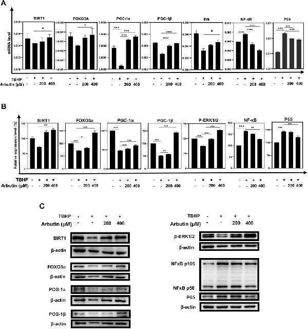
Arbutin exerted protective effects via the SIRT1/FOXO3A/PGC-1α/β and NF-κB/p65 signaling pathway. (A) qRT-PCR was used to measure transcript levels of the SIRT1/FOXO3a/PGC-1α/β pathway and NF-κB/p65 genes. TBHP decreased the expression of SIRT1, FOXO3a, and PGC-1α/β and increased the expression of NF-κB/p65, whereas mRNA levels in the groups that were pretreated with Arbutin showed reversed trend. (B) western blots were conducted to detect the proteins level of SIRT1, FOXO3a, PGC-1α/β, p-ERK, and NFKB1/P65. (C) imageJ was used to analyze the relative expression level of the proteins mentioned above (* p < 0.05, ** p < 0.01, *** p < 0.001, n = 3, bars represent SD).
Index in PubMed under a CC BY license. PMID: 36051689

Click image to see more details
Sirtinol diminished the capability of Arbutin to assist ARPE-19 cells to defend against oxidative stress. (A) (B) flow cytometric analysis showed that cells treated with only sirtinol, TBHP, cotreated with sirtinol, and TBHP displayed decreased cellular viability. However, sirtinol conduction diminished the protective capacity of Arbutin. (C) ARPE-19 cells were seeded in a 24-well plate and applied wounds at the confluence of 80%. The cells were pretreated with or without Arbutin and then subjected to TBHP (350 µM); meanwhile, cells in certain groups were incubated with sirtinol. Photos were taken at different time points post distinct treatments. (D) fluorescence images observed that ARPE-19 treated with Arbutin while subjected to sirtinol and then exposed to TBHP was unable to recuperate ΔΨm. (E) with sirtinol administration, the protein levels of the SIRT1/FOXO3a/PGC-1α/β pathway decreased in the presence of Arbutin (* p < 0.05, ** p < 0.01, *** p < 0.001, n = 3, bars represent SD).
Index in PubMed under a CC BY license. PMID: 36051689

Click image to see more details
Western blot analysis of FOXO3A using anti-FOXO3A antibody (PB9196).
Electrophoresis was performed on a 5-20% SDS-PAGE gel at 70V (Stacking gel) / 90V (Resolving gel) for 2-3 hours. The sample well of each lane was loaded with 30 ug of sample under reducing conditions.
Lane 1: human 293T whole cell lysates,
Lane 2: human MCF-7 whole cell lysates,
Lane 3: human Jurkat whole cell lysates,
Lane 4: human Hela whole cell lysates,
Lane 5: rat lung tissue lysates,
Lane 6: rat ovary tissue lysates.
After electrophoresis, proteins were transferred to a nitrocellulose membrane at 150 mA for 50-90 minutes. Blocked the membrane with 5% non-fat milk/TBS for 1.5 hour at RT. The membrane was incubated with rabbit anti-FOXO3A antigen affinity purified polyclonal antibody (Catalog # PB9196) at 0.5 μg/mL overnight at 4°C, then washed with TBS-0.1%Tween 3 times with 5 minutes each and probed with a goat anti-rabbit IgG-HRP secondary antibody at a dilution of 1:5000 for 1.5 hour at RT. The signal is developed using an Enhanced Chemiluminescent detection (ECL) kit (Catalog # EK1002) with Tanon 5200 system. A specific band was detected for FOXO3A at approximately 80-90 kDa. The expected band size for FOXO3A is at 71 kDa.

Click image to see more details
IHC analysis of FOXO3A using anti-FOXO3A antibody (PB9196).
FOXO3A was detected in a paraffin-embedded section of human pancreatic ductal adenocarcinoma tissue. Heat mediated antigen retrieval was performed in EDTA buffer (pH 8.0, epitope retrieval solution). The tissue section was blocked with 10% goat serum. The tissue section was then incubated with 2 μg/ml rabbit anti-FOXO3A Antibody (PB9196) overnight at 4°C. Peroxidase Conjugated Goat Anti-rabbit IgG was used as secondary antibody and incubated for 30 minutes at 37°C. The tissue section was developed using HRP Conjugated Rabbit IgG Super Vision Assay Kit (Catalog # SV0002) with DAB as the chromogen.

Click image to see more details
IHC analysis of FOXO3A using anti-FOXO3A antibody (PB9196).
FOXO3A was detected in a paraffin-embedded section of human colorectal adenocarcinoma tissue. Heat mediated antigen retrieval was performed in EDTA buffer (pH 8.0, epitope retrieval solution). The tissue section was blocked with 10% goat serum. The tissue section was then incubated with 2 μg/ml rabbit anti-FOXO3A Antibody (PB9196) overnight at 4°C. Peroxidase Conjugated Goat Anti-rabbit IgG was used as secondary antibody and incubated for 30 minutes at 37°C. The tissue section was developed using HRP Conjugated Rabbit IgG Super Vision Assay Kit (Catalog # SV0002) with DAB as the chromogen.

Click image to see more details
IF analysis of FOXO3A using anti-FOXO3A antibody (PB9196) and anti-Beta Tubulin antibody (M01857-3).
FOXO3A was detected in immunocytochemical section of U2OS cell. Enzyme antigen retrieval was performed using IHC enzyme antigen retrieval reagent (AR0022) for 15 mins. The cells were blocked with 10% goat serum. And then incubated with 5 μg/mL rabbit anti-FOXO3A Antibody (PB9196) and mouse anti-Beta Tubulin antibody (M01857-3) overnight at 4°C. Cy3 Conjugated Goat Anti-Rabbit IgG (BA1032) and DyLight®488 Conjugated Goat Anti-Mouse IgG (BA1126) were used as secondary antibody at 1:500 dilution and incubated for 30 minutes at 37°C. Visualize using a fluorescence microscope and filter sets appropriate for the label used.

Click image to see more details
Flow Cytometry analysis of MCF-7 cells using anti-FOXO3A antibody (PB9196).
Overlay histogram showing MCF-7 cells stained with PB9196 (Blue line). To facilitate intracellular staining, cells were fixed with 4% paraformaldehyde and permeabilized with permeabilization buffer. The cells were blocked with 10% normal goat serum. And then incubated with rabbit anti-FOXO3A Antibody (PB9196, 1 μg/1x106 cells) for 30 min at 20°C. DyLight®488 conjugated goat anti-rabbit IgG (BA1127, 5-10 μg/1x106 cells) was used as secondary antibody for 30 minutes at 20°C. Isotype control antibody (Green line) was rabbit IgG (1 μg/1x106) used under the same conditions. Unlabelled sample (Red line) was also used as a control.
Specific Publications For Anti-FOXO3A Antibody Picoband® (PB9196)
Loading publications
Recommended Resources
Here are featured tools and databases that you might find useful.
- Boster's Pathways Library
- Protein Databases
- Bioscience Research Protocol Resources
- Data Processing & Analysis Software
- Photo Editing Software
- Scientific Literature Resources
- Research Paper Management Tools
- Molecular Biology Software
- Primer Design Tools
- Bioinformatics Tools
- Phylogenetic Tree Analysis
Customer Reviews
Have you used Anti-FOXO3A Antibody Picoband®?
Share your experimental results or join a short interview to earn up to $1,000 in product credits or other rewards.
0 Reviews For Anti-FOXO3A Antibody Picoband®
Customer Q&As
Have a question?
Find answers in Q&As, reviews.
Can't find your answer?
Submit your question
5 Customer Q&As for Anti-FOXO3A Antibody Picoband®
Question
We are currently using anti-FOXO3A antibody PB9196 for rat tissue, and we are happy with the IHC results. The species of reactivity given in the datasheet says human, rat. Is it true that the antibody can work on bovine tissues as well?
Verified Customer
Verified customer
Asked: 2020-04-30
Answer
The anti-FOXO3A antibody (PB9196) has not been tested for cross reactivity specifically with bovine tissues, but there is a good chance of cross reactivity. We have an innovator award program that if you test this antibody and show it works in bovine you can get your next antibody for free. Please contact me if I can help you with anything.
Boster Scientific Support
Answered: 2020-04-30
Question
I am interested in using your anti-FOXO3A antibody for signaling by nodal studies. Has this antibody been tested with western blotting on lung cancer tissue? We would like to see some validation images before ordering.
Verified Customer
Verified customer
Asked: 2020-03-12
Answer
Thanks for your inquiry. This PB9196 anti-FOXO3A antibody is tested on rat thymus tissue, tissue lysate, nrk whole cell lysate, hepg2 whole cell lysate, hela whole cell lysate, k562 whole cell lysate, jurkat whole cell lysate, intestinal cancer tissue, lung cancer tissue. It is guaranteed to work for IHC, WB in human, rat. Our Boster guarantee will cover your intended experiment even if the sample type has not been be directly tested.
Boster Scientific Support
Answered: 2020-03-12
Question
Our lab used your anti-FOXO3A antibody for IHC on cervix carcinoma in the past. I am using rat, and We want to use the antibody for WB next. My question regards examining cervix carcinoma as well as stomach in our next experiment. Could you please give me some suggestion on which antibody would work the best for WB?
Verified Customer
Verified customer
Asked: 2018-09-28
Answer
I looked at the website and datasheets of our anti-FOXO3A antibody and it seems that PB9196 has been validated on rat in both IHC and WB. Thus PB9196 should work for your application. Our Boster satisfaction guarantee will cover this product for WB in rat even if the specific tissue type has not been validated. We do have a comprehensive range of products for WB detection and you can check out our website bosterbio.com to find out more information about them.
Boster Scientific Support
Answered: 2018-09-28
Question
We have been able to see staining in human stomach. Any tips? Is anti-FOXO3A antibody supposed to stain stomach positively?
Verified Customer
Verified customer
Asked: 2017-09-28
Answer
From literature stomach does express FOXO3. From Uniprot.org, FOXO3 is expressed in trabecular bone tissue, rhabdomyosarcoma, stomach, muscle placenta, cervix carcinoma, leukemic t-cell, cervix carcinoma erythroleukemia, among other tissues. Regarding which tissues have FOXO3 expression, here are a few articles citing expression in various tissues:
Cervix carcinoma, Pubmed ID: 18220336, 18669648
Cervix carcinoma, and Erythroleukemia, Pubmed ID: 23186163
Leukemic T-cell, Pubmed ID: 19690332
Muscle, and Placenta, Pubmed ID: 15489334
Rhabdomyosarcoma, Pubmed ID: 9479491
Stomach, Pubmed ID: 14702039
Boster Scientific Support
Answered: 2017-09-28
Question
My colleagues were content with the WB result of your anti-FOXO3A antibody. However we have observed positive staining in cervix carcinoma cytosol using this antibody. Is that expected? Could you tell me where is FOXO3 supposed to be expressed?
J. Roberts
Verified customer
Asked: 2015-05-14
Answer
According to literature, cervix carcinoma does express FOXO3. Generally FOXO3 expresses in cytoplasm, cytosol. Regarding which tissues have FOXO3 expression, here are a few articles citing expression in various tissues:
Cervix carcinoma, Pubmed ID: 18220336, 18669648
Cervix carcinoma, and Erythroleukemia, Pubmed ID: 23186163
Leukemic T-cell, Pubmed ID: 19690332
Muscle, and Placenta, Pubmed ID: 15489334
Rhabdomyosarcoma, Pubmed ID: 9479491
Stomach, Pubmed ID: 14702039
Boster Scientific Support
Answered: 2015-05-14
