Click image to see more details
-

-
-
-
-
+2
Product Info Summary
| SKU: | A05334 |
|---|---|
| Size: | 0.1 mg |
| Reactive Species: | Human, Mouse, Rat |
| Host: | Rabbit |
| Application: | ELISA, IF, IHC-P, WB |
Customers Who Bought This Also Bought

Product info
Product Name
Anti-GLS2 Antibody
SKU/Catalog Number
A05334
Size
0.1 mg
Form
Liquid
Description
Boster Bio Anti-GLS2 Antibody (Catalog # A05334). Tested in ELISA, WB, IHC-P, IF applications. This antibody reacts with Human, Mouse, Rat.
Storage & Handling
GLS2 antibody can be stored at 4°C up to one year. Antibodies should not be exposed to prolonged high temperatures.
Cite This Product
Anti-GLS2 Antibody (Boster Biological Technology, Pleasanton CA, USA, Catalog # A05334)
Host
Rabbit
Contents
GLS2 Antibody is supplied in PBS containing 0.02% sodium azide.
Clonality
Polyclonal
Isotype
IgG
Immunogen
GLS2 antibody was raised against an 18 amino acid synthetic peptide near the center terminus of human GLS2. The immunogen is located within amino acids 300 - 350 of GLS2.
Cross-reactivity
Multiple isoforms of GLS2 are known to exist.
Reactive Species
A05334 is reactive to GLS2 in Human, Mouse, Rat
Observed Molecular Weight
68 kDa
Calculated molecular weight
66.3 kDa
Background of GLS2
Phosphate-activated glutaminase, also known as Glutaminase 2 (GLS2), was initially isolated from rat liver, although it has been shown to be expressed in other tissues. Like the functionally similar, larger kidney glutaminase, GLS2 catalyzes the hydrolysis of glutamine to stoichiometric amounts of glutamate and ammonia. Expression of GLS2 is increased by p53 under both stressed and nonstressed conditions, resulting in increased levels of glutamate and alpha-ketoglutarate, which in turn results in enhanced mitochondrial respiration and ATP generation. GLS2 also regulates antioxidant defense function in cells by increasing reduced glutathione levels and decreasing ROS-levels, suggesting that GLS2 acts as a mediator of p53's role in antioxidant defense in addition to its role in energy metabolism.
Antibody Validation
Boster validates all antibodies on WB, IHC, ICC, Immunofluorescence, and ELISA with known positive control and negative samples to ensure specificity and high affinity, including thorough antibody incubations.
Application & Images
Applications
A05334 is guaranteed for ELISA, IF, IHC-P, WB Boster Guarantee
Recommend Dilution
| Application | Dilution | Species |
|---|---|---|
| Antibody validated: Western Blot in human | mouse and rat samples; Immunohistochemistry in human | mouse, and rat samples; Immunofluorescence in mouse samples. All other applications and species not yet tested. |
Validation Images & Assay Conditions
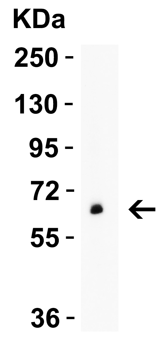
Click image to see more details
WB Validation in Human Pancreas
Loading: 10 μg of lysate
Antibodies: GLS2, A05334, 2 μ g/mL , 1 h incubation at RT in 5% NFDM/TBST.
Secondary: Goat Anti-Rabbit IgG HRP conjugate at 1:10000 dilution.

Click image to see more details
WB Validation in Mouse and Rat Brain
Loading: 15 μg of lysate
Antibodies: GLS2, A05334, 1 μg/mL , 1 h incubation at RT in 5% NFDM/TBST.
Secondary: Goat Anti-Rabbit IgG HRP conjugate at 1:10000 dilution.

Click image to see more details
Immunohistochemistry Validation of GLS2 in Human Liver
Immunohistochemical analysis of paraffin-embedded human liver tissue using anti-GLS2 antibody (A05334) at 1 μg/ml. Tissue was fixed with formaldehyde and blocked with 10% serum for 1 h at RT; antigen retrieval was by heat mediation with a citrate buffer (pH6). Samples were incubated with primary antibody overnight at 4˚C. A goat anti-rabbit IgG H&L (HRP) at 1/250 was used as secondary. Counter stained with Hematoxylin.

Click image to see more details
Immunohistochemistry Validation of GLS2 in Mouse Liver
Immunohistochemical analysis of paraffin-embedded mouse liver tissue using anti-GLS2 antibody (A05334) at 1 μg/ml. Tissue was fixed with formaldehyde and blocked with 10% serum for 1 h at RT; antigen retrieval was by heat mediation with a citrate buffer (pH6). Samples were incubated with primary antibody overnight at 4˚C. A goat anti-rabbit IgG H&L (HRP) at 1/250 was used as secondary. Counter stained with Hematoxylin.

Click image to see more details
Immunohistochemistry Validation of GLS2 in Rat Brain
Immunohistochemical analysis of paraffin-embedded rat brain tissue using anti-GLS2 antibody (A05334) at 2 μg/ml. Tissue was fixed with formaldehyde and blocked with 10% serum for 1 h at RT; antigen retrieval was by heat mediation with a citrate buffer (pH6). Samples were incubated with primary antibody overnight at 4˚C. A goat anti-rabbit IgG H&L (HRP) at 1/250 was used as secondary. Counter stained with Hematoxylin.

Click image to see more details
Immunofluorescence Validation of GLS2 in Mouse Brain
Immunofluorescent analysis of 4% paraformaldehyde-fixed mouse brain labeling GLS2 with A05334 at 20 μg g/mL, followed by goat anti-rabbit IgG secondary antibody at 1/500 dilution (green) and DAPI antibody (blue).
Specific Publications For Anti-GLS2 Antibody (A05334)
Loading publications
Recommended Resources
Here are featured tools and databases that you might find useful.
- Boster's Pathways Library
- Protein Databases
- Bioscience Research Protocol Resources
- Data Processing & Analysis Software
- Photo Editing Software
- Scientific Literature Resources
- Research Paper Management Tools
- Molecular Biology Software
- Primer Design Tools
- Bioinformatics Tools
- Phylogenetic Tree Analysis
Customer Reviews
Have you used Anti-GLS2 Antibody?
Share your experimental results or join a short interview to earn up to $1,000 in product credits or other rewards.
0 Reviews For Anti-GLS2 Antibody
Customer Q&As
Have a question?
Find answers in Q&As, reviews.
Can't find your answer?
Submit your question
