Click image to see more details
-

-
-
-
-
+4
Product Info Summary
| SKU: | PB9205 |
|---|---|
| Size: | 100 μg/vial |
| Reactive Species: | Human, Mouse, Rat |
| Host: | Rabbit |
| Application: | Flow Cytometry, IF, IHC, IHC-F, ICC, WB |
Customers Who Bought This Also Bought

Product info
Product Name
Anti-Ionotropic Glutamate receptor 2/GRIA2 Antibody Picoband®
SKU/Catalog Number
PB9205
Size
100 μg/vial
Form
Lyophilized
Description
Boster Bio Anti-Ionotropic Glutamate receptor 2/GRIA2 Antibody Picoband® catalog # PB9205. Tested in Flow Cytometry, IF, IHC, IHC-F, ICC, WB applications. This antibody reacts with Human, Mouse, Rat. The brand Picoband indicates this is a premium antibody that guarantees superior quality, high affinity, and strong signals with minimal background in Western blot applications. Only our best-performing antibodies are designated as Picoband, ensuring unmatched performance.
Storage & Handling
Store at -20˚C for one year from date of receipt. After reconstitution, at 4˚C for one month. It can also be aliquotted and stored frozen at -20˚C for six months. Avoid repeated freeze-thaw cycles.
Cite This Product
Anti-Ionotropic Glutamate receptor 2/GRIA2 Antibody Picoband® (Boster Biological Technology, Pleasanton CA, USA, Catalog # PB9205)
Host
Rabbit
Contents
Each vial contains antibody formulated with stabilizing components, 0.9 mg NaCl, 0.2 mg Na2HPO4, and 0.05 mg NaN3.
*This antibody is supplied in a stabilized formulation.
Compatibility with conjugation reactions depends on the chemistry of the conjugation method used.
For conjugation methods that are not compatible with the stabilizing components present in this formulation, a carrier-free antibody format is required.
Clonality
Polyclonal
Isotype
Rabbit IgG
Immunogen
E.coli-derived human GRIA2 recombinant protein (Position: N25-I360). Human GRIA2 shares 99% amino acid (aa) sequence identity with both mouse and rat GRIA2.
Cross-reactivity
No cross-reactivity with other proteins
Reactive Species
PB9205 is reactive to GRIA2 in Human, Mouse, Rat
Observed Molecular Weight
110 kDa
Calculated molecular weight
98.8 kDa
Background of GRIA2
Glutamate receptor 2, also known as GLUR2, is a protein that in humans is encoded by the GRIA2 gene. This gene product belongs to a family of glutamate receptors that are sensitive to alpha-amino-3-hydroxy-5-methyl-4-isoxazole propionate (AMPA), and function as ligand-activated cation channels. GLUR2's cytogenetic location is 4q32.1. The crystal structures of the GLUR2 ligand-binding core in the apo state and in the presence of the antagonist DNQX, the partial agonist kainate, and the full agonists AMPA and glutamate.GLUR2 plays a major role in depression at synapses in which glutamate remains in the synaptic cleft for prolonged periods of time during normal operation of the synapse. The overexpression of GLUR2 increases dendritic spine size and density in hippocampal neurons, and more remarkably, induces spine formation in GABA-releasing interneurons that normally lack spines.
Antibody Validation
Boster validates all antibodies on WB, IHC, ICC, Immunofluorescence, and ELISA with known positive control and negative samples to ensure specificity and high affinity, including thorough antibody incubations.
Application & Images
Applications
PB9205 is guaranteed for Flow Cytometry, IF, IHC, IHC-F, ICC, WB Boster Guarantee
Recommend Dilution
| Application | Dilution | Species |
|---|---|---|
| Western blot | 0.1-0.5μg/ml | Mouse, Rat, Human |
| Immunohistochemistry (Paraffin-embedded Section) | 0.5-1μg/ml | Mouse, Rat, Human |
| Immunohistochemistry (Frozen Section) | 0.5-1μg/ml | Rat |
| Immunocytochemistry | 0.5-1μg/ml | Human |
| Immunofluorescence | 2μg/ml | Mouse |
| Flow Cytometry (Fixed) | 1-3μg/1x106 cells | Human |
Tested application
Suggested blocking solution with 5% non-fat milk or BSA; (*)Recommended protein loading: 20-40 µg per lane
Use TE buffer pH 9.0 for antigen retrieval; (*) citrate buffer pH 6.0 is an alternative.
Validation Images & Assay Conditions
Click image to see more details
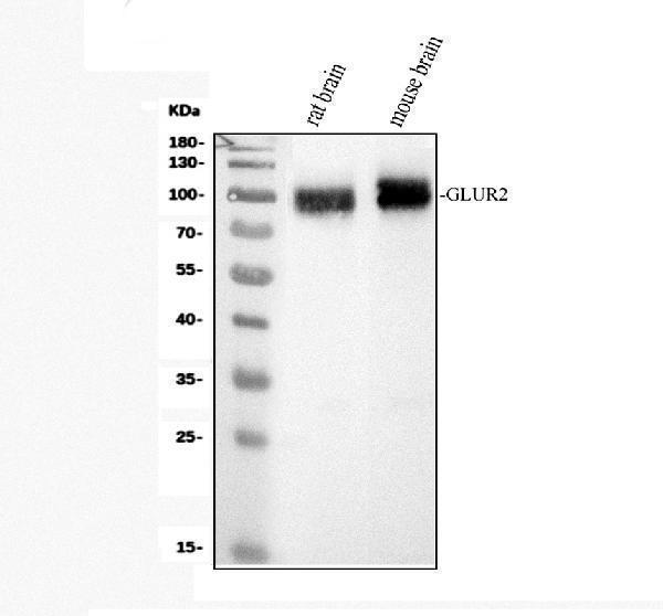
Western blot analysis of GRIA2 using anti-GRIA2 antibody (PB9205).
Electrophoresis was performed on a 10% SDS-PAGE gel at 80V (Stacking gel) / 120V (Resolving gel) for 2 hours. The sample well of each lane was loaded with 30 ug of sample under reducing conditions.
Lane 1: rat brain tissue lysates,
Lane 2: mouse brain tissue lysates.
After electrophoresis, proteins were transferred to a nitrocellulose membrane at 150 mA for 50-90 minutes. Blocked the membrane with 5% non-fat milk/TBS for 1.5 hour at RT. The membrane was incubated with rabbit anti-GRIA2 antigen affinity purified polyclonal antibody (PB9205) at 0.5 μg/mL overnight at 4°C, then washed with TBS-0.1%Tween 3 times with 5 minutes each and probed with a goat anti-rabbit IgG-HRP secondary antibody (Catalog # BA1054) at a dilution of 1:5000 for 1.5 hour at RT. The signal is developed using an ECL Plus Western Blotting Substrate (Catalog # AR1196-200) with Tanon 5200 system. A specific band was detected for GRIA2 at approximately 99 kDa. The expected band size for GRIA2 is at 99 kDa.

Click image to see more details
IHC analysis of Glutamate receptor 2/GRIA2 using anti-Glutamate receptor 2/GRIA2 antibody (PB9205).
Glutamate receptor 2/GRIA2 was detected in a paraffin-embedded section of human brain tissue. Heat mediated antigen retrieval was performed in EDTA buffer (pH 8.0, epitope retrieval solution). The tissue section was blocked with 10% goat serum. The tissue section was then incubated with 2 μg/ml rabbit anti-Glutamate receptor 2/GRIA2 Antibody (PB9205) overnight at 4°C. Peroxidase Conjugated Goat Anti-rabbit IgG was used as secondary antibody and incubated for 30 minutes at 37°C. The tissue section was developed using HRP Conjugated Rabbit IgG Super Vision Assay Kit (Catalog # SV0002) with DAB as the chromogen.

Click image to see more details
IHC analysis of GRIA2 using anti-GRIA2 antibody (PB9205).
GRIA2 was detected in paraffin-embedded section of Mouse Brain Tissue. Heat mediated antigen retrieval was performed in citrate buffer (pH6, epitope retrieval solution) for 20 mins. The tissue section was blocked with 10% goat serum. The tissue section was then incubated with 1μg/ml rabbit anti-GRIA2 Antibody (PB9205) overnight at 4°C. Biotinylated goat anti-rabbit IgG was used as secondary antibody and incubated for 30 minutes at 37°C. The tissue section was developed using Strepavidin-Biotin-Complex (SABC)(Catalog # SA1022) with DAB as the chromogen.

Click image to see more details
IHC analysis of GRIA2 using anti-GRIA2 antibody (PB9205).
GRIA2 was detected in paraffin-embedded section of Rat Brain Tissue. Heat mediated antigen retrieval was performed in citrate buffer (pH6, epitope retrieval solution) for 20 mins. The tissue section was blocked with 10% goat serum. The tissue section was then incubated with 1μg/ml rabbit anti-GRIA2 Antibody (PB9205) overnight at 4°C. Biotinylated goat anti-rabbit IgG was used as secondary antibody and incubated for 30 minutes at 37°C. The tissue section was developed using Strepavidin-Biotin-Complex (SABC)(Catalog # SA1022) with DAB as the chromogen.

Click image to see more details
IF analysis of GRIA2 using anti-GRIA2 antibody (PB9205)
GRIA2 was detected in paraffin-embedded section of mouse brain tissues. Heat mediated antigen retrieval was performed in citrate buffer (pH6, epitope retrieval solution ) for 20 mins. The tissue section was blocked with 10% goat serum. The tissue section was then incubated with 2μg/mL rabbit anti-GRIA2 Antibody (PB9205) overnight at 4°C. Biotin conjugated goat anti-rabbit IgG (BA1003) was used as secondary antibody and incubated for 30 minutes at 37°C. The tissue section was developed using DyLight®488 Conjugated Avidin (BA1128). The section was counterstained with DAPI. Visualize using a fluorescence microscope and filter sets appropriate for the label used.

Click image to see more details
IHC analysis of GRIA2 using anti-GRIA2 antibody (PB9205).
GRIA23 was detected in a frozen section of rat brain tissue. The tissue section was blocked with 10% goat serum. The tissue section was then incubated with 1 μg/ml rabbit anti-GRIA2 Antibody (PB9205) overnight at 4°C. Biotinylated goat anti-rabbit IgG was used as secondary antibody and incubated for 30 minutes at 37°C. The tissue section was developed using Strepavidin-Biotin-Complex (SABC) (Catalog # SA1022) with DAB as the chromogen.

Click image to see more details
IF analysis of GRIA2 using anti-GRIA2 antibody (PB9205).
GRIA2 was detected in an immunocytochemical section of T-47D cells. Enzyme antigen retrieval was performed using IHC enzyme antigen retrieval reagent (AR0022) for 15 mins. The cells were blocked with 10% goat serum. And then incubated with 1 μg/mL rabbit anti-GRIA2 Antibody (PB9205) overnight at 4°C. DyLight®488 Conjugated Goat Anti-Rabbit IgG (BA1127) was used as secondary antibody at 1:100 dilution and incubated for 30 minutes at 37°C. The section was counterstained with DAPI. Visualize using a fluorescence microscope and filter sets appropriate for the label used.

Click image to see more details
Flow Cytometry analysis of U-87MG cells using anti-GRIA2 antibody (PB9205).
Overlay histogram showing U-87MG cells stained with PB9205 (Blue line). To facilitate intracellular staining, cells were fixed with 4% paraformaldehyde and permeabilized with permeabilization buffer. The cells were blocked with 10% normal goat serum. And then incubated with rabbit anti-GRIA2 Antibody (PB9205,1μg/1x106 cells) for 30 min at 20°C. DyLight®488 conjugated goat anti-rabbit IgG (BA1127, 5-10μg/1x106 cells) was used as secondary antibody for 30 minutes at 20°C. Isotype control antibody (Green line) was rabbit IgG (1μg/1x106) used under the same conditions. Unlabelled sample without incubation with primary antibody and secondary antibody (Red line) was used as a blank control.
Specific Publications For Anti-Ionotropic Glutamate receptor 2/GRIA2 Antibody Picoband® (PB9205)
Loading publications
Recommended Resources
Here are featured tools and databases that you might find useful.
- Boster's Pathways Library
- Protein Databases
- Bioscience Research Protocol Resources
- Data Processing & Analysis Software
- Photo Editing Software
- Scientific Literature Resources
- Research Paper Management Tools
- Molecular Biology Software
- Primer Design Tools
- Bioinformatics Tools
- Phylogenetic Tree Analysis
Customer Reviews
Have you used Anti-Ionotropic Glutamate receptor 2/GRIA2 Antibody Picoband®?
Share your experimental results or join a short interview to earn up to $1,000 in product credits or other rewards.
0 Reviews For Anti-Ionotropic Glutamate receptor 2/GRIA2 Antibody Picoband®
Customer Q&As
Have a question?
Find answers in Q&As, reviews.
Can't find your answer?
Submit your question
1 Customer Q&As for Anti-Ionotropic Glutamate receptor 2/GRIA2 Antibody Picoband®
Question
We are currently using anti-Ionotropic Glutamate receptor 2/GRIA2 antibody PB9205 for human tissue, and we are satisfied with the ICC results. The species of reactivity given in the datasheet says human, mouse, rat. Is it likely that the antibody can work on horse tissues as well?
Verified Customer
Verified customer
Asked: 2019-12-31
Answer
The anti-Ionotropic Glutamate receptor 2/GRIA2 antibody (PB9205) has not been tested for cross reactivity specifically with horse tissues, though there is a good chance of cross reactivity. We have an innovator award program that if you test this antibody and show it works in horse you can get your next antibody for free. Please contact me if I can help you with anything.
Boster Scientific Support
Answered: 2019-12-31
