Click image to see more details
Product Info Summary
| SKU: | A01230 |
|---|---|
| Size: | 100ug |
| Reactive Species: | Human, Mouse |
| Host: | Rabbit |
| Application: | ELISA, IF, IHC, WB |
Customers Who Bought This Also Bought

Product info
Product Name
Anti-HDAC-5 (internal) Antibody
SKU/Catalog Number
A01230
Size
100ug
Form
Liquid (sterile filtered)
Description
Boster Bio Anti-HDAC-5 (internal) Antibody (Catalog # A01230). Tested in Dot blot, ELISA, WB applications. This antibody reacts with Human, Mouse.
Storage & Handling
Store vial at -20°C prior to opening. Aliquot contents and freeze at -20°C or below for extended storage. Avoid cycles of freezing and thawing. Centrifuge product if not completely clear after standing at room temperature. This product is stable for several weeks at 4°C as an undiluted liquid. Dilute only prior to immediate use. Expiration date is six (6) months from date of opening. (Ship on dry ice.)
Cite This Product
Anti-HDAC-5 (internal) Antibody (Boster Biological Technology, Pleasanton CA, USA, Catalog # A01230)
Host
Rabbit
Contents
0.02 M Potassium Phosphate, 0.15 M Sodium Chloride, pH 7.2, 50% (v/v) Glycerol
Clonality
Polyclonal
Isotype
IgG
Immunogen
HDAC5 affinity purified antibody was prepared from whole rabbit serum produced by repeated immunizations with a synthetic peptide corresponding to the internal region surrounding phosphoserine 661 of human HDAC5.
Reactive Species
A01230 is reactive to HDAC5 in Human, Mouse
Observed Molecular Weight
42 kDa
Calculated molecular weight
122.0 kDa
Background of HDAC5
HDAC5 is a member of the class II mammalian histone deacetylase family, which is structurally related to yeast HDA1. Human HDAC5 is composed of 1122 amino acid residues. The deacetylase domain of HDAC5 is located at the C-terminal half of the molecule. The N-terminal non-deacetylase domain does not show any significant homology with any published sequence. Both domains are required for HDAC5-mediated repression of gene transcription. HDAC5 interacts with a growing number of transcriptional factors including MEF2A as well as other HDAC proteins. The interacting complexes bind to specific regions of chromatin and regulate gene transcription in these regions. Anti-HDAC5 antibodies are ideal for researchers interested in Breast Cancer, Cancer, Cell Cycle and Replication, Chromatin Research, Epigenetics, and Histone Deacetylases research.
Antibody Validation
Boster validates all antibodies on WB, IHC, ICC, Immunofluorescence, and ELISA with known positive control and negative samples to ensure specificity and high affinity, including thorough antibody incubations.
Application & Images
Applications
A01230 is guaranteed for ELISA, IF, IHC, WB Boster Guarantee
Recommend Dilution
| Application | Dilution | Species |
|---|---|---|
| ELISA: 1:20 | 000 - 1:60 | 000 |
| Anti-HDAC5 antibody has been tested in ELISA | dot blot | and Western Blot. Specific conditions for reactivity should be optimized by the end user. Expect a band approximately ~124 kDa corresponding to the appropriate cell lysate or extract. |
Validation Images & Assay Conditions
Click image to see more details
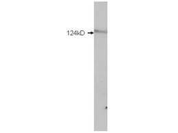
Western Blot of Rabbit anti-HDAC5 antibody. Lane 1: mouse brain extract. Load: 5 µg per lane. Primary antibody: HDAC5 antibody at 0.2µg/mL for overnight at 4°C. Secondary antibody: IRDye800™ rabbit secondary antibody at 1:10,000 for 45 min at RT. Block: 5% BLOTTO overnight at 4°C. Predicted/Observed size: 124 kDa for HDAC5.

Click image to see more details
Dot blot for Rabbit Anti-HDAC5 (internal) Antibody. Lane 1: HDAC-4 (internal). Lane 2: HDAC-5 . Lane 3: HDAC-5 (600-401-J68). Lane 4: HDAC-7 . Load: 100, 10, and 1 picomoles of peptide. Primary antibody: HDAC-5 antibody at 1:1000 for 45 min at 4°C. Secondary antibody: Dylight™488 rabbit secondary antibody at 1:10,000 for 45 min at RT. Block: 5% BLOTTO overnight at 4°C.

Click image to see more details
Western Blot of Rabbit anti-HDAC5 antibody. Marker: Opal Pre-stained ladder . Lane 1: HEK293 lysate . Lane 2: HeLa Lysate . Lane 3: MCF-7 Lysate . Lane 4: Jurkat Lysate . Lane 5: A431 Lysate . Lane 6: A549 Lysate . Lane 7: LNCap Lysate . Lane 8: MOLT-4 Lysate . Lane 9: Ramos Lysate . Lane 10: Raji Lysate . Lane 11: A-172 Lysate . Lane 12: NIH/3T3 Lysate . Load: 35 µg per lane. Primary antibody: HDAC5 antibody at 1ug/mL overnight at 4C. Secondary antibody: Peroxidase rabbit secondary antibody at 1:30,000 for 60 min at RT. Blocking Buffer: 1% Casein-TTBS for 30 min at RT. Predicted/Observed size: 124kDa for HDAC5.
Specific Publications For Anti-HDAC-5 (internal) Antibody (A01230)
Loading publications
Recommended Resources
Here are featured tools and databases that you might find useful.
- Boster's Pathways Library
- Protein Databases
- Bioscience Research Protocol Resources
- Data Processing & Analysis Software
- Photo Editing Software
- Scientific Literature Resources
- Research Paper Management Tools
- Molecular Biology Software
- Primer Design Tools
- Bioinformatics Tools
- Phylogenetic Tree Analysis
Customer Reviews
Have you used Anti-HDAC-5 (internal) Antibody?
Share your experimental results or join a short interview to earn up to $1,000 in product credits or other rewards.
0 Reviews For Anti-HDAC-5 (internal) Antibody
Customer Q&As
Have a question?
Find answers in Q&As, reviews.
Can't find your answer?
Submit your question
