Click image to see more details
Product Info Summary
| SKU: | PA1350 |
|---|---|
| Size: | 100 μg/vial |
| Reactive Species: | Human, Mouse, Rat |
| Host: | Rabbit |
| Application: | WB |
Customers Who Bought This Also Bought

Product info
Product Name
Anti-Histone deacetylase 2 HDAC2 Antibody Picoband®
SKU/Catalog Number
PA1350
BA0915 is an alternative SKU for this antibody, used in previous lots.
Size
100 μg/vial
Form
Lyophilized
Description
Boster Bio Anti-Histone deacetylase 2 HDAC2 Antibody catalog # PA1350. Tested in WB applications. This antibody reacts with Human, Mouse, Rat. The brand Picoband indicates this is a premium antibody that guarantees superior quality, high affinity, and strong signals with minimal background in Western blot applications. Only our best-performing antibodies are designated as Picoband, ensuring unmatched performance.
Storage & Handling
Store at -20˚C for one year from date of receipt. After reconstitution, at 4˚C for one month. It can also be aliquotted and stored frozen at -20˚C for six months. Avoid repeated freeze-thaw cycles.
Cite This Product
Anti-Histone deacetylase 2 HDAC2 Antibody Picoband® (Boster Biological Technology, Pleasanton CA, USA, Catalog # PA1350)
Host
Rabbit
Contents
Each vial contains 4 mg Trehalose, 0.9 mg NaCl and 0.2 mg Na2HPO4.
Clonality
Polyclonal
Isotype
Rabbit IgG
Immunogen
A synthetic peptide corresponding to a sequence at the C-terminus of human HDAC2, identical to the related rat sequence, and different from the related mouse sequence by one amino acid.
Cross-reactivity
No cross-reactivity with other proteins
Reactive Species
PA1350 is reactive to HDAC2 in Human, Mouse, Rat
Observed Molecular Weight
60 kDa
Calculated molecular weight
55.4 kDa
Background of HDAC2
Histone deacetylase 2 is an enzyme that in humans is encoded by the HDAC2 gene. This gene product belongs to the histone deacetylase family. Histone deacetylases act via the formation of large multiprotein complexes and are responsible for the deacetylation of lysine residues on the N-terminal region of the core histones (H2A, H2B, H3 and H4). This protein also forms transcriptional repressor complexes by associating with many different proteins, including YY1, a mammalian zinc-finger transcription factor. Thus it plays an important role in transcriptional regulation, cell cycle progression and developmental events. Betz et al. (1998) performed PCR using HDAC2-specific primers to screen a somatic cell hybrid mapping panel. They mapped the HDAC2 gene to human chromosome 6q21, a region of the genome altered in some cancers, including retinoblastoma.
Antibody Validation
Boster validates all antibodies on WB, IHC, ICC, Immunofluorescence, and ELISA with known positive control and negative samples to ensure specificity and high affinity, including thorough antibody incubations.
Application & Images
Applications
PA1350 is guaranteed for WB Boster Guarantee
Recommend Dilution
| Application | Dilution | Species |
|---|---|---|
| Western blot | 0.1-0.5μg/ml | Human, Mouse, Rat |
Tested application
Suggested blocking solution with 5% non-fat milk or BSA; (*)Recommended protein loading: 20-40 µg per lane
Validation Images & Assay Conditions
Click image to see more details
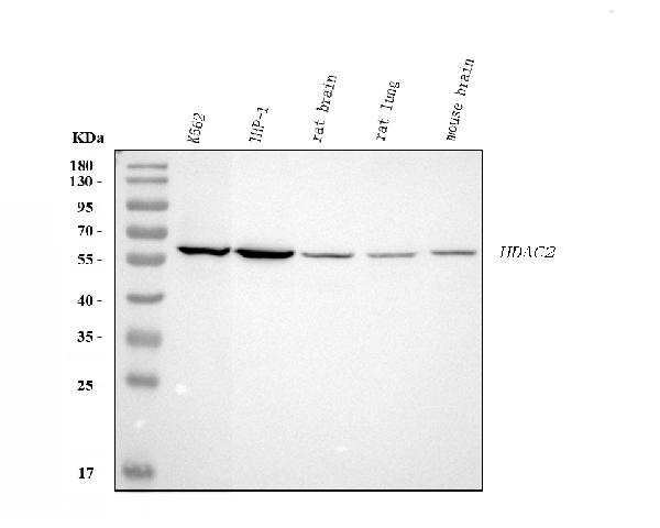
Western blot analysis of HDAC2 using anti-HDAC2 antibody (PA1350).
Electrophoresis was performed on a 5-20% SDS-PAGE gel at 70V (Stacking gel) / 90V (Resolving gel) for 2-3 hours. The sample well of each lane was loaded with 30 ug of sample under reducing conditions.
Lane 1: human K562 whole cell lysates,
Lane 2: human THP-1 whole cell lysates,
Lane 3: rat brian tissue lysates,
Lane 4: rat lung tissue lysates,
Lane 5: mouse brain tissue lysates.
After electrophoresis, proteins were transferred to a nitrocellulose membrane at 150 mA for 50-90 minutes. Blocked the membrane with 5% non-fat milk/TBS for 1.5 hour at RT. The membrane was incubated with rabbit anti-HDAC2 antigen affinity purified polyclonal antibody (Catalog # PA1350) at 0.5 μg/mL overnight at 4°C, then washed with TBS-0.1%Tween 3 times with 5 minutes each and probed with a goat anti-rabbit IgG-HRP secondary antibody at a dilution of 1:5000 for 1.5 hour at RT. The signal is developed using an Enhanced Chemiluminescent detection (ECL) kit (Catalog # EK1002) with Tanon 5200 system. A specific band was detected for HDAC2 at approximately 60 kDa. The expected band size for HDAC2 is at 55 kDa.
Specific Publications For Anti-Histone deacetylase 2 HDAC2 Antibody Picoband® (PA1350)
Loading publications
Recommended Resources
Here are featured tools and databases that you might find useful.
- Boster's Pathways Library
- Protein Databases
- Bioscience Research Protocol Resources
- Data Processing & Analysis Software
- Photo Editing Software
- Scientific Literature Resources
- Research Paper Management Tools
- Molecular Biology Software
- Primer Design Tools
- Bioinformatics Tools
- Phylogenetic Tree Analysis
Customer Reviews
Have you used Anti-Histone deacetylase 2 HDAC2 Antibody Picoband®?
Share your experimental results or join a short interview to earn up to $1,000 in product credits or other rewards.
0 Reviews For Anti-Histone deacetylase 2 HDAC2 Antibody Picoband®
Customer Q&As
Have a question?
Find answers in Q&As, reviews.
Can't find your answer?
Submit your question
