Click image to see more details
Product Info Summary
| SKU: | M00838 |
|---|---|
| Size: | 100ug |
| Reactive Species: | Human, Mouse, Rat |
| Host: | Mouse |
| Application: | ELISA, IP, IF, WB |
Customers Who Bought This Also Bought

Product info
Product Name
Anti-HEF1 NEDD9 Monoclonal Antibody
SKU/Catalog Number
M00838
Size
100ug
Form
Liquid (sterile filtered)
Description
Boster Bio Anti-HEF1 NEDD9 Monoclonal Antibody (Catalog # M00838). Tested in IF, IP, WB applications. This antibody reacts with Human, Mouse, Rat.
Storage & Handling
Store vial at -20°C prior to opening. Aliquot contents and freeze at -20°C or below for extended storage. Avoid cycles of freezing and thawing. Centrifuge product if not completely clear after standing at room temperature. This product is stable for several weeks at 4°C as an undiluted liquid. Dilute only prior to immediate use. Expiration date is one (1) year from date of opening. (Ship on dry ice.)
Cite This Product
Anti-HEF1 NEDD9 Monoclonal Antibody (Boster Biological Technology, Pleasanton CA, USA, Catalog # M00838)
Host
Mouse
Contents
0.02 M Potassium Phosphate, 0.15 M Sodium Chloride, pH 7.2, 0.01% (w/v) Sodium Azide
Clonality
Monoclonal
Clone Number
Clone: 2G9
Isotype
IgG1 kappa
Immunogen
Anti-HEF1 monoclonal antibody was produced by repeated immunizations with a synthetic peptide corresponding to amino acid residues 82-398 of human HEF1 protein (hHEF1, 843 aa, predicted MW 92.8 kDa).
Reactive Species
M00838 is reactive to NEDD9 in Human, Mouse, Rat
Observed Molecular Weight
42 kDa
Calculated molecular weight
92.9 kDa
Background of NEDD9
HEF1, also known as Enhancer of filamentation 1, CRK-associated substrate-related protein, CAS-L, CasL, p105 and Neural precursor cell expressed developmentally down-regulated 9 is the product of the NEDD9 (CASGL) gene. HEF1 functions as a docking protein that plays a central coordinating role for tyrosine-kinase-based signaling related to cell adhesion. HEF1 may also function in transmitting growth control signals between focal adhesions at the cell periphery and the mitotic spindle in response to adhesion or growth factor signals initiating cell proliferation. HEF1 may also play an important role in integrin beta-1 or B cell antigen receptor (BCR) mediated signaling in B- and T-cells. Integrin beta-1 stimulation leads to recruitment of various proteins including CRK, NCK and SHPTP2 to the tyrosine phosphorylated form. HEF1 forms a homodimer and can heterodimerize with HLH proteins ID2, E12, E47 and also with p130cas. HEF1 also forms complexes in vivo with related adhesion focal tyrosine kinase (RAFTK), adapter protein CRKL and LYN kinase and also interacts with MICAL and TXNL4/DIM1. This protein localizes to both the cell nucleus and the cell periphery and is differently localized in fibroblasts and epithelial cells. In fibroblasts, it is predominantly nuclear and in some cells is present in the Golgi apparatus. In epithelial cells, it is localized predominantly in the cell periphery with particular concentration in lamellipodia, but it is also found in the nucleus. HEF1 is widely expressed although higher levels are detected in kidney, lung, and placental tissue. HEF1 is also detected in T-cells, B-cells and diverse cell lines. HEF1 is activated upon induction of cell growth. Cell cycle-regulated processing produces four isoforms: p115, p105, p65, and p55. Isoform p115 arises from p105 phosphorylation and appears later in the cell cycle. Isoform p55 arises from p105 as a result of cleavage at a caspase cleavage-related site and it appears specifically at mitosis. The p65 isoform is poorly detected. Isoforms p105 and p115 are predominantly cytoplasmic and associate with focal adhesions while p55 associates with the mitotic spindle.
Antibody Validation
Boster validates all antibodies on WB, IHC, ICC, Immunofluorescence, and ELISA with known positive control and negative samples to ensure specificity and high affinity, including thorough antibody incubations.
Application & Images
Applications
M00838 is guaranteed for ELISA, IP, IF, WB Boster Guarantee
Recommend Dilution
| Application | Dilution | Species |
|---|---|---|
| ELISA: 1:5 | 000 - 1:20 | 000 |
| IP: 1:1 | 000 | |
| WB: 1:5 | 000 | |
| This monoclonal antibody has been tested for use in western blotting | immunoprecipitation and immunofluorescence. Specific conditions for reactivity should be optimized by the end user. Expect bands approximately 115 and 105 in size corresponding to isoforms of HEF1 protein by western blotting in the appropriate cell lysate or extract. This antibody does not recognize p130Cas. Sin1 has not been tested. IF was performed using 4% PFA fixed cells. This monoclonal antibody mostly detects HEF1 localized at the focal adhesion sites. |
Validation Images & Assay Conditions
Click image to see more details
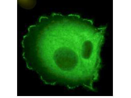
Immunofluorescence microscopy using Boster's Monoclonal anti-HEF1 antibody (clone 2G9) shows detection of HEF1 localized at focal adhesion sites. The antibody was used at a 1:500 dilution with a 3-sec exposure time. Personal Communication. Elena Pugacheva, Fox Chase Cancer Center, Philadelphia, PA.

Click image to see more details
Western blot using Boster's monoclonal anti-HEF1 antibody (clone 2G9) antibody shows detection of a ~92 kDa band corresponding to HEF1 in MCF7 lysate [arrowhead]. Approximately 35 µg of lysate was loaded for SDS-PAGE followed by transfer onto nitrocellulose and reaction with a 1:1,000 dilution of anti-HEF1 antibody. Detection occurred using a 1:5,000 dilution of IRDye®800 conjugated Sh-a-Mouse IgG [H&L] for 45 min at room temperature (800 nm channel, green). Molecular weight estimation was made by comparison to prestained MW markers (indicated at left). IRDye®800 fluorescence image was captured using the Odyssey® Infrared Imaging System developed by LI-COR. IRDye is a trademark of LI-COR, Inc. Other detection systems will yield similar results.

Click image to see more details
Western blotting using Boster's monoclonal anti-HEF1 antibody (clone 2G9) shows detection of endogenous HEF1 present in various cell lines [MCF7, HeLa, CHO, 3Y1]. Panel A shows detection using a 15 min exposure. Panel B is the same blot exposed for 2 min. The doublet represents p105 and p115 staining. The lower MW band represents p55. 3Y1 cells are derived from rat fibroblast cells. Mouse 3T3 cells are also reactive (not shown). To date no staining has been noted on CHO cells. Personal Communication. Elena Pugacheva, Fox Chase Cancer Center, Philadelphia, PA.
Specific Publications For Anti-HEF1 NEDD9 Monoclonal Antibody (M00838)
Loading publications
Recommended Resources
Here are featured tools and databases that you might find useful.
- Boster's Pathways Library
- Protein Databases
- Bioscience Research Protocol Resources
- Data Processing & Analysis Software
- Photo Editing Software
- Scientific Literature Resources
- Research Paper Management Tools
- Molecular Biology Software
- Primer Design Tools
- Bioinformatics Tools
- Phylogenetic Tree Analysis
Customer Reviews
Have you used Anti-HEF1 NEDD9 Monoclonal Antibody?
Share your experimental results or join a short interview to earn up to $1,000 in product credits or other rewards.
0 Reviews For Anti-HEF1 NEDD9 Monoclonal Antibody
Customer Q&As
Have a question?
Find answers in Q&As, reviews.
Can't find your answer?
Submit your question
