Click image to see more details
-

-
-
-
-
+5
Product Info Summary
| SKU: | A01389 |
|---|---|
| Size: | 100 μg/vial |
| Reactive Species: | Human, Mouse, Rat |
| Host: | Rabbit |
| Application: | Flow Cytometry, IHC, WB |
Customers Who Bought This Also Bought

Product info
Product Name
Anti-Hexokinase II/HK2 Antibody Picoband®
SKU/Catalog Number
A01389
Size
100 μg/vial
Form
Lyophilized
Description
Boster Bio Anti-Hexokinase II/HK2 Antibody Picoband® catalog # A01389. Tested in Flow Cytometry, IHC, WB applications. This antibody reacts with Human, Mouse, Rat. The brand Picoband indicates this is a premium antibody that guarantees superior quality, high affinity, and strong signals with minimal background in Western blot applications. Only our best-performing antibodies are designated as Picoband, ensuring unmatched performance.
Storage & Handling
Store at -20˚C for one year from date of receipt. After reconstitution, at 4˚C for one month. It can also be aliquotted and stored frozen at -20˚C for six months. Avoid repeated freeze-thaw cycles.
Cite This Product
Anti-Hexokinase II/HK2 Antibody Picoband® (Boster Biological Technology, Pleasanton CA, USA, Catalog # A01389)
Host
Rabbit
Contents
Each vial contains 4 mg Trehalose, 0.9 mg NaCl and 0.2 mg Na2HPO4.
Clonality
Polyclonal
Isotype
Rabbit IgG
Immunogen
A synthetic peptide corresponding to a sequence in the middle region of human Hexokinase II, different from the related mouse and rat sequences by four amino acids.
Cross-reactivity
No cross-reactivity with other proteins.
Reactive Species
A01389 is reactive to HK2 in Human, Mouse, Rat
Observed Molecular Weight
102 kDa
Calculated molecular weight
102.4 kDa
Background of HK2
Hexokinase 2, also known as HK2, is an enzyme which in humans is encoded by the HK2 gene on chromosome 2. Hexokinases phosphorylate glucose to produce glucose-6-phosphate, the first step in most glucose metabolism pathways. This gene encodes hexokinase 2, the predominant form found in skeletal muscle. It localizes to the outer membrane of mitochondria. Expression of this gene is insulin-responsive, and studies in rat suggest that it is involved in the increased rate of glycolysis seen in rapidly growing cancer cells.
Antibody Validation
Boster validates all antibodies on WB, IHC, ICC, Immunofluorescence, and ELISA with known positive control and negative samples to ensure specificity and high affinity, including thorough antibody incubations.
Application & Images
Applications
A01389 is guaranteed for Flow Cytometry, IHC, WB Boster Guarantee
Recommend Dilution
| Application | Dilution | Species |
|---|---|---|
| Western blot | 0.1-0.5μg/ml | Human, Mouse, Rat |
| Immunohistochemistry (Paraffin-embedded Section) | 2-5μg/ml | Human, Mouse, Rat |
| Flow Cytometry(Fixed) | 1-3 μg/1x106 cells | Human |
Tested application
Suggested blocking solution with 5% non-fat milk or BSA; (*)Recommended protein loading: 20-40 µg per lane
Use TE buffer pH 9.0 for antigen retrieval; (*) citrate buffer pH 6.0 is an alternative.
Validation Images & Assay Conditions
Click image to see more details
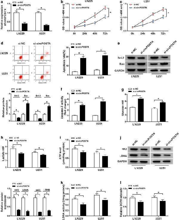
The influences of circPOSTN silencing on proliferation, apoptosis and aerobic glycolysis of glioma cells. a – l LN229 and U251 cells were transfected with si-circPOSTN or si-NC. a The interference efficiency of si-circPOSTN was analyzed with RT-qPCR assay in LN229 and U251 cells. b , c Effect of circPOSTN silencing on the cell viability of LN229 and U251 cells was assessed with MTT assay. d The apoptosis rate was computed with flow cytometry assay in transfected LN229 and U251 cells. e The western blot assay showed the expression levels of Bcl-2 and Bax in LN229 and U251 cells. f The caspase-3 activity was measured with a caspase-3 assay kit. g – i The concentration of glucose and lactate in the culture medium, as well as ATP production level were measured with a series of kits, respectively. j The protein expression levels of HK2 and LDHA were determined with western blot assay in transfected LN229 and U251 cells. k – l LDHA enzyme activity and ROS accumulation were evaluated in LN229 and U251 cells post-transfection with lactate dehydrogenase activity detection kit and reactive oxygen species assay kit, respectively. * P < 0.05
Index in PubMed under a CC BY license. PMID: 32774168

Click image to see more details
CircPOSTN silencing inhibited aerobic glycolysis of glioma cells via regulating miR-361-5p. a – l LN229 and U251 cells were transfected with si-NC, si-circPOSTN, si-circPOSTN + anti-miR-NC, or si-circPOSTN + anti-miR-361-5p. a – f The concentration of glucose and lactate, as well as cellular ATP level were detected with different kits. g , h The protein expression levels of HK2 and LDHA in LN229 and U251 cells were measured with western blot assay. i – l The enzyme activity of LDHA and ROS level were measured in transfected LN229 and U251 cells. * P < 0.05
Index in PubMed under a CC BY license. PMID: 32774168

Click image to see more details
TPX2 regulated proliferation, apoptosis, and aerobic glycolysis in glioma cells. a – l LN229 and U251 cells were introduced with si-NC or si-TPX2. a The transfection efficiency of si-TPX2 was checked with RT-qPCR assay in LN229 and U251 cells. b , c The cell viability of LN229 and U251 cells was determined with MTT assay. d The apoptosis rate of transfected LN229 and U251 cells was represented by flow cytometry assay. e The western blot assay was used to assay the expression levels of Bcl-2 and Bax in LN229 and U251 cells. f The activity of caspase-3 was detected with a caspase-3 assay kit. g – i The glucose, lactate, and ATP production levels were shown. j The protein expression levels of HK2 and LDHA were estimated by western blot assay in LN229 and U251 cells. k , l LDHA enzyme activity and ROS content were evaluated in LN229 and U251 cells post-transfection. * P < 0.05
Index in PubMed under a CC BY license. PMID: 32774168

Click image to see more details
Western blot analysis of Hexokinase 2/HK2 using anti-Hexokinase 2/HK2 antibody (A01389).
Electrophoresis was performed on a 8% SDS-PAGE gel at 80V (Stacking gel) / 120V (Resolving gel) for 2 hours. The sample well of each lane was loaded with 30 ug of sample under reducing conditions.
Lane 1: human Hela- WT whole cell lysates,
Lane 2: human Hela-HK2 KO whole cell lysates.
After electrophoresis, proteins were transferred to a nitrocellulose membrane at 150 mA for 50-90 minutes. Blocked the membrane with 5% non-fat milk/TBS for 1.5 hour at RT. The membrane was incubated with rabbit anti-Hexokinase 2/HK2 antigen affinity purified polyclonal antibody (A01389) at 0.5 μg/mL overnight at 4°C, then washed with TBS-0.1%Tween 3 times with 5 minutes each and probed with a goat anti-rabbit IgG-HRP secondary antibody at a dilution of 1:5000 for 1.5 hour at RT. The signal is developed using an ECL Plus Western Blotting Substrate (Catalog # AR1196-200) with Tanon 5200 system. A specific band was detected for Hexokinase 2/HK2 at approximately 102 kDa. The expected band size for Hexokinase 2/HK2 is at 102 kDa.

Click image to see more details
Western blot analysis of Hexokinase II using anti-Hexokinase II antibody (A01389).
Electrophoresis was performed on a 8% SDS-PAGE gel at 80V (Stacking gel) / 120V (Resolving gel) for 2 hours. The sample well of each lane was loaded with 30 ug of sample under reducing conditions.
Lane 1: human Hela whole cell lysates,
Lane 2: human MCF-7 whole cell lysates,
Lane 3: human HepG2 whole cell lysates,
Lane 4: rat skeletal muscle tissue lysates,
Lane 5: rat heart tissue lysates,
Lane 6: mouse skeletal muscle tissue lysates,
Lane 7: mouse heart tissue lysates.
After electrophoresis, proteins were transferred to a nitrocellulose membrane at 150 mA for 50-90 minutes. Blocked the membrane with 5% non-fat milk/TBS for 1.5 hour at RT. The membrane was incubated with rabbit anti-Hexokinase II antigen affinity purified polyclonal antibody (A01389) at 0.5 μg/mL overnight at 4°C, then washed with TBS-0.1%Tween 3 times with 5 minutes each and probed with a goat anti-rabbit IgG-HRP secondary antibody (Catalog # BA1054) at a dilution of 1:5000 for 1.5 hour at RT. The signal is developed using an ECL Plus Western Blotting Substrate (Catalog # AR1196-200) with Tanon 5200 system. A specific band was detected for Hexokinase II at approximately 102 kDa. The expected band size for Hexokinase II is at 102 kDa.

Click image to see more details
IHC analysis of Hexokinase II using anti-Hexokinase II antibody (A01389).
Hexokinase II was detected in a paraffin-embedded section of human breast cancer tissue. Heat mediated antigen retrieval was performed in EDTA buffer (pH 8.0, epitope retrieval solution). The tissue section was blocked with 10% goat serum. The tissue section was then incubated with 2 μg/ml rabbit anti-Hexokinase II Antibody (A01389) overnight at 4°C. Peroxidase Conjugated Goat Anti-rabbit IgG was used as secondary antibody and incubated for 30 minutes at 37°C. The tissue section was developed using HRP Conjugated Rabbit IgG Super Vision Assay Kit (Catalog # SV0002) with DAB as the chromogen.

Click image to see more details
IHC analysis of Hexokinase II using anti-Hexokinase II antibody (A01389).
Hexokinase II was detected in a paraffin-embedded section of mouse skeletal muscle tissue. Heat mediated antigen retrieval was performed in EDTA buffer (pH 8.0, epitope retrieval solution). The tissue section was blocked with 10% goat serum. The tissue section was then incubated with 2 μg/ml rabbit anti-Hexokinase II Antibody (A01389) overnight at 4°C. Peroxidase Conjugated Goat Anti-rabbit IgG was used as secondary antibody and incubated for 30 minutes at 37°C. The tissue section was developed using HRP Conjugated Rabbit IgG Super Vision Assay Kit (Catalog # SV0002) with DAB as the chromogen.

Click image to see more details
IHC analysis of Hexokinase II using anti-Hexokinase II antibody (A01389).
Hexokinase II was detected in a paraffin-embedded section of rat skeletal muscle tissue. Heat mediated antigen retrieval was performed in EDTA buffer (pH 8.0, epitope retrieval solution). The tissue section was blocked with 10% goat serum. The tissue section was then incubated with 2 μg/ml rabbit anti-Hexokinase II Antibody (A01389) overnight at 4°C. Peroxidase Conjugated Goat Anti-rabbit IgG was used as secondary antibody and incubated for 30 minutes at 37°C. The tissue section was developed using HRP Conjugated Rabbit IgG Super Vision Assay Kit (Catalog # SV0002) with DAB as the chromogen.

Click image to see more details
Flow Cytometry analysis of HepG2 cells using anti-Hexokinase II antibody (A01389).
Overlay histogram showing HepG2 cells stained with A01389 (Blue line). To facilitate intracellular staining, cells were fixed with 4% paraformaldehyde and permeabilized with permeabilization buffer. The cells were blocked with 10% normal goat serum. And then incubated with rabbit anti-Hexokinase II Antibody (A01389, 1 μg/1x106 cells) for 30 min at 20°C. DyLight®488 conjugated goat anti-rabbit IgG (BA1127, 5-10 μg/1x106 cells) was used as secondary antibody for 30 minutes at 20°C. Isotype control antibody (Green line) was rabbit IgG (1 μg/1x106) used under the same conditions. Unlabelled sample without incubation with primary antibody and secondary antibody (Red line) was used as a blank control.
Specific Publications For Anti-Hexokinase II/HK2 Antibody Picoband® (A01389)
Loading publications
Recommended Resources
Here are featured tools and databases that you might find useful.
- Boster's Pathways Library
- Protein Databases
- Bioscience Research Protocol Resources
- Data Processing & Analysis Software
- Photo Editing Software
- Scientific Literature Resources
- Research Paper Management Tools
- Molecular Biology Software
- Primer Design Tools
- Bioinformatics Tools
- Phylogenetic Tree Analysis
Customer Reviews
Have you used Anti-Hexokinase II/HK2 Antibody Picoband®?
Share your experimental results or join a short interview to earn up to $1,000 in product credits or other rewards.
0 Reviews For Anti-Hexokinase II/HK2 Antibody Picoband®
Customer Q&As
Have a question?
Find answers in Q&As, reviews.
Can't find your answer?
Submit your question
1 Customer Q&As for Anti-Hexokinase II/HK2 Antibody Picoband®
Question
We are currently using anti-Hexokinase II/HK2 antibody A01389 for mouse tissue, and we are satisfied with the WB results. The species of reactivity given in the datasheet says human, mouse, rat. Is it possible that the antibody can work on zebrafish tissues as well?
Verified Customer
Verified customer
Asked: 2019-10-24
Answer
The anti-Hexokinase II/HK2 antibody (A01389) has not been tested for cross reactivity specifically with zebrafish tissues, though there is a good chance of cross reactivity. We have an innovator award program that if you test this antibody and show it works in zebrafish you can get your next antibody for free. Please contact me if I can help you with anything.
Boster Scientific Support
Answered: 2019-10-24
