Click image to see more details
-

-
-
-
-
+4
Product Info Summary
| SKU: | RP1094 |
|---|---|
| Size: | 100 μg/vial |
| Reactive Species: | Human, Mouse, Rat |
| Host: | Rabbit |
| Application: | IF, IHC, IHC-F, WB |
Customers Who Bought This Also Bought

Product info
Product Name
Anti-HSD11B2 Antibody Picoband®
SKU/Catalog Number
RP1094
PB0747 is an alternative SKU for this antibody, used in previous lots.
Size
100 μg/vial
Form
Lyophilized
Description
Boster Bio Anti-HSD11B2 Antibody catalog # RP1094. Tested in IF, IHC, IHC-F, WB applications. This antibody reacts with Human, Mouse, Rat. The brand Picoband indicates this is a premium antibody that guarantees superior quality, high affinity, and strong signals with minimal background in Western blot applications. Only our best-performing antibodies are designated as Picoband, ensuring unmatched performance.
Storage & Handling
Store at -20˚C for one year from date of receipt. After reconstitution, at 4˚C for one month. It can also be aliquotted and stored frozen at -20˚C for six months. Avoid repeated freeze-thaw cycles.
Cite This Product
Anti-HSD11B2 Antibody Picoband® (Boster Biological Technology, Pleasanton CA, USA, Catalog # RP1094)
Host
Rabbit
Contents
Each vial contains antibody formulated with stabilizing components, 0.9 mg NaCl, 0.2 mg Na2HPO4, and 0.05 mg NaN3.
*This antibody is supplied in a stabilized formulation.
Compatibility with conjugation reactions depends on the chemistry of the conjugation method used.
For conjugation methods that are not compatible with the stabilizing components present in this formulation, a carrier-free antibody format is required.
Clonality
Polyclonal
Isotype
Rabbit IgG
Immunogen
A synthetic peptide corresponding to a sequence at the C-terminus of human HSD11B2, different from the related mouse sequence by five amino acids, and from the related rat sequence by three amino acids.
Cross-reactivity
No cross-reactivity with other proteins
Reactive Species
RP1094 is reactive to HSD11B2 in Human, Mouse, Rat
Observed Molecular Weight
43 kDa
Calculated molecular weight
44.1 kDa
Background of HSD11B2
Corticosteroid 11-β-dehydrogenase isozyme 2, also known as 11-β-hydroxysteroid dehydrogenase 2, is an enzyme that in humans is encoded by the HSD11B2 gene. There are at least two isozymes of the corticosteroid 11-beta-dehydrogenase, a microsomal enzyme complex responsible for the interconversion of cortisol and cortisone. The type I isozyme has both 11-beta-dehydrogenase (cortisol to cortisone) and 11-oxoreductase (cortisone to cortisol) activities. The type II isozyme, encoded by this gene, has only 11-beta-dehydrogenase activity. In aldosterone-selective epithelial tissues such as the kidney, the type II isozyme catalyzes the glucocorticoid cortisol to the inactive metabolite cortisone, thus preventing illicit activation of the mineralocorticoid receptor. In tissues that do not express the mineralocorticoid receptor, such as the placenta and testis, it protects cells from the growth-inhibiting and/or pro-apoptotic effects of cortisol, particularly during embryonic development. Mutations in this gene cause the syndrome of apparent mineralocorticoid excess and hypertension.
Antibody Validation
Boster validates all antibodies on WB, IHC, ICC, Immunofluorescence, and ELISA with known positive control and negative samples to ensure specificity and high affinity, including thorough antibody incubations.
Application & Images
Applications
RP1094 is guaranteed for IF, IHC, IHC-F, WB Boster Guarantee
Recommend Dilution
| Application | Dilution | Species |
|---|---|---|
| Western blot | 0.1-0.5μg/ml | Human, Mouse, Rat |
| Immunohistochemistry (Paraffin-embedded Section) | 0.5-1μg/ml | Human, Mouse, Rat |
| Immunohistochemistry(Frozen Section) | 2-5 μg/ml | Human, Mouse, Rat |
| Immunofluorescence | 5 μg/ml | Human |
Tested application
Suggested blocking solution with 5% non-fat milk or BSA; (*)Recommended protein loading: 20-40 µg per lane
Use TE buffer pH 9.0 for antigen retrieval; (*) citrate buffer pH 6.0 is an alternative.
Validation Images & Assay Conditions
Click image to see more details
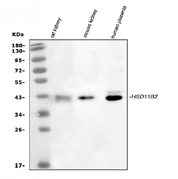
Western blot analysis of HSD11B2 using anti-HSD11B2 antibody (RP1094).
Electrophoresis was performed on a 5-20% SDS-PAGE gel at 70V (Stacking gel) / 90V (Resolving gel) for 2-3 hours. The sample well of each lane was loaded with 30 ug of sample under reducing conditions.
Lane 1: rat kidney tissue lysates,
Lane 2: mouse kidney tissue lysates,
Lane 3: human placenta tissue lysates.
After electrophoresis, proteins were transferred to a nitrocellulose membrane at 150 mA for 50-90 minutes. Blocked the membrane with 5% non-fat milk/TBS for 1.5 hour at RT. The membrane was incubated with rabbit anti-HSD11B2 antigen affinity purified polyclonal antibody (Catalog # RP1094) at 0.5 μg/mL overnight at 4°C, then washed with TBS-0.1%Tween 3 times with 5 minutes each and probed with a goat anti-rabbit IgG-HRP secondary antibody at a dilution of 1:5000 for 1.5 hour at RT. The signal is developed using an Enhanced Chemiluminescent detection (ECL) kit (Catalog # EK1002) with Tanon 5200 system. A specific band was detected for HSD11B2 at approximately 43 kDa. The expected band size for HSD11B2 is at 44 kDa.

Click image to see more details
IHC analysis of HSD11B2 using anti-HSD11B2 antibody (RP1094).
HSD11B2 was detected in a paraffin-embedded section of Mouse Pancreas tissue. Heat mediated antigen retrieval was performed in EDTA buffer (pH 8.0, epitope retrieval solution). The tissue section was blocked with 10% goat serum. The tissue section was then incubated with 1 μg/ml rabbit anti-HSD11B2 Antibody (RP1094) overnight at 4°C. Peroxidase Conjugated Goat Anti-rabbit IgG was used as secondary antibody and incubated for 30 minutes at 37°C. The tissue section was developed using HRP Conjugated Rabbit IgG Super Vision Assay Kit (Catalog # SV0002) with DAB as the chromogen.

Click image to see more details
IHC analysis of HSD11B2 using anti-HSD11B2 antibody (RP1094).
HSD11B2 was detected in a paraffin-embedded section of Rat Pancreas tissue. Heat mediated antigen retrieval was performed in EDTA buffer (pH 8.0, epitope retrieval solution). The tissue section was blocked with 10% goat serum. The tissue section was then incubated with 1 μg/ml rabbit anti-HSD11B2 Antibody (RP1094) overnight at 4°C. Peroxidase Conjugated Goat Anti-rabbit IgG was used as secondary antibody and incubated for 30 minutes at 37°C. The tissue section was developed using HRP Conjugated Rabbit IgG Super Vision Assay Kit (Catalog # SV0002) with DAB as the chromogen.

Click image to see more details
IHC analysis of HSD11B2 using anti-HSD11B2 antibody (RP1094).
HSD11B2 was detected in a paraffin-embedded section of Human Placenta tissue. Heat mediated antigen retrieval was performed in EDTA buffer (pH 8.0, epitope retrieval solution). The tissue section was blocked with 10% goat serum. The tissue section was then incubated with 1 μg/ml rabbit anti-HSD11B2 Antibody (RP1094) overnight at 4°C. Peroxidase Conjugated Goat Anti-rabbit IgG was used as secondary antibody and incubated for 30 minutes at 37°C. The tissue section was developed using HRP Conjugated Rabbit IgG Super Vision Assay Kit (Catalog # SV0002) with DAB as the chromogen.

Click image to see more details
IHC analysis of HSD11B2 using anti-HSD11B2 antibody (RP1094).
HSD11B2 was detected in a frozen section of human placenta tissue. The tissue section was blocked with 10% goat serum. The tissue section was then incubated with 5 μg/ml rabbit anti-HSD11B2 Antibody (RP1094) overnight at 4°C. Peroxidase Conjugated Goat Anti-rabbit IgG was used as secondary antibody and incubated for 30 minutes at 37°C. The tissue section was developed using HRP Conjugated Rabbit IgG Super Vision Assay Kit (Catalog # SV0002) with DAB as the chromogen.

Click image to see more details
IHC analysis of HSD11B2 using anti-HSD11B2 antibody (RP1094).
HSD11B2 was detected in a frozen section of mouse kidney tissue. The tissue section was blocked with 10% goat serum. The tissue section was then incubated with 2 μg/ml rabbit anti-HSD11B2 Antibody (RP1094) overnight at 4°C. Peroxidase Conjugated Goat Anti-rabbit IgG was used as secondary antibody and incubated for 30 minutes at 37°C. The tissue section was developed using HRP Conjugated Rabbit IgG Super Vision Assay Kit (Catalog # SV0002) with DAB as the chromogen.

Click image to see more details
IHC analysis of HSD11B2 using anti-HSD11B2 antibody (RP1094).
HSD11B2 was detected in a frozen section of rat kidney tissue. The tissue section was blocked with 10% goat serum. The tissue section was then incubated with 2 μg/ml rabbit anti-HSD11B2 Antibody (RP1094) overnight at 4°C. Peroxidase Conjugated Goat Anti-rabbit IgG was used as secondary antibody and incubated for 30 minutes at 37°C. The tissue section was developed using HRP Conjugated Rabbit IgG Super Vision Assay Kit (Catalog # SV0002) with DAB as the chromogen.

Click image to see more details
IF analysis of HSD11B2 using anti-HSD11B2 antibody (RP1094).
HSD11B2 was detected in a paraffin-embedded section of human placenta tissue. Heat mediated antigen retrieval was performed in EDTA buffer (pH 8.0, epitope retrieval solution). The tissue section was blocked with 10% goat serum. The tissue section was then incubated with 5 μg/mL rabbit anti-HSD11B2 Antibody (RP1094) overnight at 4°C. DyLight®488 Conjugated Goat Anti-Rabbit IgG (BA1127) was used as secondary antibody at 1:500 dilution and incubated for 30 minutes at 37°C. The section was counterstained with DAPI. Visualize using a fluorescence microscope and filter sets appropriate for the label used.
Specific Publications For Anti-HSD11B2 Antibody Picoband® (RP1094)
Loading publications
Recommended Resources
Here are featured tools and databases that you might find useful.
- Boster's Pathways Library
- Protein Databases
- Bioscience Research Protocol Resources
- Data Processing & Analysis Software
- Photo Editing Software
- Scientific Literature Resources
- Research Paper Management Tools
- Molecular Biology Software
- Primer Design Tools
- Bioinformatics Tools
- Phylogenetic Tree Analysis
Customer Reviews
Have you used Anti-HSD11B2 Antibody Picoband®?
Share your experimental results or join a short interview to earn up to $1,000 in product credits or other rewards.
0 Reviews For Anti-HSD11B2 Antibody Picoband®
Customer Q&As
Have a question?
Find answers in Q&As, reviews.
Can't find your answer?
Submit your question
4 Customer Q&As for Anti-HSD11B2 Antibody Picoband®
Question
I see that the anti-HSD11B2 antibody RP1094 works with WB, what is the protocol used to produce the result images on the product page?
Verified Customer
Verified customer
Asked: 2020-04-20
Answer
You can find protocols for WB on the "support/technical resources" section of our navigation menu. If you have any further questions, please send an email to support@bosterbio.com
Boster Scientific Support
Answered: 2020-04-20
Question
My question regards to test anti-HSD11B2 antibody RP1094 on mouse placenta for research purposes, then I may be interested in using anti-HSD11B2 antibody RP1094 for diagnostic purposes as well. Is the antibody suitable for diagnostic purposes?
Verified Customer
Verified customer
Asked: 2019-05-31
Answer
The products we sell, including anti-HSD11B2 antibody RP1094, are only intended for research use. They would not be suitable for use in diagnostic work. If you have the means to develop a product into diagnostic use, and are interested in collaborating with us and develop our product into an IVD product, please contact us for more discussions.
Boster Scientific Support
Answered: 2019-05-31
Question
Thanks for helping with my inquiry over the phone. Here are the WB image, lot number and protocol we used for placenta using anti-HSD11B2 antibody RP1094. Let me know if you need anything else.
Verified Customer
Verified customer
Asked: 2018-10-29
Answer
Thanks for the data. You have provided everything we needed. Our lab team are working to resolve your inquiry as quickly as possible, and we appreciate your patience and understanding! Please let me know if there is anything you need in the meantime.
Boster Scientific Support
Answered: 2018-10-29
Question
We are currently using anti-HSD11B2 antibody RP1094 for human tissue, and we are content with the WB results. The species of reactivity given in the datasheet says human, mouse, rat. Is it possible that the antibody can work on canine tissues as well?
M. Johnson
Verified customer
Asked: 2013-09-20
Answer
The anti-HSD11B2 antibody (RP1094) has not been tested for cross reactivity specifically with canine tissues, though there is a good chance of cross reactivity. We have an innovator award program that if you test this antibody and show it works in canine you can get your next antibody for free. Please contact me if I can help you with anything.
Boster Scientific Support
Answered: 2013-09-20
