Click image to see more details
Product Info Summary
| SKU: | M00458-2 |
|---|---|
| Size: | 0.1 mg |
| Reactive Species: | Human |
| Host: | Mouse |
| Application: | Flow Cytometry, IP, IHC-P, IHC-F, WB |
Customers Who Bought This Also Bought

Product info
Product Name
Anti-Hu CD18 Purified ITGB2 Monoclonal Antibody
SKU/Catalog Number
M00458-2
Size
0.1 mg
Form
Liquid
Description
Boster Bio Anti-Hu CD18 Purified ITGB2 Monoclonal Antibody (Catalog# M00458-2). Tested in Flow Cytometry, IP, WB, IHC-P, IHC-F application(s). This antibody reacts with Human.
Storage & Handling
Store at 2-8°C. Do not freeze.
Cite This Product
Anti-Hu CD18 Purified ITGB2 Monoclonal Antibody (Boster Biological Technology, Pleasanton CA, USA, Catalog # M00458-2)
Host
Mouse
Contents
Phosphate buffered saline (PBS), pH 7.4, 15 mM sodium azide
Clonality
Monoclonal
Clone Number
MEM-48
Isotype
Mouse IgG1
Immunogen
Leukocytes of a patient suffering from a LGL-type leukemia. The antibody MEM-48 recognizes an extracellular epitope involving residues 534-546 in cysteine-rich repeat 3 of the CD18 antigen (integrin beta2 subunit; beta2 integrin). CD18 is a 90-95 kDa type I transmembrane protein expressed on all leukocytes.
Reactive Species
M00458-2 is reactive to ITGB2 in Human
Observed Molecular Weight
90-100 kDa
Calculated molecular weight
84.8 kDa
Background of ITGB2
CD18, integrin beta2 subunit, forms heterodimers with four types of CD11 molecule to constitute leukocyte (beta2) integrins: alphaLbeta2 (CD11a/CD18, LFA-1), alphaMbeta2 (CD11b/CD18, Mac-1, CR3), alphaXbeta2 (CD11c/CD18) and alphaDbeta2 (CD11d/CD18). In most cases, the response mediated by the integrin is a composite of the functions of its individual subunits. These integrins are essential for proper leukocyte migration, mediating intercellular contacts. Absence of CD18 leads to leukocyte adhesion deficiency-1; severe reduction of CD18 expression leads to the development of a psoriasiform skin disease. CD18 is also a target of Mannheimia (Pasteurella) haemolytica leukotoxin and is sufficient to mediate leukotoxin-mediated cytolysis.
Antibody Validation
Boster validates all antibodies on WB, IHC, ICC, Immunofluorescence, and ELISA with known positive control and negative samples to ensure specificity and high affinity, including thorough antibody incubations.
Application & Images
Applications
M00458-2 is guaranteed for Flow Cytometry, IP, IHC-P, IHC-F, WB Boster Guarantee
Recommend Dilution
| Application | Dilution | Species |
|---|---|---|
| Immunohistochemistry (paraffin sections): Recommended dilution: 10 μg/ml; positive tissue: spleen | microglia. |
Validation Images & Assay Conditions
Click image to see more details
Separation of human leukocytes (red-filled) from CD18 negative blood debris (black-dashed) in flow cytometry analysis (surface staining) of human peripheral whole blood stained using anti-human CD18 (MEM-48) purified antibody (concentration in sample 1 µg/ml, GAM APC).

Click image to see more details
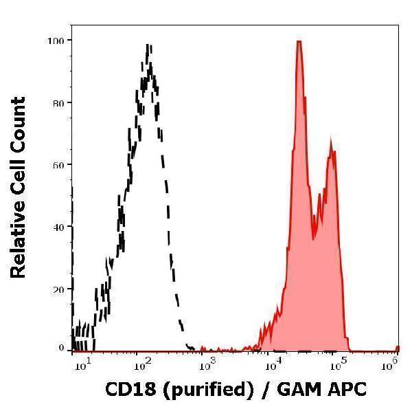
Flow cytometry surface staining pattern of human peripheral whole blood stained using anti-human CD18 (MEM-48) purified antibody (concentration in sample 1 µg/ml, GAM APC).

Click image to see more details
Immunohistochemistry staining of human spleen (paraffin sections) using anti-CD18 (MEM-48). Commercially tested by LifeSpan BioSciences.

Click image to see more details
Anti-Hu CD18 Purified (clone MEM-48) reactivity pattern in WB application.
The reactivity of MEM-48 antibody was assessed by comparing binding signals in a panel of various human cell samples.
Western blotting analysis was performed on RIPA buffer extracts of THP-1, Kg1a, U937, HPB-ALL, Raji, Jurkat, leukocytes, and HeLa cells, mixed and heated (100°C, 5 min) with reducing (2-mercaptoethanol) SDS-loading buffer. Samples were resolved using 10% SDS-PAGE gel.
Nitrocellulose membrane blot was probed with mouse IgG1 monoclonal antibody MEM-48 (2 µg/ml), followed by IRDye 800CW Goat-anti-Mouse IgG (green). For multiplex fluorescent Western blot detection, mouse anti-GAPDH monoclonal antibody FF26A conjugated with DyLight 680 (0.1 µg/ml) was used as the loading control (red).
CD18 was detected at ~90-100 kDa in the respective cell lines.
Specific Publications For Anti-Hu CD18 Purified ITGB2 Monoclonal Antibody (M00458-2)
Loading publications
Recommended Resources
Here are featured tools and databases that you might find useful.
- Boster's Pathways Library
- Protein Databases
- Bioscience Research Protocol Resources
- Data Processing & Analysis Software
- Photo Editing Software
- Scientific Literature Resources
- Research Paper Management Tools
- Molecular Biology Software
- Primer Design Tools
- Bioinformatics Tools
- Phylogenetic Tree Analysis
Customer Reviews
Have you used Anti-Hu CD18 Purified ITGB2 Monoclonal Antibody?
Share your experimental results or join a short interview to earn up to $1,000 in product credits or other rewards.
0 Reviews For Anti-Hu CD18 Purified ITGB2 Monoclonal Antibody
Customer Q&As
Have a question?
Find answers in Q&As, reviews.
Can't find your answer?
Submit your question
