Click image to see more details
-

-
-
-
-
+3
Product Info Summary
| SKU: | RP1009 |
|---|---|
| Size: | 100 μg/vial |
| Reactive Species: | Human |
| Host: | Rabbit |
| Application: | ELISA, IHC, WB |
Customers Who Bought This Also Bought

Product info
Product Name
Anti-Interleukin-4 IL4 Antibody Picoband®
SKU/Catalog Number
RP1009
BA14809 is an alternative SKU for this antibody, used in previous lots.
Size
100 μg/vial
Form
Lyophilized
Description
Boster Bio Anti-Interleukin-4 IL4 Antibody catalog # RP1009. Tested in ELISA, IHC, WB applications. This antibody reacts with Human. The brand Picoband indicates this is a premium antibody that guarantees superior quality, high affinity, and strong signals with minimal background in Western blot applications. Only our best-performing antibodies are designated as Picoband, ensuring unmatched performance.
Storage & Handling
Store at -20˚C for one year from date of receipt. After reconstitution, at 4˚C for one month. It can also be aliquotted and stored frozen at -20˚C for six months. Avoid repeated freeze-thaw cycles.
Cite This Product
Anti-Interleukin-4 IL4 Antibody Picoband® (Boster Biological Technology, Pleasanton CA, USA, Catalog # RP1009)
Host
Rabbit
Contents
Each vial contains 0.9mg NaCl, 0.2mg Na2HPO4, 0.05mg NaN3. Carrier free (No BSA) form available in stock. If you want this antibody carrier free please specify "Carrier Free" or "No BSA" in your order note.
Clonality
Polyclonal
Isotype
Rabbit IgG
Immunogen
E. coli-derived human IL-4 recombinant protein (Position: H25-S153).
Cross-reactivity
No cross-reactivity with other proteins
Reactive Species
RP1009 is reactive to IL4 in Human
Observed Molecular Weight
14 kDa
Calculated molecular weight
17.5 kDa
Background of IL4
Interleukin-4 (IL-4), also knowns as a B-cell stimulatory factor1 (BSF1), is an immunomodulatory cytokine, which can inhibit the growth of tumour cells.1 The human cDNA contains a single open reading frame encoding a protein of 153 amino acids, including a putative signal peptide. IL-4 may act as an autocrine growth factor in pancreatic cancer cells and also give rise to the possibility that cancer-derived IL-4 may suppress cancer-directed immunosurveillance in vivo in addition to its growth-promoting effects, thereby facilitating pancreatic tumor growth and metastasis.1 The mouse and human genes and their protein products show structural and functional similarities. The human IL-4 gene, which occurs as a single copy in the haploid genome, is mapped on chromosome 5.2 The standard product used in this kit is recombinant human IL-4, consisting of 130 amino acids with the molecular mass of 14KDa.
Antibody Validation
Boster validates all antibodies on WB, IHC, ICC, Immunofluorescence, and ELISA with known positive control and negative samples to ensure specificity and high affinity, including thorough antibody incubations.
Application & Images
Applications
RP1009 is guaranteed for ELISA, IHC, WB Boster Guarantee
Recommend Dilution
| Application | Dilution | Species |
|---|---|---|
| Immunohistochemistry (Paraffin-embedded Section) | 0.5-1μg/ml | Human |
| ELISA | 0.1-0.5μg/ml | - |
| Western blot | 0.1-0.5μg/ml | Human |
Validation Images & Assay Conditions
Click image to see more details
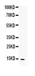
Figure. Western blot analysis of IL-4 using anti-IL-4 antibody (RP1009).
Electrophoresis was performed on a 5-20% SDS-PAGE gel at 70V (Stacking gel) / 90V (Resolving gel) for 2-3 hours. The sample well of each lane was loaded with 50ug of sample under reducing conditions.
Lane: Recombinant Human IL-4 Protein 0.5ng,
After Electrophoresis, proteins were transferred to a Nitrocellulose membrane at 150mA for 50-90 minutes. Blocked the membrane with 5% Non-fat Milk/ TBS for 1.5 hour at RT. The membrane was incubated with rabbit anti-IL-4 antigen affinity purified polyclonal antibody (Catalog # RP1009) at 0.5 μg/mL overnight at 4°C, then washed with TBS-0.1%Tween 3 times with 5 minutes each and probed with a goat anti-rabbit IgG-HRP secondary antibody at a dilution of 1:10000 for 1.5 hour at RT. The signal is developed using an Enhanced Chemiluminescent detection (ECL) kit (Catalog # EK1002) with Tanon 5200 system. A specific band was detected for IL-4 at approximately 14KD. The expected band size for IL-4 is at 14KD.

Click image to see more details
Protective effect of M2 macrophages induced by hUCMSCs-EVs on OA chondrocytes in vitro. A IL-1β-induced OA chondrocytes were co-cultured with the supernatant of M2 macrophages (M2S) induced by hUCMSCs-EVs, or platelet-rich plasma (PRP) for 48 h, relative mRNA expression of the key genes TNF-α, MMP13, SOX9, and ACAN was measured by quantitative RT-PCR analysis; the experiment was performed triplicate; *p < 0.05, **p < 0.01, ***p < 0.001. B Western blot was performed to evaluate the expression of TNF-α, MMP13, and IL-4 proteins in PBS, M2S, or PRP-treated OA chondrocytes; GAPDH was employed as the loading control; *p < 0.05, **p < 0.01, ***p < 0.001. C The influence of M2S or PRP on the viability of chondrocytes was detected by the cell live/death experiment; green represents live cells while red represents dead cells; Scale bar: 1 mm
Index in PubMed under a CC BY license. PMID: 35057811

Click image to see more details
Serum levels of IP-10, IFN-γ, IL-4, and TGF-β1 as well as the IFN-γ/IL-4 ratio in chronic hepatitis B patients with or without fibrosis. The levels of serum IP-10, IFN-γ, IL-4, and TGF-β1 in CHB patients with or without liver fibrosis were determined by ELISA, and the IFN-γ/IL-4 ratio was calculated. ♦♦♦ Differs from controls (the F0 group), P < 0.05; ★ ★ ★ differs from mild or minimal fibrosis (the F1–2 group), P < 0.05; ▴ ▴ ▴ differs from moderate fibrosis (the F3–4 group), P < 0.05. ( a ) IP-10; ( b ) IFN-γ; ( c ) IL-4; ( d ) TGF-β1; ( e ) the IFN-γ/IL-4 ratio.
Index in PubMed under a CC BY license. PMID: 28067328

Click image to see more details
Statistical analysis of the correlation between the serum IP-10 level or the IFN-γ/IL-4 ratio with liver fibrosis among chronic hepatitis B patients. Spearman’s correlation analysis of the association between ( a ) IP-10; ( b ) IFN-γ; ( c ) the IFN-γ/IL-4 ratio and TGF-β1. Spearman’s correlation analysis of the association between serum ( d ) IFN-γ; ( e ) IL-4; ( f ) the IFN-γ/IL-4 ratio and IP-10.
Index in PubMed under a CC BY license. PMID: 28067328

Click image to see more details
ROC curve analysis for evaluating the sensitivity and specificity of the IP-10 level, IFN-γ/IL-4 ratio, or their combination to predict significant fibrosis among CHB patients. ( a ) ROC curve analysis for serum IP-10 (with the cut-of f value of 300 pg/mL), the serum IFN-γ/IL-4 ratio (with the cut off value of 1.8), and the combination of IP-10 and the IFN-γ/IL-4 ratio; ( b ) Specificity and sensitivity for IP-10, the IFN-γ/IL-4 ratio, and their combination to predict significant liver fibrosis among patients with CHB.
Index in PubMed under a CC BY license. PMID: 28067328

Click image to see more details
Intrahepatic mRNA levels of IP-10, IFN-γ, and IL-4 in chronic hepatitis B patients with or without fibrosis. Real-time qRT-PCR was conducted to quantify the mRNA levels of intrahepatic IP-10, IFN-γ, and IL-4 in the CHB patients without or with fibrosis as described in the Materials and Methods section. The relative mRNA levels of intrahepatic IP-10, IFN-γ, and IL-4 were calculated by comparative Ct analysis after normalization for the quantity of GAPDH in the same samples and were represented as 2 - △ △ Ct values for controls (the F0 group), which were set equal 1. ♦♦♦ Differs from controls (the F0 group), P < 0.05; ★ ★ ★ differs from mild or minimal fibrosis (the F1–2 group), P < 0.05; ▴ ▴ ▴ differs from moderate fibrosis (the F3–4 group), P < 0.05. (a) IP-10; (b) IFN-γ; (c) IL-4.
Index in PubMed under a CC BY license. PMID: 28067328

Click image to see more details
Intrahepatic protein expression of IP-10, IFN-γ, IL-4, TGF-β1, and α-SMA as well as the IFN-γ/IL-4 ratio in chronic hepatitis B patients with or without fibrosis. The protein expression of intrahepatic (a, b, c, and d) IP-10, (e, f, g, and h) IFN-γ, and (i, g, k, and l) IL-4. In addition, the protein levels of intrahepatic IP-10, IFN-γ, IL-4, TGF-β1, and α-SMA were quantified based on the value of integrated optical density (IOD) and represented as histograms, from which the IFN-γ/IL-4 ratio was calculated. ♦♦♦ Differs from controls (the F0 group), P < 0.05; ★ ★ ★ differs from mild or minimal fibrosis (the F1–2 group), P < 0.05; ▴ ▴ ▴ differs from moderate fibrosis (the F3–4 group), P < 0.05.
Index in PubMed under a CC BY license. PMID: 28067328
Specific Publications For Anti-Interleukin-4 IL4 Antibody Picoband® (RP1009)
Loading publications
Recommended Resources
Here are featured tools and databases that you might find useful.
- Boster's Pathways Library
- Protein Databases
- Bioscience Research Protocol Resources
- Data Processing & Analysis Software
- Photo Editing Software
- Scientific Literature Resources
- Research Paper Management Tools
- Molecular Biology Software
- Primer Design Tools
- Bioinformatics Tools
- Phylogenetic Tree Analysis
Customer Reviews
Have you used Anti-Interleukin-4 IL4 Antibody Picoband®?
Share your experimental results or join a short interview to earn up to $1,000 in product credits or other rewards.
0 Reviews For Anti-Interleukin-4 IL4 Antibody Picoband®
Customer Q&As
Have a question?
Find answers in Q&As, reviews.
Can't find your answer?
Submit your question
4 Customer Q&As for Anti-Interleukin-4 IL4 Antibody Picoband®
Question
My boss were content with the WB result of your anti-IL4 antibody. However we have observed positive staining in oocyte secreted. using this antibody. Is that expected? Could you tell me where is IL4 supposed to be expressed?
Verified Customer
Verified customer
Asked: 2019-12-30
Answer
According to literature, oocyte does express IL4. Generally IL4 expresses in secreted. Regarding which tissues have IL4 expression, here are a few articles citing expression in various tissues:
Blood, Pubmed ID: 15489334
Boster Scientific Support
Answered: 2019-12-30
Question
We bought anti-IL4 antibody for ELISA on oocyte a few years ago. I am using human, and We are going to use the antibody for IHC next. you antibody examining oocyte as well as blood in our next experiment. Could you please give me some suggestion on which antibody would work the best for IHC?
Verified Customer
Verified customer
Asked: 2018-04-16
Answer
I have checked the website and datasheets of our anti-IL4 antibody and it appears that RP1009 has been validated on human in both ELISA and IHC. Thus RP1009 should work for your application. Our Boster satisfaction guarantee will cover this product for IHC in human even if the specific tissue type has not been validated. We do have a comprehensive range of products for IHC detection and you can check out our website bosterbio.com to find out more information about them.
Boster Scientific Support
Answered: 2018-04-16
Question
We have been able to see staining in human blood. Are there any suggestions? Is anti-IL4 antibody supposed to stain blood positively?
Verified Customer
Verified customer
Asked: 2017-11-23
Answer
Based on literature blood does express IL4. Based on Uniprot.org, IL4 is expressed in oocyte, blood, among other tissues. Regarding which tissues have IL4 expression, here are a few articles citing expression in various tissues:
Blood, Pubmed ID: 15489334
Boster Scientific Support
Answered: 2017-11-23
Question
We are currently using anti-IL4 antibody RP1009 for human tissue, and we are content with the WB results. The species of reactivity given in the datasheet says human. Is it possible that the antibody can work on dog tissues as well?
K. Krishna
Verified customer
Asked: 2014-06-19
Answer
The anti-IL4 antibody (RP1009) has not been validated for cross reactivity specifically with dog tissues, but there is a good chance of cross reactivity. We have an innovator award program that if you test this antibody and show it works in dog you can get your next antibody for free. Please contact me if I can help you with anything.
Boster Scientific Support
Answered: 2014-06-19
