Click image to see more details
-

-
-
-
-
+1
Product Info Summary
| SKU: | A03286-2 |
|---|---|
| Size: | 0.1 mg |
| Reactive Species: | Human |
| Host: | Rabbit |
| Application: | ELISA, IF, IHC-P, WB |
Customers Who Bought This Also Bought

Product info
Product Name
Anti-IL-32 Antibody
SKU/Catalog Number
A03286-2
Size
0.1 mg
Form
Liquid
Description
Boster Bio Anti-IL-32 Antibody (Catalog # A03286-2). Tested in ELISA, WB, IHC-P, IF applications. This antibody reacts with Human.
Storage & Handling
IL-32 antibody can be stored at 4°C for three months and -20°C, stable for up to one year. Avoid repeated freeze-thaw cycles. Antibodies should not be exposed to prolonged high temperatures.
Cite This Product
Anti-IL-32 Antibody (Boster Biological Technology, Pleasanton CA, USA, Catalog # A03286-2)
Host
Rabbit
Contents
IL-32 Antibody is supplied in PBS containing 0.02% sodium azide.
Clonality
Polyclonal
Isotype
IgG
Immunogen
Anti-IL-32 antibody was raised against a peptide corresponding to 15 amino acids near the carboxy terminus of human IL-32. The immunogen is located within the last 50 amino acids of IL-32.
Cross-reactivity
This antibody detects all isoforms of IL-32.
Reactive Species
A03286-2 is reactive to IL32 in Human
Observed Molecular Weight
68 kDa
Calculated molecular weight
26.7 kDa
Background of IL32
Interleukin-32 (IL-32) was initially identified as a transcript (NK4) that is selectively expressed in lymphocytes and NK cells and whose expression is increased following activation by IL-2. It was later re-isolated from an IL-18-treated lung carcinoma cell line and re-named IL-32. IL-32 is unusual in that it does not share sequence homology with known cytokine families and is highly expressed in immune tissues, existing in at least four differentially spliced isoforms. Because treatment of human monocytic and mouse macrophage cells with IL-32 induces several proinflammatory cytokines such as TNF-α, IL-8 and MIP-2, and because it is also induced in human peripheral lymphocyte cells after mitogen stimulation and in epithelial cells by IFNγ, it has been suggested that IL-32 may play a role in autoimmune and inflammatory diseases such as rheumatoid arthritis.
Antibody Validation
Boster validates all antibodies on WB, IHC, ICC, Immunofluorescence, and ELISA with known positive control and negative samples to ensure specificity and high affinity, including thorough antibody incubations.
Application & Images
Applications
A03286-2 is guaranteed for ELISA, IF, IHC-P, WB Boster Guarantee
Recommend Dilution
WB: 0.5-4 μg/mL; IHC: 2-10 μg/mL; IF: 20 μg/mL.
Antibody validated: Western Blot in human samples; Immunohistochemistry in human and mouse samples; Immunofluorescence in human samples. All other applications and species not yet tested.
Optimal dilutions for each application should be determined by the researcher.
Validation Images & Assay Conditions
Click image to see more details
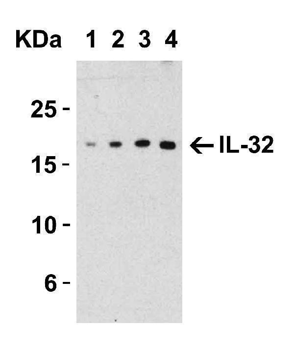
Western Blot Validation with Recombinant Protein
Loading: 30 ng of human IL-32 recombinant protein per lane.
Antibodies: IL-32, A03286-2 (Lane 1: 0.5 μg/mL, Lane 2: 1 μg/mL, Lane 3: 2 μg/mL and Lane 4: 4 μg/mL), 1h incubation at RT in 5% NFDM/TBST.
Secondary: Goat anti-rabbit IgG HRP conjugate at 1:10000 dilution.
Observed at around 17kD.

Click image to see more details
Western Blot Validation in Human Spleen Lysate, showing two isoforms of IL-32
Loading: 15 μg of lysates per lane.
Antibodies: IL-32, A03286-2 (lane 1: 2 μg/mL and lane 2: 4 μg/mL), 1h incubation at RT in 5% NFDM/TBST.
Secondary: Goat anti-rabbit IgG HRP conjugate at 1:10000 dilution.

Click image to see more details
Immunohistochemistry Validation of IL-32 in Human Spleen Tissue
Immunohistochemical analysis of paraffin-embedded human spleen tissue using anti-IL-32 antibody (A03286-2) at 10 μg/ml. Tissue was fixed with formaldehyde and blocked with 10% serum for 1 h at RT; antigen retrieval was by heat mediation with a citrate buffer (pH6). Samples were incubated with primary antibody overnight at 4˚C. A goat anti-rabbit IgG H&L (HRP) at 1/250 was used as secondary. Counter stained with Hematoxylin.

Click image to see more details
Immunofluorescence Validation of IL-32 in Human Spleen Cells
Immunofluorescent analysis of 4% paraformaldehyde-fixed human spleen cells labeling IL-32 with A03286-2 at 20 μg/mL, followed by goat anti-rabbit IgG secondary antibody at 1/500 dilution (red).

Click image to see more details
Immunohistochemistry Validation of IL-32 in Mouse Spleen Tissue
Immunohistochemical analysis of paraffin-embedded mouse spleen tissue using anti-IL-32 antibody (A03286-2) at 2 μg/ml. Tissue was fixed with formaldehyde and blocked with 10% serum for 1 h at RT; antigen retrieval was by heat mediation with a citrate buffer (pH6). Samples were incubated with primary antibody overnight at 4˚C. A goat anti-rabbit IgG H&L (HRP) at 1/250 was used as secondary. Counter stained with Hematoxylin.
Specific Publications For Anti-IL-32 Antibody (A03286-2)
Loading publications
Recommended Resources
Here are featured tools and databases that you might find useful.
- Boster's Pathways Library
- Protein Databases
- Bioscience Research Protocol Resources
- Data Processing & Analysis Software
- Photo Editing Software
- Scientific Literature Resources
- Research Paper Management Tools
- Molecular Biology Software
- Primer Design Tools
- Bioinformatics Tools
- Phylogenetic Tree Analysis
Customer Reviews
Have you used Anti-IL-32 Antibody?
Share your experimental results or join a short interview to earn up to $1,000 in product credits or other rewards.
0 Reviews For Anti-IL-32 Antibody
Customer Q&As
Have a question?
Find answers in Q&As, reviews.
Can't find your answer?
Submit your question
