Click image to see more details
-

-
-
-
-
+1
Product Info Summary
| SKU: | A01152-3 |
|---|---|
| Size: | 100 μg/vial |
| Reactive Species: | Mouse, Rat |
| Host: | Rabbit |
| Application: | ELISA, IHC, WB |
Customers Who Bought This Also Bought

Product info
Product Name
Anti-IL12B Antibody Picoband®
SKU/Catalog Number
A01152-3
Size
100 μg/vial
Form
Lyophilized
Description
Boster Bio Anti-IL12B Antibody Picoband® catalog # A01152-3. Tested in ELISA, IHC, WB applications. This antibody reacts with Mouse, Rat. The brand Picoband indicates this is a premium antibody that guarantees superior quality, high affinity, and strong signals with minimal background in Western blot applications. Only our best-performing antibodies are designated as Picoband, ensuring unmatched performance.
Storage & Handling
Store at -20˚C for one year from date of receipt. After reconstitution, at 4˚C for one month. It can also be aliquotted and stored frozen at -20˚C for six months. Avoid repeated freeze-thaw cycles.
Cite This Product
Anti-IL12B Antibody Picoband® (Boster Biological Technology, Pleasanton CA, USA, Catalog # A01152-3)
Host
Rabbit
Contents
Each vial contains 4mg Trehalose, 0.9mg NaCl, 0.2mg Na2HPO4, 0.05mg NaN3.
Clonality
Polyclonal
Isotype
Rabbit IgG
Immunogen
E. coli-derived rat IL12B recombinant protein (Position: M23-E250).
Cross-reactivity
No cross-reactivity with other proteins.
Reactive Species
A01152-3 is reactive to IL12B in Mouse, Rat
Observed Molecular Weight
37-45 kDa
Background of IL12B
Subunit beta of interleukin 12 (IL12B), also known as interleukin 12B, or interleukin-12 subunit p40, is a subunit of human interleukin 12. It is mapped to 5q33.3. This cytokine is expressed by activated macrophages that serve as an essential inducer of Th1 cells development. This cytokine has been found to be important for sustaining a sufficient number of memory/effector Th1 cells to mediate long-term protection to an intracellular pathogen. Overexpression of this gene was observed in the central nervous system of patients with multiple sclerosis (MS), suggesting a role of this cytokine in the pathogenesis of the disease.
Antibody Validation
Boster validates all antibodies on WB, IHC, ICC, Immunofluorescence, and ELISA with known positive control and negative samples to ensure specificity and high affinity, including thorough antibody incubations.
Application & Images
Applications
A01152-3 is guaranteed for ELISA, IHC, WB Boster Guarantee
Assay Dilutions Recommendation
The recommendations below provide a starting point for assay optimization. The actual working concentration varies and should be decided by the user.
Western blot, 0.1-0.5μg/ml
Immunohistochemistry (Paraffin-embedded Section), 0.5-1μg/ml
ELISA, 0.1-0.5μg/ml
Positive Control
WB: rat kidney tissue
IHC: rat spleen tissue
Validation Images & Assay Conditions
Click image to see more details
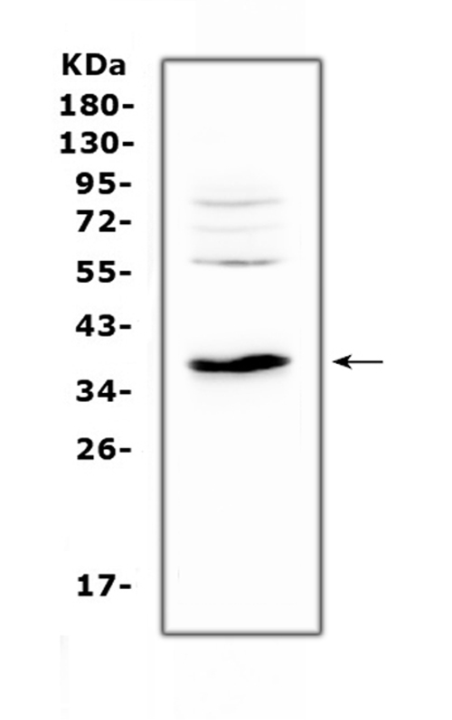
Western blot analysis of IL12B using anti-IL12B antibody (A01152-3).
Electrophoresis was performed on a 5-20% SDS-PAGE gel at 70V (Stacking gel) / 90V (Resolving gel) for 2-3 hours. The sample well of each lane was loaded with 50ug of sample under reducing conditions.
Lane 1: rat kidney tissue lysates.
After Electrophoresis, proteins were transferred to a Nitrocellulose membrane at 150mA for 50-90 minutes. Blocked the membrane with 5% Non-fat Milk/ TBS for 1.5 hour at RT. The membrane was incubated with rabbit anti-IL12B antigen affinity purified polyclonal antibody (Catalog # A01152-3) at 0.5 ug/mL overnight at 4 then washed with TBS-0.1%Tween 3 times with 5 minutes each and probed with a goat anti-rabbit IgG-HRP secondary antibody at a dilution of 1:10000 for 1.5 hour at RT. The signal is developed using an Enhanced Chemiluminescent detection (ECL) kit (Catalog # EK1002) with Tanon 5200 system. A specific band was detected for IL12B at approximately 37-45KD. The expected band size for IL12B is at 37KD.

Click image to see more details
IL-25 inhibited the production of IL-12, IL-23 in amouse model of of neutrophilia-dominant airway inflammation. ( A - D ) Measurement of Il12a , Il12b , Il23a and Il25 mRNA levels in mouse lung tissue using RT-PCR. ( E ) Representative images of IL-12B and CD80 immunofluorescence staining in mouse lung sections. Scale bar, 50 μm. ( F ) The proportion of CD80 staining-positive cells in mouse lung sections in different groups. ( G ) The proportion of IL-12B staining-positive cells in mouse lung sections in different groups. ( H ) The proportion of CD80 and IL-12B staining-positive cells in mouse lung sections in different groups. There were 4–6 mice in each group. One-way ANOVA was used for statistical analysis (* P < 0.05; ** P < 0.01; *** P < 0.001)
Index in PubMed under a CC BY license. PMID: 37898756

Click image to see more details
Exogenous IL-25 inhibited LPS-induced M1 polarization and the expression of IL-12 and IL-23 in mouse pulmonary cells. ( A ) Representative Western blots showing IL-12 A and β-Tubulin protein in primary culture of mouse pulmonary macrophages. ( B ) Quantitative analysis of IL-12 A in mouse pulmonary macrophages using ImageJ. Values are expressed in arbitrary units (a.u.). ( C ) Representative Western blots showing IL-12B, IL-23 A, and β-Tubulin protein in mouse pulmonary macrophages. ( D - E ) Quantitative analysis IL-12B, and IL-23A in mouse pulmonary macrophages using ImageJ. ( F - J ) Detection of Il12a, Il12b, Il23a, Cd80 , and Il-1β mRNA level in mouse pulmonary macrophages using RT-PCR. The experiment was repeated 3 times independently, and a similar trend was obtained. One-way ANOVA was used for statistical analysis (* P < 0.05; ** P < 0.01; *** P < 0.001)
Index in PubMed under a CC BY license. PMID: 37898756

Click image to see more details
The expression of IL-25 was decreased whereas IL-12, IL-23, and M1 macrophage markers were increased in severe of non-eosinophilic asthmatics. ( A ) IL-25 protein levels were decreased in sputum from a cohort of non-smoker severe asthma in the U-BIOPRED study. Based on the analysis of the data provided by Takahashi et al. [ ], 158 downregulated and 187 upregulated proteins were identified in the supernatant of induced sputum from non-smoker severe asthma patients (n = 37) compared to controls (n = 18) by proteomic assay and were shown in the volcano plot. The protein level of IL-25 (indicated by the red arrow) was decreased in the supernatant of induced sputum from non-smoker severe asthma patients compared to controls (log2 of fold change = -0.274, P = 0.019). ( B ) Detection of IL-25 mRNA level in airway brushings of eosinophilic asthma (n = 20) and non-eosinophilic asthma (n = 14) by RT-PCR. ( C - E ) Detection of IL-12 A, IL-12B and IL-23 A mRNA level in induced sputum of eosinophilic asthma (n = 17) and non-eosinophilic asthma (n = 12) by RT-PCR. ( F - J ) Detection of I L-1β, IFN-γ, CD80, iNOS , and CD206 mRNA level in induced sputum of eosinophilic asthma (n = 17) and non-eosinophilic asthma (n = 12) by RT-PCR. ( K ) Representative images of IL-12B and CD80 immunofluorescence staining in BALF cells of control, eosinophilic asthma, and non-eosinophilic asthma. Scale bar, 5 μm. ( L ) Detection of IL-25 mRNA level in induced sputum of eosinophilic asthma (n = 17) and non-eosinophilic asthma (n = 12) by RT-PCR. One way ANOVA was used for statistical analysis. (* P < 0.05; ** P < 0.01; *** P < 0.001)
Index in PubMed under a CC BY license. PMID: 37898756

Click image to see more details
IHC analysis of IL12B using anti-IL12B antibody (A01152-3).
IL12B was detected in paraffin-embedded section of rat spleen tissue. Heat mediated antigen retrieval was performed in citrate buffer (pH6, epitope retrieval solution) for 20 mins. The tissue section was blocked with 10% goat serum. The tissue section was then incubated with 1ug/ml rabbit anti-IL12B Antibody (A01152-3) overnight at 4 Biotinylated goat anti-rabbit IgG was used as secondary antibody and incubated for 30 minutes at 37 The tissue section was developed using Strepavidin-Biotin-Complex (SABC)(Catalog # SA1022) with DAB as the chromogen.
Specific Publications For Anti-IL12B Antibody Picoband® (A01152-3)
Loading publications
Recommended Resources
Here are featured tools and databases that you might find useful.
- Boster's Pathways Library
- Protein Databases
- Bioscience Research Protocol Resources
- Data Processing & Analysis Software
- Photo Editing Software
- Scientific Literature Resources
- Research Paper Management Tools
- Molecular Biology Software
- Primer Design Tools
- Bioinformatics Tools
- Phylogenetic Tree Analysis
Customer Reviews
Have you used Anti-IL12B Antibody Picoband®?
Share your experimental results or join a short interview to earn up to $1,000 in product credits or other rewards.
0 Reviews For Anti-IL12B Antibody Picoband®
Customer Q&As
Have a question?
Find answers in Q&As, reviews.
Can't find your answer?
Submit your question
