Click image to see more details
Product Info Summary
| SKU: | A01097-1 |
|---|---|
| Size: | 100 μg/vial |
| Reactive Species: | Human, Mouse, Rat |
| Host: | Rabbit |
| Application: | ELISA, WB |
Customers Who Bought This Also Bought

Product info
Product Name
Anti-IL23/IL23A Antibody Picoband®
SKU/Catalog Number
A01097-1
Size
100 μg/vial
Form
Lyophilized
Description
Boster Bio Anti-IL23/IL23A Antibody Picoband® catalog # A01097-1. Tested in ELISA, WB applications. This antibody reacts with Human, Mouse, Rat. The brand Picoband indicates this is a premium antibody that guarantees superior quality, high affinity, and strong signals with minimal background in Western blot applications. Only our best-performing antibodies are designated as Picoband, ensuring unmatched performance.
Storage & Handling
Store at -20˚C for one year from date of receipt. After reconstitution, at 4˚C for one month. It can also be aliquotted and stored frozen at -20˚C for six months. Avoid repeated freeze-thaw cycles.
Cite This Product
Anti-IL23/IL23A Antibody Picoband® (Boster Biological Technology, Pleasanton CA, USA, Catalog # A01097-1)
Host
Rabbit
Contents
Each vial contains 4mg Trehalose, 0.9mg NaCl, 0.2mg Na2HPO4, 0.05mg NaN3.
Clonality
Polyclonal
Isotype
Rabbit IgG
Immunogen
E. coli-derived human IL23 recombinant protein (Position: R20-R178).
Cross-reactivity
No cross-reactivity with other proteins.
Reactive Species
A01097-1 is reactive to IL23A in Human, Mouse, Rat
Observed Molecular Weight
19 kDa
Calculated molecular weight
20.7 kDa
Background of IL23A
Interleukin-23 subunit alpha is a protein that in humans is encoded by the IL23A gene. IL-23, also known as Interleukin-23 subunit alphainin (IL23A), is a heterodimeric cytokine consisting of two subunits, one called p40, which is shared with another cytokine, IL-12, and another called p19 (the IL-23 alpha subunit). The International Radiation Hybrid Mapping Consortium mapped the IL-23 gene to chromosome 12. IL-23 is an important part of the inflammatory response against infection. It promotes upregulation of the matrix metalloprotease MMP9, increases angiogenesis and reduces CD8+ T-cell infiltration.
Antibody Validation
Boster validates all antibodies on WB, IHC, ICC, Immunofluorescence, and ELISA with known positive control and negative samples to ensure specificity and high affinity, including thorough antibody incubations.
Application & Images
Applications
A01097-1 is guaranteed for ELISA, WB Boster Guarantee
Recommend Dilution
| Application | Dilution | Species |
|---|---|---|
| Western blot | 0.1-0.5μg/ml | |
| ELISA | 0.1-0.5μg/ml |
Tested application
Suggested blocking solution with 5% non-fat milk or BSA; (*)Recommended protein loading: 20-40 µg per lane
Validation Images & Assay Conditions
Click image to see more details
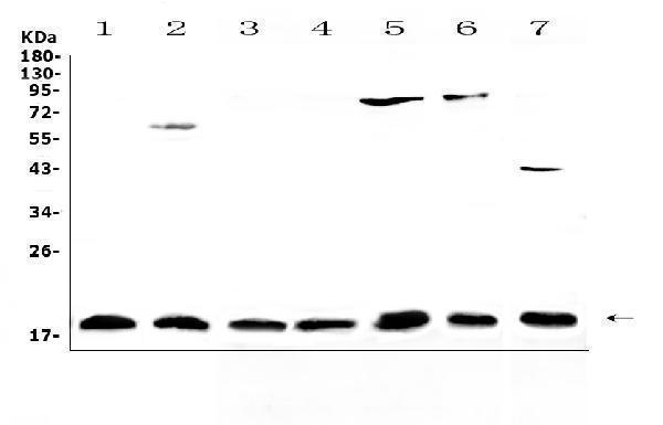
Western blot analysis of IL23 using anti-IL23 antibody (A01097-1).
Electrophoresis was performed on a 5-20% SDS-PAGE gel at 70V (Stacking gel) / 90V (Resolving gel) for 2-3 hours. The sample well of each lane was loaded with 50ug of sample under reducing conditions.
Lane 1: human placenta tissue lysates,
Lane 2: human PANC-1 cell lysates,
Lane 3: rat lymph tissue lysates,
Lane 4: rat small intestine tissue lysates,
Lane 5: rat testis tissue lysates,
Lane 6: rat ovary tissue lysates,
Lane 7: mouse testis tissue lysates.
After Electrophoresis, proteins were transferred to a Nitrocellulose membrane at 150mA for 50-90 minutes. Blocked the membrane with 5% Non-fat Milk/ TBS for 1.5 hour at RT. The membrane was incubated with rabbit anti-IL23 antigen affinity purified polyclonal antibody (Catalog # A01097-1) at 0.5 μg/mL overnight at 4°C, then washed with TBS-0.1%Tween 3 times with 5 minutes each and probed with a goat anti-rabbit IgG-HRP secondary antibody at a dilution of 1:10000 for 1.5 hour at RT. The signal is developed using an Enhanced Chemiluminescent detection (ECL) kit (Catalog # EK1002) with Tanon 5200 system. A specific band was detected for IL23 at approximately 19KD. The expected band size for IL23 is at 21KD.

Click image to see more details
Neutrophils, lung function and BALF IL-23/IL-17A levels between patients with stable COPD (con-COPD, n = 33) and patients with P. aeruginosa -infected COPD (PA-COPD, n = 34). PA-COPD patients had higher absolute and percent numbers of neutrophils in BALF, but not in blood (A, B) . Forced expiratory volums (C) , expiratory flows (D) and forced oscillation tests (E) were compared between con-COPD and PA-COPD patients. IL-23 and IL-17A levels were significantly elevated in patients with PA-COPD (F) . Data were presented as mean ± SD. *P < 0.05, ** P < 0.01, ns, non-significant. Correlation analysis showed that there was a notably positive correlation between the two cytokines (G) , and further regression analysis revealed that IL-23 might be an important factor contributing to elevation of IL-17A (H) . There was also a significantly positive correlation between IL-17A levels and absolute neutrophil numbers in BALF (I) . The spirometry results, including FEV 1.0 % predicted (J) , FEV 1.0 /FVC% (K) and MMEF (L) were negatively correlated with the levels of IL-17A. BALF, bronchoalveolar lavage fluid; COPD, chronic obstructive pulmonary disease; IL, interleukin; FEV 1.0 , forced expiratory volume in one second; FVC, forced vital capacity; MMEF, maximal mid-expiratory flow.
Index in PubMed under a CC BY license. PMID: 35095906

Click image to see more details
P. aeruginosa infection increased IL-23/IL-17 axis signaling in the lungs of COPD mouse models. C57BL/6 mice were exposed to ozone twice a week for 6 weeks to establish COPD models, and then were intrabronchially inoculated with sterile agar beads (con-COPD) or 1.0 × 10 5 CFU agar-entrapped P. aeruginosa (PA-COPD). Lungs were excised at one day post inoculation, and were sectioned and stained with masson trichrome to measure tissue fibrosis ( A , upper row), and PAS to quantify glycogen [ (A) , bottom row]. Blue areas around airways were identified as collagen deposition (indicated by black arrows), and purple areas within tracheal cavity were identified as mucus production and goblet cell hyperpasia (indicated by blue arrows). Internal scale bar = 100 μm. Quantitative real-time PCR, semiquantitative RT-PCR, and western-blot analysis of IL-23 and its receptor (B) , and IL-17A and its receptor (C) were performed using lung tissues. β-actin serves as the loading control. Data were presented as mean ± SD ( n = 5 per group). ** P < 0.01. The lungs were processed for immunofluorescent analysis (D) , and the sections were stained with anti-IL-17RA (red) and anti-Ly6G (green), and DAPI (blue) for nuclei. Internal scale bar = 100 μm. Two independent experiments were performed. Correlation analysis showed that there was a significantly negative correlation between the numbers of Ly6G + IL-17RA + cells and the spirometry results (E) . COPD, chronic obstructive pulmonary disease; CFU, colony-forming units; IL, interleukin; FVC, forced vital capacity; FEV 50 , volume expired in the first 50 ms of fast expiration.
Index in PubMed under a CC BY license. PMID: 35095906

Click image to see more details
Adjunctive therapy of IL-17A–neutralizing antibody to antibiotic improved the outcome of P. aeruginosa infection in COPD mouse models. COPD mouse models were intrabronchially inoculated with 1.0 × 10 5 CFU agar-entrapped P. aeruginosa . Oral ciprofloxacin (5 mg/kg, every 12 h) was applied concurrently with intraperitoneally administered IL-17A–neutralizing antibody (2 mg/kg, every 4 h) or IgG starting at 16 h post inoculation. Mouse lungs were excised, sectioned and stained with hematoxylin and eosin (internal scale bar = 100 μm) after treatment (A) , and the infiltrate scores (B) and MPO activity (C) were determined on each day. Bacterial plate counting (D) , protein levels of IL-17A (E) , IL-23 (F) , and RBP4 (G) , and spirometry tests, including FVC (H) , FEV 50 (I) , MMEF (J) , FEF 50 (K) , and FEF 75 (L) , were performed and compared between groups treated with and without IL-17A block. Data are representative of three independent experiments, and are presented as mean ± SD ( n = 3 per group). * P < 0.05. COPD, chronic obstructive pulmonary disease; Cip, ciprofloxacin; CFU, colony-forming units; MPO, myeloperoxidase; FVC, forced vital capacity; FEV50, volume expired in the first 50 ms of fast expiration; MMEF, maximal mid-expiratory flow; FEF 50 , forced expiratory flow at 50% FVC; FEF 75 , forced expiratory flow at 75% FVC.
Index in PubMed under a CC BY license. PMID: 35095906
Specific Publications For Anti-IL23/IL23A Antibody Picoband® (A01097-1)
Loading publications
Recommended Resources
Here are featured tools and databases that you might find useful.
- Boster's Pathways Library
- Protein Databases
- Bioscience Research Protocol Resources
- Data Processing & Analysis Software
- Photo Editing Software
- Scientific Literature Resources
- Research Paper Management Tools
- Molecular Biology Software
- Primer Design Tools
- Bioinformatics Tools
- Phylogenetic Tree Analysis
Customer Reviews
Have you used Anti-IL23/IL23A Antibody Picoband®?
Share your experimental results or join a short interview to earn up to $1,000 in product credits or other rewards.
0 Reviews For Anti-IL23/IL23A Antibody Picoband®
Customer Q&As
Have a question?
Find answers in Q&As, reviews.
Can't find your answer?
Submit your question
