Click image to see more details
-

-
-
-
-
+6
Product Info Summary
| SKU: | M00254-8 |
|---|---|
| Size: | 100 μg/vial |
| Reactive Species: | Human |
| Host: | Mouse |
| Application: | Flow Cytometry, IF, IHC, ICC |
Customers Who Bought This Also Bought

Product info
Product Name
Anti-Ki67 Antibody (monoclonal, 5E12)
SKU/Catalog Number
M00254-8
Size
100 μg/vial
Form
Lyophilized
Description
Boster Bio Anti-Ki67 Antibody (monoclonal, 5E12) catalog # M00254-8. Tested in Flow Cytometry, IF, IHC, ICC applications. This antibody reacts with Human.
Storage & Handling
Store at -20˚C for one year from date of receipt. After reconstitution, at 4˚C for one month. It can also be aliquotted and stored frozen at -20˚C for six months. Avoid repeated freeze-thaw cycles.
Cite This Product
Anti-Ki67 Antibody (monoclonal, 5E12) (Boster Biological Technology, Pleasanton CA, USA, Catalog # M00254-8)
Host
Mouse
Contents
Each vial contains 4mg Trehalose, 0.9mg NaCl and 0.2mg Na2HPO4.
Clonality
Monoclonal
Clone Number
5E12
Isotype
Mouse IgG2b
Immunogen
E. coli-derived human Ki67 recombinant protein (Position: K2860-I3256).
Cross-reactivity
No cross-reactivity with other proteins.
Reactive Species
M00254-8 is reactive to MKI67 in Human
Calculated molecular weight
358.7 kDa
Background of MKI67
Ki-67 (Proliferation-related Ki-67 antigen), also known as MKI67 or KIA, is a protein that in humans is encoded by the MKI67 gene. From study of a panel of human-rodent somatic cell hybrids, it has been demonstrated that a gene involved in the expression of the MKI67 antigen is located on chromosome 10. By in situ hybridization, Fonatsch et al. (1991) regionalized the MKI67 gene to chromosome 10q25-qter. By FISH, Traut et al. (1998) mapped the mouse Mki67 gene to chromosome 7F3-F5. Antigen KI-67 is a nuclear protein that is associated with and may be necessary for cellular proliferation. Furthermore it is associated with ribosomal RNA transcription. Inactivation of antigen KI-67 leads to inhibition of ribosomal RNA synthesis.
Antibody Validation
Boster validates all antibodies on WB, IHC, ICC, Immunofluorescence, and ELISA with known positive control and negative samples to ensure specificity and high affinity, including thorough antibody incubations.
Application & Images
Applications
M00254-8 is guaranteed for Flow Cytometry, IF, IHC, ICC Boster Guarantee
Assay Dilutions Recommendation
The recommendations below provide a starting point for assay optimization. The actual working concentration varies and should be decided by the user.
Immunohistochemistry (Paraffin-embedded Section), 2-5μg/ml, Human
Immunocytochemistry/Immunofluorescence, 5μg/ml, Human
Immunofluorescence, 5μg/ml, Human
Flow Cytometry (Fixed), 1-3μg/1x106 cells, Human
Positive Control
Use TE buffer pH 9.0 for antigen retrieval; (*) citrate buffer pH 6.0 is an alternative.
Validation Images & Assay Conditions
Click image to see more details
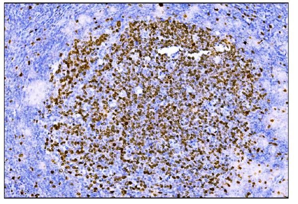
IHC analysis of Ki67 using anti-Ki67 antibody (M00254-8).
Ki67 was detected in paraffin-embedded section of human tonsil tissue. Heat mediated antigen retrieval was performed in EDTA buffer (pH8.0, epitope retrieval solution). The tissue section was blocked with 10% goat serum. The tissue section was then incubated with 2μg/ml mouse anti-Ki67 Antibody (M00254-8) overnight at 4°C. Biotinylated goat anti-mouse IgG was used as secondary antibody and incubated for 30 minutes at 37°C. The tissue section was developed using Strepavidin-Biotin-Complex (SABC) (Catalog # SA1021) with DAB as the chromogen.

Click image to see more details
IL-6 was closely associated with osimertinib resistance. a High-throughput transcriptome sequencing in 3 paired cell strains sensitive/resistant to osimertinib, respectively, as indicated. Left: The Venn diagram of the commonly and exclusively differentially expressed genes (DEGs), and the overall overlapping site refers to the DEGs commonly found in all groups. Right: The protein–protein interaction network of 233 DEGs. Red and green nodes represent upregulated and downregulated genes after TKI resistance, respectively. The size of nodes refers to the degree of interaction with other genes. GR, gefitinib resistance; OR, osimertinib resistance. b ELISA assay of IL-6 levels in culture medium (pg/ml) or cell lysates (pg/100 μg protein) from paired osimertinib-sensitive and osimertinib-resistant cells, as indicated ( n = 3 biologically independent experiments). Data are shown as mean ± SEM. *, p < 0.01 as compared to each parental cell strains. c and d Kaplan–Meier (KM) estimates of PFS in NSCLC patients treated with gefitinib, or osimertinib, respectively, according to high (greater than 7 mg/L) or low baseline IL-6 levels; e paired plasma IL-6 levels at baseline and disease progression of patients treated with gefitinib ( n = 126) or osimertinib ( n = 39). Lines indicate median and interquartile range of each group. P -value was calculated using a Mann–Whitney test. BL, baseline; PD, progression of disease. f gefitinib resistance mechanisms in low IL-6 group ( n = 39) and high IL-6 group ( n = 49), respectively. g Osimertinib resistance mechanisms in low IL-6 group ( n = 9) and high IL-6 group ( n = 17), respectively. h Cell viability CCK-8 assay for cells with different treatments as indicated. HCC827 cells, PC-9 cells, or PC-9GR cells were treated with IL-6 (20 ng/ml) together with increasing concentrations of osimertinib for 48 h. IL-6-GFP PC-9 cells, IL-6-GFP PC-9GR cells, and IL-6-GFP HCC827 cells were treated with indicated concentrations of osimertinib for 48 h. Data are shown as mean ± SEM ( n = 3 biologically independent experiments). i Ki67 incorporation assay on PC-9 cells and IL-6-GFP PC-9 cells with different treatments as indicated. IL-6 (20 ng/ml) or osimertinib (1 μM) were added to the culture medium for 48 h. Cells were counterstained with DAPI. Scale bars: 100 μm.
Index in PubMed under a CC BY license. PMID: 35197546

Click image to see more details
Macrophages were widely activated in the liver of patients with acute-on-chronic liver failure. A: Representative microscopic images of the double immunofluorescence staining of Ki67 and CD68 in the liver (400 fold). The red fluorescence signal represented Ki67; the green fluorescence signal represented CD68; and the blue fluorescence signal represented DAPI; B: Ki67 immunofluorescence intensity in the liver of healthy donors ( n = 10) and patients with acute-on-chronic liver failure ( n = 12); C: CD68 immunofluorescence intensity in the liver; D: The mean number of Ki67 + CD68 + -positive cells in five × 200 fields; E: Chemokine C-C motif ligand 2 levels in liver homogenates. Data with normal distribution were compared using unpaired student’s t -test, and the Mann-Whitney test was used for non-normal data. ACLF: Acute-on-chronic liver failure; HD: Healthy donor.
Index in PubMed under a CC BY license. PMID: 38516248

Click image to see more details
IHC analysis of Ki67 using anti-Ki67 antibody (M00254-8).
Ki67 was detected in paraffin-embedded section of human cervical cancer tissue. Heat mediated antigen retrieval was performed in EDTA buffer (pH8.0, epitope retrieval solution). The tissue section was blocked with 10% goat serum. The tissue section was then incubated with 2μg/ml mouse anti-Ki67 Antibody (M00254-8) overnight at 4°C. Biotinylated goat anti-mouse IgG was used as secondary antibody and incubated for 30 minutes at 37°C. The tissue section was developed using Strepavidin-Biotin-Complex (SABC) (Catalog # SA1021) with DAB as the chromogen.

Click image to see more details
IHC analysis of Ki67 using anti-Ki67 antibody (M00254-8).
Ki67 was detected in paraffin-embedded section of human esophageal squamous carcinoma tissue. Heat mediated antigen retrieval was performed in EDTA buffer (pH8.0, epitope retrieval solution). The tissue section was blocked with 10% goat serum. The tissue section was then incubated with 2μg/ml mouse anti-Ki67 Antibody (M00254-8) overnight at 4°C. Biotinylated goat anti-mouse IgG was used as secondary antibody and incubated for 30 minutes at 37°C. The tissue section was developed using Strepavidin-Biotin-Complex (SABC) (Catalog # SA1021) with DAB as the chromogen.

Click image to see more details
IHC analysis of Ki67 using anti-Ki67 antibody (M00254-8).
Ki67 was detected in paraffin-embedded section of human liver tissue. Heat mediated antigen retrieval was performed in EDTA buffer (pH8.0, epitope retrieval solution). The tissue section was blocked with 10% goat serum. The tissue section was then incubated with 2μg/ml mouse anti-Ki67 Antibody (M00254-8) overnight at 4°C. Biotinylated goat anti-mouse IgG was used as secondary antibody and incubated for 30 minutes at 37°C. The tissue section was developed using Strepavidin-Biotin-Complex (SABC) (Catalog # SA1021) with DAB as the chromogen.

Click image to see more details
IHC analysis of Ki67 using anti-Ki67 antibody (M00254-8).
Ki67 was detected in paraffin-embedded section of human lung cancer tissue. Heat mediated antigen retrieval was performed in EDTA buffer (pH8.0, epitope retrieval solution). The tissue section was blocked with 10% goat serum. The tissue section was then incubated with 2μg/ml mouse anti-Ki67 Antibody (M00254-8) overnight at 4°C. Biotinylated goat anti-mouse IgG was used as secondary antibody and incubated for 30 minutes at 37°C. The tissue section was developed using Strepavidin-Biotin-Complex (SABC) (Catalog # SA1021) with DAB as the chromogen.

Click image to see more details
IF analysis of Ki67 using anti-Ki67 antibody (M00254-8).
Ki67 was detected in immunocytochemical section of A431 cells. Enzyme antigen retrieval was performed using IHC enzyme antigen retrieval reagent (AR0022) for 15 mins. The cells were blocked with 10% goat serum. And then incubated with 5μg/mL mouse anti-Ki67 Antibody (M00254-8) overnight at 4°C. DyLight®488 Conjugated Goat Anti-Mouse IgG (BA1126) was used as secondary antibody at 1:100 dilution and incubated for 30 minutes at 37°C. The section was counterstained with DAPI. Visualize using a fluorescence microscope and filter sets appropriate for the label used.

Click image to see more details
IF analysis of Ki67 using anti-Ki67 antibody (M00254-8).
Ki67 was detected in paraffin-embedded section of human tonsil tissue. Heat mediated antigen retrieval was performed in EDTA buffer (pH8.0, epitope retrieval solution). The tissue section was blocked with 10% goat serum. The tissue section was then incubated with 5μg/mL mouse anti-Ki67 Antibody (M00254-8) overnight at 4°C. Biotin conjugated goat anti-mouse IgG (BA1001) was used as secondary antibody and incubated for 30 minutes at 37°C. The tissue section was developed using DyLight®488 Conjugated Avidin (BA1128). The section was counterstained with DAPI. Visualize using a fluorescence microscope and filter sets appropriate for the label used.

Click image to see more details
Flow Cytometry analysis of Jurkat cells using anti-Ki67 antibody (M00254-8).
Overlay histogram showing Jurkat cells stained with M00254-8 (Blue line).The cells were blocked with 10% normal goat serum. And then incubated with mouse anti-Ki67 Antibody (M00254-8, 1μg/1x106 cells) for 30 min at 20°C. DyLight®488 conjugated goat anti-mouse IgG (BA1126, 5-10μg/1x106 cells) was used as secondary antibody for 30 minutes at 20°C. Isotype control antibody (Green line) was mouse IgG (1μg/1x106) used under the same conditions. Unlabelled sample (Red line) was also used as a control.
Specific Publications For Anti-Ki67 Antibody (monoclonal, 5E12) (M00254-8)
Loading publications
Recommended Resources
Here are featured tools and databases that you might find useful.
- Boster's Pathways Library
- Protein Databases
- Bioscience Research Protocol Resources
- Data Processing & Analysis Software
- Photo Editing Software
- Scientific Literature Resources
- Research Paper Management Tools
- Molecular Biology Software
- Primer Design Tools
- Bioinformatics Tools
- Phylogenetic Tree Analysis
Customer Reviews
Have you used Anti-Ki67 Antibody (monoclonal, 5E12)?
Share your experimental results or join a short interview to earn up to $1,000 in product credits or other rewards.
0 Reviews For Anti-Ki67 Antibody (monoclonal, 5E12)
Customer Q&As
Have a question?
Find answers in Q&As, reviews.
Can't find your answer?
Submit your question
