
Click image to see more details
-

-
-
-
-
+8
Product Info Summary
| SKU: | PB9026 |
|---|---|
| Size: | 100 μg/vial |
| Reactive Species: | Human |
| Host: | Rabbit |
| Application: | IF, IHC, ICC, WB |
Customers Who Bought This Also Bought

Product info
Product Name
Anti-Ki67/MKI67 Antibody Picoband®
SKU/Catalog Number
PB9026
Size
100 μg/vial
Form
Lyophilized
Description
Boster Bio Anti-Ki67/MKI67 Antibody Picoband® catalog # PB9026. Tested in IF, IHC, ICC/IF, ICC, WB applications. This antibody reacts with Human. The brand Picoband indicates this is a premium antibody that guarantees superior quality, high affinity, and strong signals with minimal background in Western blot applications. Only our best-performing antibodies are designated as Picoband, ensuring unmatched performance.
Storage & Handling
Store at -20˚C for one year from date of receipt. After reconstitution, at 4˚C for one month. It can also be aliquotted and stored frozen at -20˚C for six months. Avoid repeated freeze-thaw cycles.
Cite This Product
Anti-Ki67/MKI67 Antibody Picoband® (Boster Biological Technology, Pleasanton CA, USA, Catalog # PB9026)
Host
Rabbit
Contents
Each vial contains 4 mg Trehalose, 0.9 mg NaCl and 0.2 mg Na2HPO4.
Clonality
Polyclonal
Isotype
Rabbit IgG
Immunogen
E.coli-derived human Ki67 recombinant protein (Position: K2860-I3256).
Cross-reactivity
No cross-reactivity with other proteins
Reactive Species
PB9026 is reactive to MKI67 in Human
Observed Molecular Weight
358 kDa
Calculated molecular weight
358.7 kDa
Background of MKI67
Ki-67 (Proliferation-related Ki-67 antigen), also known as MKI67 or KIA, is a protein that in humans is encoded by the MKI67 gene. From study of a panel of human-rodent somatic cell hybrids, it has been demonstrated that a gene involved in the expression of the MKI67 antigen is located on chromosome 10. By in situ hybridization, Fonatsch et al. (1991) regionalized the MKI67 gene to chromosome 10q25-qter. By FISH, Traut et al. (1998) mapped the mouse Mki67 gene to chromosome 7F3-F5. Antigen KI-67 is a nuclear protein that is associated with and may be necessary for cellular proliferation. Furthermore it is associated with ribosomal RNA transcription. Inactivation of antigen KI-67 leads to inhibition of ribosomal RNA synthesis.
Antibody Validation
Boster validates all antibodies on WB, IHC, ICC, Immunofluorescence, and ELISA with known positive control and negative samples to ensure specificity and high affinity, including thorough antibody incubations.
Application & Images
Applications
PB9026 is guaranteed for IF, IHC, ICC, WB Boster Guarantee
Recommend Dilution
| Application | Dilution | Species |
|---|---|---|
| Western blot | 0.1-0.5μg/ml | Human |
| Immunohistochemistry (Paraffin-embedded Section) | 0.5-1μg/ml | Human |
| Immunocytochemistry | 0.5-1μg/ml | Human, - |
| Immunofluorescence | 5μg/ml | Human |
Tested application
Suggested blocking solution with 5% non-fat milk or BSA; (*)Recommended protein loading: 20-40 µg per lane
Use TE buffer pH 9.0 for antigen retrieval; (*) citrate buffer pH 6.0 is an alternative.
Validation Images & Assay Conditions
Click image to see more details
IHC analysis of Ki67 using anti-Ki67 antibody (PB9026).
Ki67 was detected in paraffin-embedded section of human tonsil tissues. Heat mediated antigen retrieval was performed in citrate buffer (pH6, epitope retrieval solution) for 20 mins. The tissue section was blocked with 10% goat serum. The tissue section was then incubated with 1μg/ml rabbit anti-Ki67 Antibody (PB9026) overnight at 4°C. Biotinylated goat anti-rabbit IgG was used as secondary antibody and incubated for 30 minutes at 37°C. The tissue section was developed using Strepavidin-Biotin-Complex (SABC)(Catalog # SA1022) with DAB as the chromogen.

Click image to see more details
Western blot analysis of Ki67 using anti-Ki67 antibody (PB9026).
Electrophoresis was performed on a 5-20% SDS-PAGE gel at 70V (Stacking gel) / 90V (Resolving gel) for 2-3 hours. The sample well of each lane was loaded with 50ug of sample under reducing conditions.
Lane 1: HELA Whole Cell Lysate,
Lane 2: MCF-7 Whole Cell Lysate,
Lane 3: COLO320 Whole Cell Lysate,
Lane 4: HEPG2 Whole Cell Lysate,
Lane 5: SKOV Whole Cell Lysate.
After Electrophoresis, proteins were transferred to a Nitrocellulose membrane at 150mA for 50-90 minutes. Blocked the membrane with 5% Non-fat Milk/ TBS for 1.5 hour at RT. The membrane was incubated with rabbit anti-Ki67 antigen affinity purified polyclonal antibody (Catalog # PB9026) at 0.5 μg/mL overnight at 4°C, then washed with TBS-0.1%Tween 3 times with 5 minutes each and probed with a goat anti-rabbit IgG-HRP secondary antibody at a dilution of 1:10000 for 1.5 hour at RT. The signal is developed using an Enhanced Chemiluminescent detection (ECL) kit (Catalog # EK1002) with Tanon 5200 system. A specific band was detected for Ki67 at approximately 358KD. The expected band size for Ki67 is at 358KD.

Click image to see more details
Therapeutic potentials of Pg-Fe-hMSC in both monocellular and multicellular humanized fibrotic models. a Schematic illustration and representative image of EpCAM immunostaining in primary human lung epithelial cells (hLECs). Scale bar, 50 μm. b Mitochondrial transfer rates from the indicated hMSC to the primary hLEC ( n = 3 biologically independent cells). c Relative intracellular ROS levels ( n = 3 biologically independent cells), d TGF- β expression levels ( n = 4 biologically independent cells), and e Viability of BLM-treated hLEC after the indicated treatment using different engineered hMSCs ( n = 3 biologically independent cells). f Schematic illustration showing the preparation of the 3D multicellular human fibrotic model. g Representative immunostaining images showing the expression of α -smooth muscle actin ( α -SMA), vimentin, collagen-I, and Ki-67 in the healthy and fibrotic multicellular human spheroid models. Blue fluorescent signals indicate the cell nuclei and green signals indicate the biomarkers. Scale bar, 20 μm. h Mitochondrial transfer rates of different engineered hMSC in fibrotic human lung spheroids ( n = 3 biologically independent experiments). i Relative intracellular ROS levels ( n = 3 biologically independent experiments) and j TGF- β expression levels of fibrotic human lung spheroids after the indicated treatment using different engineered hMSCs ( n = 4 biologically independent experiments). k Viability of fibrotic human lung spheroids after the indicated treatment using different engineered hMSCs ( n = 3 biologically independent experiments). Data are presented as means ± SD. Statistical significance was analyzed using ordinary one-way ANOVA. ECs epithelial cells.
Index in PubMed under a CC BY license. PMID: 37723135

Click image to see more details
IF analysis of Ki67 using anti-Ki67 antibody (PB9026).
Ki67 was detected in paraffin-embedded section of human colon organoid tissue. Heat mediated antigen retrieval was performed in citrate buffer (pH6, epitope retrieval solution) for 20 mins. The tissue section was blocked with 10% goat serum. The tissue section was then incubated with 5μg/mL rabbit anti-Ki67 Antibody (PB9026) overnight at 4°C. DyLight®488 Conjugated Goat Anti-Rabbit IgG (BA1127) was used as secondary antibody at 1:100 dilution and incubated for 30 minutes at 37°C. The section was counterstained with DAPI. Visualize using a fluorescence microscope and filter sets appropriate for the label used.

Click image to see more details
miR-134-3p overexpression inhibits HuOCSC activity both in vitro and in vivo . (A) Proliferation inhibition rate assay (n = 3). ** P < 0.01 vs. miR-mut. t test. (B) Cell cycle assay (n = 3). * P < 0.05 vs. miR-mut; ** P < 0.01 vs. miR-mut. t test. (C) Transwell assay of migration ability of HuOCSCs (n = 3). ** P < 0.01 vs. miR-mut. t test. (D) Angiogenesis assay to detect the ability of formation of tubular 3D structures (n = 3). ** P < 0.01 vs. miR-mut. t test. (E) In vitro graft tumor assay. * P < 0.05 vs. miR-mut. t test. (F) The weight and volume of graft tumors. * P < 0.05 vs. miR-mut. t test. (G) HE staining (2× magnification). (H) Immunofluorescence labeling of Ki67 (400× magnification).
Index in PubMed under a CC BY license. PMID: 38021163

Click image to see more details
ICC analysis of Ki67 using anti-Ki67 antibody (PB9026).
Ki67 was detected in immunocytochemical section of Hela cells. Enzyme antigen retrieval was performed using IHC enzyme antigen retrieval reagent (AR0022) for 15 mins. The cells were blocked with 10% goat serum. And then incubated with 2μg/ml rabbit anti-Ki67 Antibody (PB9026) overnight at 4°C. Biotinylated goat anti-rabbit IgG was used as secondary antibody and incubated for 30 minutes at 37°C. The section was developed using Strepavidin-Biotin-Complex (SABC)(Catalog # SA1022) with DAB as the chromogen.

Click image to see more details
Silencing TFAP2A inhibits proliferation of TNBC cells. Expression of TFAP2A was knocked down by siRNAs in MDA-MB-231 and BT-549 cells, cell proliferation was assessed by A MTS assay, B Colony formation assay, and C EdU assay. D Western blot analysis showing the expression levels of Ki67, TFAP2A, and the phosphorylation levels of ERK and AKT in MDA-MB-231 cells and E BT-549 cells with or without TFAP2A knockdown
Index in PubMed under a CC BY license. PMID: 38890750

Click image to see more details
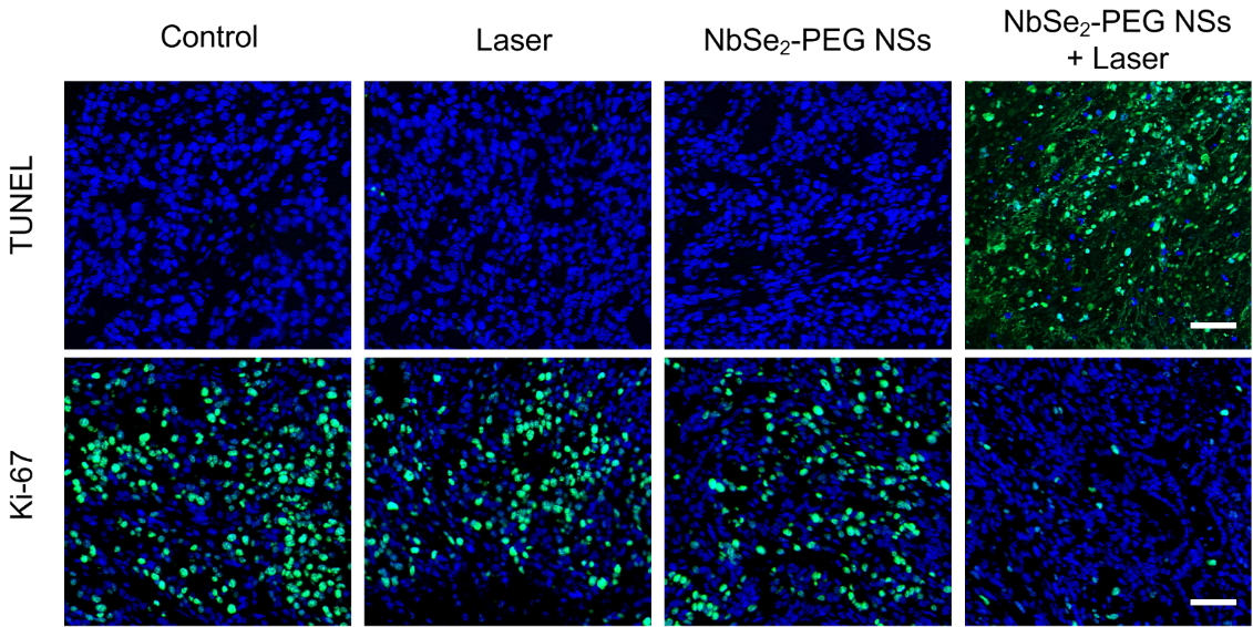
miR-8072 suppresses TNBC tumor progression and predicts favorable prognosis. ( A ) Serum levels of miR-8072 in breast cancer, benign breast disease, and non-cancer samples were analyzed using data from GEO (GSE73002) (** P < 0.01) . B The relationships between miR-8072 level and overall survival (OS) in breast cancer patients were analyzed using public database Kaplan–Meier plotter ( ). 1062 breast cancer patients were included, 525 cases were in miR-8072 low-expression cohort, and 537 cases were in miR-8072 high-expression cohort. The median survival for the high-expression cohort of miR-8072 is 115.4 months, while it is 148.53 months for the low-expression cohort of miR-8072 (upper panel). 97 TNBC patients were included for analysis. Among them, 28 cases were in miR-8072 low-expression cohort, and 69 cases were in miR-8072 high-expression cohort. The upper quartile survival for cohorts with high-expression of miR-8072 is 98.83 months, while it is 36.43 months for cohorts with low-expression of miR-8072 (lower panel). C MDA-MB-231 cells with stably overexpressed miR-8072 or control (Ctrl) cells were subcutaneously implanted in nude mice, tumor volume were measured every 7 days. D Tumor nodules were collected and weighed 35 days later after implantation. E Expression of Ki67 in the tumor nodules was detected by immunohistochemistry
Index in PubMed under a CC BY license. PMID: 38890750

Click image to see more details
IF analysis of Ki67 using anti-Ki67 antibody (PB9026).
Ki67 was detected in a paraffin-embedded section ofNude mouse tumor tissue. Heat mediated antigen retrieval was performed in EDTA buffer (pH 8.0, epitope retrieval solution). The tissue section was blocked with 10% goat serum. The tissue section was then incubated with 1:200 rabbit anti-Ki67 Antibody (PB9026) overnight at 4°C. DyLight®488 Conjugated Goat Anti-Rabbit IgG (BA1127) was used as secondary antibody at 1:500 dilution and incubated for 30 minutes at 37°C. The section was counterstained with DAPI. Visualize using a fluorescence microscope and filter sets appropriate for the label used.

Click image to see more details
ICC/IF analysis of Ki67 using anti-Ki67 antibody (PB9026).
Ki67 was detected in an immunocytochemical section of human Hela cells. The cells were fixed with 4% paraformaldehyde for 10 minutes and then treated with a membrane permeabilization agent (AR0205) for 5 minutes.The cells were blocked with 10% goat serum. And then incubated with rabbit anti-Ki67 Antibody (PB9026) at a dilution of 1:50 overnight at 4°C. DyLight®488 conjugated goat anti-rabbit IgG (BA1127) was used as secondary antibody at 1:500 dilution and incubated for 30 minutes at 37°C. The section was counterstained with DAPI. Visualize using a fluorescence microscope and filter sets appropriate for the label used.

Click image to see more details
ICC/IF analysis of Ki67 using anti-Ki67 antibody (PB9026).
Ki67 was detected in an immunocytochemical section of human SIHA cells. The cells were fixed with 4% paraformaldehyde for 10 minutes and then treated with a membrane permeabilization agent (AR0205) for 5 minutes.The cells were blocked with 10% goat serum. And then incubated with rabbit anti-Ki67 Antibody (PB9026) at a dilution of 1:50 overnight at 4°C. DyLight®488 conjugated goat Anti-rabbit IgG (BA1127) was used as secondary antibody at 1:500 dilution and incubated for 30 minutes at 37°C. The section was counterstained with DAPI. Visualize using a fluorescence microscope and filter sets appropriate for the label used.

Click image to see more details
IHC analysis of Ki67 using anti-Ki67 antibody (PB9026).
Ki67 was detected in paraffin-embedded section of nude mouse tumor tissues. Heat mediated antigen retrieval was performed in citrate buffer (pH6, epitope retrieval solution) for 20 mins. The tissue section was blocked with 10% goat serum. The tissue section was then incubated with 1:1000 rabbit anti-Ki67 Antibody (PB9026) overnight at 4°C. Ready-to-use SABC-POD kit (rabbit IgG) was used as secondary antibody and incubated for 30 minutes at 37°C. The tissue section was developed using Strepavidin-Biotin-Complex (SABC)(Catalog # SA1022) with DAB as the chromogen.
Specific Publications For Anti-Ki67/MKI67 Antibody Picoband® (PB9026)
Loading publications
Recommended Resources
Here are featured tools and databases that you might find useful.
- Boster's Pathways Library
- Protein Databases
- Bioscience Research Protocol Resources
- Data Processing & Analysis Software
- Photo Editing Software
- Scientific Literature Resources
- Research Paper Management Tools
- Molecular Biology Software
- Primer Design Tools
- Bioinformatics Tools
- Phylogenetic Tree Analysis
Customer Reviews
Have you used Anti-Ki67/MKI67 Antibody Picoband®?
Share your experimental results or join a short interview to earn up to $1,000 in product credits or other rewards.
3 Reviews For Anti-Ki67/MKI67 Antibody Picoband®
In IHC using Ki67 antibody (Cat# PB9026), strong and specific nuclear staining was observed in nude mouse tumor tissue, confirming high Ki67 expression and active cell proliferation.
Excellent

| SKU | PB9026 |
|---|---|
| Application | Western Blot |
| Sample | Nude mouse tumor tissue |
| Sample Processing Description | Paraffin-embedded tumor tissue sections. |
| Primary Antibody | Ki67/MKI67 Antibody Picoband® |
| Primary Incubation | 1:1000, overnight at 4 ℃ |
| Secondary Antibody | HRP-conjugated Anti-Rabbit IgG Secondary Antibody |
| Secondary Incubation | 1 hour in room temperature |
| Detection | Substrate: ECL reagent, Imaging system:ChemiDoc MP |
| Results Summary | This antibody is highly specific and efficient, with a clean background and no nonspecific bands. The target band has sharp and well-defined edges. |
Jin Sun, Tangdu Hospital, Xi’an, China
Verified customer
Submitted 2026-02-26
BOSTER’s Ki-67 antibody can effectively mark the proliferative activity of tumor cells. After treatment, a significant decrease in Ki-67 expression in tumor tissues can be clearly observed, indicating that the proliferative activity of tumor cells is mark
Excellent
| SKU | PB9026 |
|---|---|
| Application | Immunofluorenscence |
| Sample | Cells in nude mouse tumor tissue |
| Sample Processing Description | Sections of nude mouse tumor tissues fixed and embedded under different treatment conditions (Scale Bar = 100 μm) |
| Primary Antibody | Anti-Ki67/MKI67 Antibody Picoband® |
| Primary Incubation | 1:200, overnight at 4 ℃ |
| Blocking Agent | Goat serum |
| Secondary Antibody | DyLight 488-conjugated goat anti-rabbit antibody. |
| Secondary Incubation | Incubate at room temperature for 1 hour |
| Detection | Fluorescence microscope |
| Results Summary | The antibodies used in the experiment demonstrated good sensitivity and high cost-effectiveness, providing strong support for the smooth progress of the study. |
Zhibin Li, Shenzhen Second People's Hospital
Verified customer
Submitted 2025-10-17
Immunocytochemistry forAnti-P53/TP53 Antibody
Excellent

| SKU | PB9026 |
|---|---|
| Application | Immunohistochemistry (Paraffin-embedded) |
| Blocking step | 5% BSA as a blocking agent for 30 min at 37°C |
| Sample | Human tonsil |
| Fixative | Fixed with 4% paraformaldehyde |
| Primary Ab Incubation | 37°C for 30 minutes |
| Primary Ab Incubation diluent | 5% BSA in TBS |
| Primary Ab Concentration | 2ug/ml |
| Secondary Antibody | SABC kit from Boster Bio, (SA1022) |
| Secondary Ab Dilution | The kit was ready to use, no dilution needed |
| Secondary Ab Incubation | 4°C overnight |
Verified Customer
Verified customer
Submitted 2019-07-19
Customer Q&As
Have a question?
Find answers in Q&As, reviews.
Can't find your answer?
Submit your question
3 Customer Q&As for Anti-Ki67/MKI67 Antibody Picoband®
Question
We are currently using anti-Ki67/MKI67 antibody PB9026 for human tissue, and we are satisfied with the IHC-P results. The species of reactivity given in the datasheet says human. Is it possible that the antibody can work on goat tissues as well?
Verified Customer
Verified customer
Asked: 2019-07-17
Answer
The anti-Ki67/MKI67 antibody (PB9026) has not been validated for cross reactivity specifically with goat tissues, though there is a good chance of cross reactivity. We have an innovator award program that if you test this antibody and show it works in goat you can get your next antibody for free. Please contact me if I can help you with anything.
Boster Scientific Support
Answered: 2019-07-17
Question
Is PB9026 suitable to stain ki67 with mouse and rat sample IHC-P sections?
Verified customer
Asked: 2019-05-15
Answer
The Anti-Ki67/MKI67 Antibody Picoband™ (PB9026) is only suitable for human samples.
Boster Scientific Support
Answered: 2019-05-16
Question
Has PB9026 been tested on mouse and rat samples for IHC-P?
Verified Customer
Verified customer
Asked: 2017-08-25
Answer
PB9026 is only suitable for human samples.
Boster Scientific Support
Answered: 2017-08-25
