Click image to see more details
Product Info Summary
| SKU: | A02739-1 |
|---|---|
| Size: | 100 µg/vial |
| Reactive Species: | Rat |
| Host: | Rabbit |
| Application: | ELISA, IHC, WB |
Customers Who Bought This Also Bought

Product info
Product Name
Anti-Klk7 Antibody Picoband®
SKU/Catalog Number
A02739-1
Size
100 µg/vial
Form
Lyophilized
Description
Boster Bio Anti-Klk7 Antibody Picoband® catalog # A02739-1. Tested in ELISA, IHC, WB applications. This antibody reacts with Rat. The brand Picoband indicates this is a premium antibody that guarantees superior quality, high affinity, and strong signals with minimal background in Western blot applications. Only our best-performing antibodies are designated as Picoband, ensuring unmatched performance.
Storage & Handling
At -20°C for one year from date of receipt. After reconstitution, at 4°C for one month. It can also be aliquotted and stored frozen at -20°C for six months. Avoid repeated freezing and thawing.
Cite This Product
Anti-Klk7 Antibody Picoband® (Boster Biological Technology, Pleasanton CA, USA, Catalog # A02739-1)
Host
Rabbit
Contents
Each vial contains 4 mg Trehalose, 0.9 mg NaCl, 0.2 mg Na2HPO4.
Clonality
Polyclonal
Isotype
Rabbit IgG
Immunogen
E.coli-derived rat Klk7 recombinant protein (Position: K30-N238).
Cross-reactivity
No cross-reactivity with other proteins.
Reactive Species
A02739-1 is reactive to Klk7 in Rat
Observed Molecular Weight
38 kDa
Calculated molecular weight
29.0 kDa
Background of Klk7
Kallikrein-related peptidase 7 (KLK7) is a serine protease that in humans is encoded by the KLK7 gene. KLK7 is a chymotrypsin-like serine protease of the member of peptidase S1 family. It is synthesized with a signal peptide of 22 amino acids (aa 1-22) and a propeptide region (aa 23-29) that are subsequently cleaved to generate the mature form. Human KLK7 displays about 40 to 60% amino acid homology with other members of the family. It cleaves proteins with aromatic side chains in the P1 position. KLK7 is abundantly expressed in the skin and is also present in the brain, mammary gland, cerebellum, spinal cord, and kidney. Lower levels of expression have been reported in in salivary glands, uterus, thymus, thyroid, placenta, trachea, and testis. Its levels are shown to be up-regulated in ovarian carcinoma, especially late-stage serous carcinoma. Less differentiated tumors of more advanced stage show higher level KLK7 expression. KLK7 activity is inhibited by zinc and copper ions
Antibody Validation
Boster validates all antibodies on WB, IHC, ICC, Immunofluorescence, and ELISA with known positive control and negative samples to ensure specificity and high affinity, including thorough antibody incubations.
Application & Images
Applications
A02739-1 is guaranteed for ELISA, IHC, WB Boster Guarantee
Recommend Dilution
| Application | Dilution | Species |
|---|---|---|
| Western blot | 0.25-0.5 μg/ml | Rat |
| Immunohistochemistry(Paraffin-embedded Section) | 2-5 μg/ml | Rat |
| ELISA | 0.1-0.5 μg/ml | Rat |
Tested application
Suggested blocking solution with 5% non-fat milk or BSA; (*)Recommended protein loading: 20-40 µg per lane
Use TE buffer pH 9.0 for antigen retrieval; (*) citrate buffer pH 6.0 is an alternative.
Validation Images & Assay Conditions
Click image to see more details
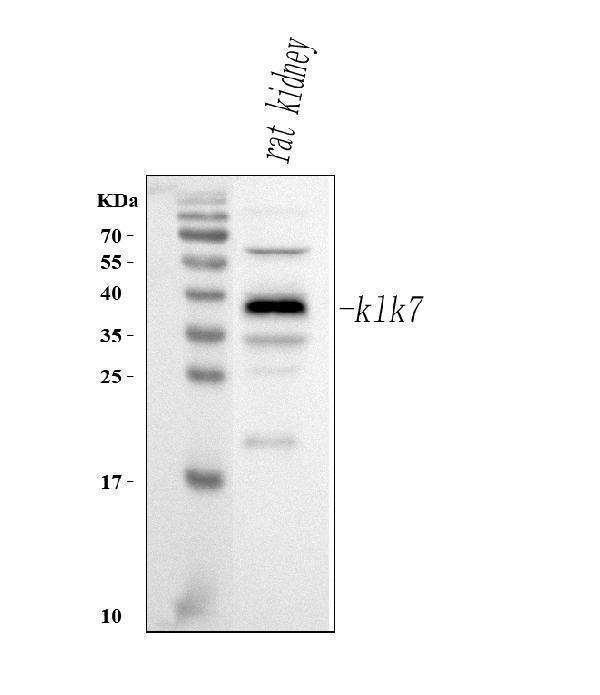
Western blot analysis of Klk7 using anti-Klk7 antibody (A02739-1).
Electrophoresis was performed on a 5-20% SDS-PAGE gel at 70V (Stacking gel) / 90V (Resolving gel) for 2-3 hours. The sample well of each lane was loaded with 30 ug of sample under reducing conditions.
Lane 1: rat kidney tissue lysates.
After electrophoresis, proteins were transferred to a nitrocellulose membrane at 150 mA for 50-90 minutes. Blocked the membrane with 5% non-fat milk/TBS for 1.5 hour at RT. The membrane was incubated with rabbit anti-Klk7 antigen affinity purified polyclonal antibody (Catalog # A02739-1) at 0.5 μg/mL overnight at 4°C, then washed with TBS-0.1%Tween 3 times with 5 minutes each and probed with a goat anti-rabbit IgG-HRP secondary antibody at a dilution of 1:5000 for 1.5 hour at RT. The signal is developed using an Enhanced Chemiluminescent detection (ECL) kit (Catalog # EK1002) with Tanon 5200 system. A specific band was detected for Klk7 at approximately 38 kDa. The expected band size for Klk7 is at 28 kDa.

Click image to see more details
IHC analysis of Klk7 using anti-Klk7 antibody (A02739-1).
Klk7 was detected in a paraffin-embedded section of rat kidney tissue. Heat mediated antigen retrieval was performed in EDTA buffer (pH 8.0, epitope retrieval solution). The tissue section was blocked with 10% goat serum. The tissue section was then incubated with 2 μg/ml rabbit anti-Klk7 Antibody (A02739-1) overnight at 4°C. Peroxidase Conjugated Goat Anti-rabbit IgG was used as secondary antibody and incubated for 30 minutes at 37°C. The tissue section was developed using HRP Conjugated Rabbit IgG Super Vision Assay Kit (Catalog # SV0002) with DAB as the chromogen.
Specific Publications For Anti-Klk7 Antibody Picoband® (A02739-1)
Loading publications
Recommended Resources
Here are featured tools and databases that you might find useful.
- Boster's Pathways Library
- Protein Databases
- Bioscience Research Protocol Resources
- Data Processing & Analysis Software
- Photo Editing Software
- Scientific Literature Resources
- Research Paper Management Tools
- Molecular Biology Software
- Primer Design Tools
- Bioinformatics Tools
- Phylogenetic Tree Analysis
Customer Reviews
Have you used Anti-Klk7 Antibody Picoband®?
Share your experimental results or join a short interview to earn up to $1,000 in product credits or other rewards.
0 Reviews For Anti-Klk7 Antibody Picoband®
Customer Q&As
Have a question?
Find answers in Q&As, reviews.
Can't find your answer?
Submit your question
