Click image to see more details
-

-
-
-
-
+7
Product Info Summary
| SKU: | PA1048 |
|---|---|
| Size: | 100 μg/vial |
| Reactive Species: | Human, Mouse, Rat |
| Host: | Rabbit |
| Application: | IP, IF, IHC, ICC, WB |
Customers Who Bought This Also Bought

Product info
Product Name
Anti-Lamin B1/LMNB1 Antibody Picoband®
SKU/Catalog Number
PA1048
BA1228 is an alternative SKU for this antibody, used in previous lots.
Size
100 μg/vial
Form
Lyophilized
Description
Boster Bio Anti-Lamin B1/LMNB1 Antibody catalog # PA1048. Tested in IP, ICC/IF, IF, IHC, WB applications. This antibody reacts with Human, Mouse, Rat. The brand Picoband indicates this is a premium antibody that guarantees superior quality, high affinity, and strong signals with minimal background in Western blot applications. Only our best-performing antibodies are designated as Picoband, ensuring unmatched performance.
Storage & Handling
Store at -20˚C for one year from date of receipt. After reconstitution, at 4˚C for one month. It can also be aliquotted and stored frozen at -20˚C for six months. Avoid repeated freeze-thaw cycles.
Cite This Product
Anti-Lamin B1/LMNB1 Antibody Picoband® (Boster Biological Technology, Pleasanton CA, USA, Catalog # PA1048)
Host
Rabbit
Contents
Each vial contains 4 mg Trehalose, 0.9 mg NaCl and 0.2 mg Na2HPO4.
Clonality
Polyclonal
Isotype
Rabbit IgG
Immunogen
A synthetic peptide corresponding to a sequence at the C-terminus of human Lamin B1, different from the related rat sequence by one amino acid, and from the related mouse sequence by three amino acids.
Cross-reactivity
No cross-reactivity with other proteins
Reactive Species
PA1048 is reactive to LMNB1 in Human, Mouse, Rat
Observed Molecular Weight
70-72 kDa
Calculated molecular weight
66.4 kDa
Background of LMNB1
Lamin-B1 is a protein that in humans is encoded by the LMNB1 gene. The nuclear lamina consists of a two-dimensional matrix of proteins located next to the inner nuclear membrane. The lamin family of proteins make up the matrix and are highly conserved in evolution. During mitosis, the lamina matrix is reversibly disassembled as the lamin proteins are phosphorylated. Lamin proteins are though to be involved in nuclear stability, chromatin structure and gene expression. Vertebrate lamins consist of two types, A and B. This gene encodes one of the two B type proteins, B1.
Antibody Validation
Boster validates all antibodies on WB, IHC, ICC, Immunofluorescence, and ELISA with known positive control and negative samples to ensure specificity and high affinity, including thorough antibody incubations.
Application & Images
Applications
PA1048 is guaranteed for IP, IF, IHC, ICC, WB Boster Guarantee
Assay Dilutions Recommendation
The recommendations below provide a starting point for assay optimization. The actual working concentration varies and should be decided by the user.
Western blot, 0.1-0.5μg/ml, Human, Mouse, Rat
Immunohistochemistry (Paraffin-embedded Section), 2-5μg/ml, Human, Mouse, Rat
Immunocytochemistry/Immunofluorescence, 5 μg/ml, Human
Immunofluorescence, 5 μg/ml, Human, Mouse, Rat
Immunoprecipitation, 0.5-2 μg/ml, Human
Positive Control
WB: human Hela whole cell, human K562 whole cell, human SH-SY5Y whole cell, rat spleen tissue, rat PC-12 whole cell, mouse RAW264.7 whole cell
IHC: human breast cancer tissue, human colon cancer tissue, human lung cancer tissue, mouse brain tissue, rat brain tissue
IF: human lung cancer tissue, rat brain tissue, mouse brain tissue
ICC/IF: U2OS cell
IP: Hela cell
Validation Images & Assay Conditions
Click image to see more details
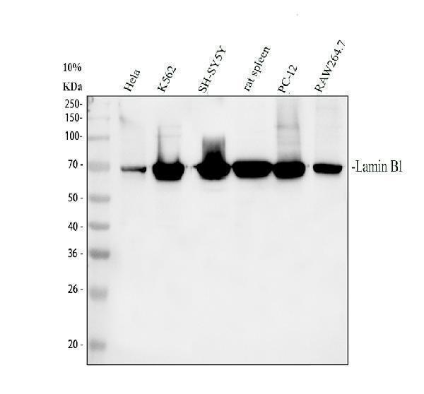
Western blot analysis of Lamin B1 using anti-Lamin B1 antibody (PA1048).
Electrophoresis was performed on a 10% SDS-PAGE gel at 80V (Stacking gel) / 120V (Resolving gel) for 2 hours. The sample well of each lane was loaded with 30 ug of sample under reducing conditions.
Lane 1: human Hela whole cell lysates,
Lane 2: human K562 whole cell lysates,
Lane 3: human SH-SY5Y whole cell lysates,
Lane 4: rat spleen tissue lysates,
Lane 5: rat PC-12 whole cell lysates,
Lane 6: mouse RAW264.7 whole cell lysates.
After electrophoresis, proteins were transferred to a nitrocellulose membrane at 150 mA for 50-90 minutes. Blocked the membrane with 5% non-fat milk/TBS for 1.5 hour at RT. The membrane was incubated with rabbit anti-Lamin B1 antigen affinity purified polyclonal antibody (Catalog # PA1048) at 0.5 μg/mL overnight at 4°C, then washed with TBS-0.1%Tween 3 times with 5 minutes each and probed with a goat anti-rabbit IgG-HRP secondary antibody at a dilution of 1:5000 for 1.5 hour at RT. The signal is developed using an ECL Plus Western Blotting Substrate (Catalog # AR1196-200) with Tanon 5200 system. A specific band was detected for Lamin B1 at approximately 70-72 kDa. The expected band size for Lamin B1 is at 66 kDa.

Click image to see more details
IHC analysis of Lamin B1 using anti-Lamin B1 antibody (PA1048).
Lamin B1 was detected in a paraffin-embedded section of human breast cancer tissue. Heat mediated antigen retrieval was performed in EDTA buffer (pH 8.0, epitope retrieval solution). The tissue section was blocked with 10% goat serum. The tissue section was then incubated with 2 μg/ml rabbit anti-Lamin B1 Antibody (PA1048) overnight at 4°C. Peroxidase Conjugated Goat Anti-rabbit IgG was used as secondary antibody and incubated for 30 minutes at 37°C. The tissue section was developed using HRP Conjugated Rabbit IgG Super Vision Assay Kit (Catalog # SV0002) with DAB as the chromogen.

Click image to see more details
IHC analysis of Lamin B1 using anti-Lamin B1 antibody (PA1048).
Lamin B1 was detected in a paraffin-embedded section of human colon cancer tissue. Heat mediated antigen retrieval was performed in EDTA buffer (pH 8.0, epitope retrieval solution). The tissue section was blocked with 10% goat serum. The tissue section was then incubated with 2 μg/ml rabbit anti-Lamin B1 Antibody (PA1048) overnight at 4°C. Peroxidase Conjugated Goat Anti-rabbit IgG was used as secondary antibody and incubated for 30 minutes at 37°C. The tissue section was developed using HRP Conjugated Rabbit IgG Super Vision Assay Kit (Catalog # SV0002) with DAB as the chromogen.

Click image to see more details
IHC analysis of Lamin B1 using anti-Lamin B1 antibody (PA1048).
Lamin B1 was detected in a paraffin-embedded section of human lung cancer tissue. Heat mediated antigen retrieval was performed in EDTA buffer (pH 8.0, epitope retrieval solution). The tissue section was blocked with 10% goat serum. The tissue section was then incubated with 2 μg/ml rabbit anti-Lamin B1 Antibody (PA1048) overnight at 4°C. Peroxidase Conjugated Goat Anti-rabbit IgG was used as secondary antibody and incubated for 30 minutes at 37°C. The tissue section was developed using HRP Conjugated Rabbit IgG Super Vision Assay Kit (Catalog # SV0002) with DAB as the chromogen.

Click image to see more details
IHC analysis of Lamin B1 using anti-Lamin B1 antibody (PA1048).
Lamin B1 was detected in a paraffin-embedded section of mouse brain tissue. Heat mediated antigen retrieval was performed in EDTA buffer (pH 8.0, epitope retrieval solution). The tissue section was blocked with 10% goat serum. The tissue section was then incubated with 2 μg/ml rabbit anti-Lamin B1 Antibody (PA1048) overnight at 4°C. Peroxidase Conjugated Goat Anti-rabbit IgG was used as secondary antibody and incubated for 30 minutes at 37°C. The tissue section was developed using HRP Conjugated Rabbit IgG Super Vision Assay Kit (Catalog # SV0002) with DAB as the chromogen.

Click image to see more details
IHC analysis of Lamin B1 using anti-Lamin B1 antibody (PA1048).
Lamin B1 was detected in a paraffin-embedded section of rat brain tissue. Heat mediated antigen retrieval was performed in EDTA buffer (pH 8.0, epitope retrieval solution). The tissue section was blocked with 10% goat serum. The tissue section was then incubated with 2 μg/ml rabbit anti-Lamin B1 Antibody (PA1048) overnight at 4°C. Peroxidase Conjugated Goat Anti-rabbit IgG was used as secondary antibody and incubated for 30 minutes at 37°C. The tissue section was developed using HRP Conjugated Rabbit IgG Super Vision Assay Kit (Catalog # SV0002) with DAB as the chromogen.

Click image to see more details
IF analysis of Lamin B1 using anti-Lamin B1 antibody (PA1048).
Lamin B1 was detected in a paraffin-embedded section of human lung cancer tissue. Heat mediated antigen retrieval was performed in EDTA buffer (pH 8.0, epitope retrieval solution). The tissue section was blocked with 10% goat serum. The tissue section was then incubated with 5 μg/mL rabbit anti-Lamin B1 Antibody (PA1048) overnight at 4°C. Cy3 Conjugated Goat Anti-Rabbit IgG (BA1032) was used as secondary antibody at 1:500 dilution and incubated for 30 minutes at 37°C. The section was counterstained with DAPI. Visualize using a fluorescence microscope and filter sets appropriate for the label used.

Click image to see more details
IF analysis of Lamin B1 using anti-Lamin B1 antibody (PA1048).
Lamin B1 was detected in a paraffin-embedded section of rat brain tissue. Heat mediated antigen retrieval was performed in EDTA buffer (pH 8.0, epitope retrieval solution). The tissue section was blocked with 10% goat serum. The tissue section was then incubated with 5 μg/mL rabbit anti-Lamin B1 Antibody (PA1048) overnight at 4°C. Cy3 Conjugated Goat Anti-Rabbit IgG (BA1032) was used as secondary antibody at 1:500 dilution and incubated for 30 minutes at 37°C. The section was counterstained with DAPI. Visualize using a fluorescence microscope and filter sets appropriate for the label used.

Click image to see more details
IF analysis of Lamin B1 using anti-Lamin B1 antibody (PA1048).
Lamin B1 was detected in a paraffin-embedded section of mouse brain tissue. Heat mediated antigen retrieval was performed in EDTA buffer (pH 8.0, epitope retrieval solution). The tissue section was blocked with 10% goat serum. The tissue section was then incubated with 5 μg/mL rabbit anti-Lamin B1 Antibody (PA1048) overnight at 4°C. Cy3 Conjugated Goat Anti-Rabbit IgG (BA1032) was used as secondary antibody at 1:500 dilution and incubated for 30 minutes at 37°C. The section was counterstained with DAPI. Visualize using a fluorescence microscope and filter sets appropriate for the label used.

Click image to see more details
IF analysis of Lamin B1 using anti-Lamin B1 antibody (PA1048).
Lamin B1 was detected in an immunocytochemical section of U2OS cells. Enzyme antigen retrieval was performed using IHC enzyme antigen retrieval reagent (AR0022) for 15 mins. The cells were blocked with 10% goat serum. And then incubated with 5 μg/mL rabbit anti-Lamin B1 Antibody (PA1048) overnight at 4°C. DyLight®488 Conjugated Goat Anti-Rabbit IgG (BA1127) was used as secondary antibody at 1:500 dilution and incubated for 30 minutes at 37°C. The section was counterstained with DAPI. Visualize using a fluorescence microscope and filter sets appropriate for the label used.

Click image to see more details
Immunoprecipitating (IP) Lamin B1 in Hela whole cell lysate.
Western blot analysis of Lamin B1 using anti-Lamin B1 antibody (PA1048);
Lane 1: Hela whole cell lysates (30ug);
Lane 2: Rabbit control IgG instead of anti-Lamin B1 antibody in Hela whole cell lysate;
Lane 3: anti-Lamin B1 antibody (2μg) + Hela whole cell lysate (500μg).
After electrophoresis, proteins were transferred to a membrane. Then the membrane was incubated with rabbit anti-Lamin B1 antigen affinity purified polyclonal antibody (PA1048) at a dilution of 0.5 μg/mL and probed with a goat anti-rabbit IgG-HRP secondary antibody (Catalog # BA1054). The signal is developed using ECL Plus Western Blotting Substrate (Catalog # AR1196-200). A specific band was detected for Lamin B1 at approximately 70-72 kDa. The expected band size for Lamin B1 is at 66 kDa.
Specific Publications For Anti-Lamin B1/LMNB1 Antibody Picoband® (PA1048)
Loading publications
Recommended Resources
Here are featured tools and databases that you might find useful.
- Boster's Pathways Library
- Protein Databases
- Bioscience Research Protocol Resources
- Data Processing & Analysis Software
- Photo Editing Software
- Scientific Literature Resources
- Research Paper Management Tools
- Molecular Biology Software
- Primer Design Tools
- Bioinformatics Tools
- Phylogenetic Tree Analysis
Customer Reviews
Have you used Anti-Lamin B1/LMNB1 Antibody Picoband®?
Share your experimental results or join a short interview to earn up to $1,000 in product credits or other rewards.
0 Reviews For Anti-Lamin B1/LMNB1 Antibody Picoband®
Customer Q&As
Have a question?
Find answers in Q&As, reviews.
Can't find your answer?
Submit your question
5 Customer Q&As for Anti-Lamin B1/LMNB1 Antibody Picoband®
Question
Can you help my question with product PA1048, anti-Lamin B1/LMNB1 antibody. I was wondering if it would be possible to conjugate this antibody with biotin. I would need it to be without BSA or sodium azide. I am planning on using a buffer exchange of sodium azide with PBS only. Would there be problems for me to conjugate the antibody and store it in -20 degrees in small aliquots?
Verified Customer
Verified customer
Asked: 2020-04-22
Answer
We do not recommend storing this antibody with PBS buffer only in -20 degrees. If you want to store it in -20 degrees it is best to add some cryoprotectant like glycerol. If you want carrier free PA1048 anti-Lamin B1/LMNB1 antibody, we can provide it to you in a special formula with trehalose and/or glycerol. These molecules will not interfere with conjugation chemistry and provide a good level of protection for the antibody from degradation. Please be sure to specify this in your purchase order.
Boster Scientific Support
Answered: 2020-04-22
Question
Will PA1048 anti-Lamin B1/LMNB1 antibody work on parafin embedded sections? If so, which fixation method do you recommend we use (PFA, paraformaldehyde, other)?
Verified Customer
Verified customer
Asked: 2020-04-09
Answer
As indicated on the product datasheet, PA1048 anti-Lamin B1/LMNB1 antibody as been tested on IHC. It is best to use PFA for fixation because it has better tissue penetration ability. PFA needs to be prepared fresh before use. Long term stored PFA turns into formalin, as the PFA molecules congregate and become formalin.
Boster Scientific Support
Answered: 2020-04-09
Question
Is a blocking peptide available for product anti-Lamin B1/LMNB1 antibody (PA1048)?
Verified Customer
Verified customer
Asked: 2020-03-31
Answer
We do provide the blocking peptide for product anti-Lamin B1/LMNB1 antibody (PA1048). If you would like to place an order for it please contact support@bosterbio.com and make a special request.
Boster Scientific Support
Answered: 2020-03-31
Question
We appreciate helping with my inquiry over the phone. Here are the WB image, lot number and protocol we used for colon carcinoma using anti-Lamin B1/LMNB1 antibody PA1048. Let me know if you need anything else.
Verified Customer
Verified customer
Asked: 2019-08-15
Answer
We appreciate the data. You have provided everything we needed. Our lab team are working to resolve your inquiry as quickly as possible, and we appreciate your patience and understanding! Please let me know if there is anything you need in the meantime.
Boster Scientific Support
Answered: 2019-08-15
Question
I was wanting to use your anti-Lamin B1/LMNB1 antibody for IHC for rat colon carcinoma on frozen tissues, but I want to know if it has been tested for this particular application. Has this antibody been tested and is this antibody a good choice for rat colon carcinoma identification?
Verified Customer
Verified customer
Asked: 2017-07-28
Answer
As indicated on the product datasheet, PA1048 anti-Lamin B1/LMNB1 antibody has been tested for IHC, ICC, WB on human, mouse, rat tissues. We have an innovator award program that if you test this antibody and show it works in rat colon carcinoma in IHC-frozen, you can get your next antibody for free.
Boster Scientific Support
Answered: 2017-07-28
