Click image to see more details
-

-
-
-
-
+6
Product Info Summary
| SKU: | A03522 |
|---|---|
| Size: | 100 μg/vial |
| Reactive Species: | Human, Mouse, Rat |
| Host: | Rabbit |
| Application: | IF, IHC, WB |
Customers Who Bought This Also Bought

Product info
Product Name
Anti-Laminin/Lamc1/Lamc2/Lamc3 Antibody Picoband®
SKU/Catalog Number
A03522
Size
100 μg/vial
Form
Lyophilized
Description
Boster Bio Anti-Laminin/Lamc1/Lamc2/Lamc3 Antibody Picoband® catalog # A03522. Tested in IF, IHC, WB applications. This antibody reacts with Human, Mouse, Rat. The brand Picoband indicates this is a premium antibody that guarantees superior quality, high affinity, and strong signals with minimal background in Western blot applications. Only our best-performing antibodies are designated as Picoband, ensuring unmatched performance.
Storage & Handling
Store at -20˚C for one year from date of receipt. After reconstitution, at 4˚C for one month. It can also be aliquotted and stored frozen at -20˚C for six months. Avoid repeated freeze-thaw cycles.
Cite This Product
Anti-Laminin/Lamc1/Lamc2/Lamc3 Antibody Picoband® (Boster Biological Technology, Pleasanton CA, USA, Catalog # A03522)
Host
Rabbit
Contents
Each vial contains 4mg Trehalose, 0.9mg NaCl, 0.2mg Na2HPO4, 0.05mg NaN3.
Clonality
Polyclonal
Isotype
Rabbit IgG
Immunogen
Peptide mixture of laminin gamma1,2,3 (NKLNEIEGSLNKAKDEMKAS; DLEERVRRQRNHLHLLETSI; LQLDSHGALHHKLRQLEEES). Laminin gamma has only three subtypes of antibody to gamma1-3 reactive with all isoforms of laminin.
Cross-reactivity
No cross-reactivity with other proteins.
Reactive Species
A03522 is reactive to Lamc1 in Human, Mouse, Rat
Observed Molecular Weight
150, 220-250 kDa
Calculated molecular weight
177.3 kDa
Background of Lamc1
Tumor necrosis factor ligand superfamily member 14 is a protein that in humans is encoded by the TNFSF14 gene. TNFSF14 has also been designated as CD258, as well as LIGHT. It was mapped on chromosome 19p13.3. The protein encoded by this gene is a member of the tumor necrosis factor(TNF) ligand family. This protein may function as a costimulatory factor for the activation of lymphoid cells and as a deterrent to infection by herpesvirus. It has been shown to stimulate the proliferation of T cells, and trigger apoptosis of various tumor cells. This protein is also reported to prevent tumor necrosis factor alpha mediated apoptosis in primary hepatocyte. Two alternatively spliced transcript variant encoding distinct isoforms have been reported.
Antibody Validation
Boster validates all antibodies on WB, IHC, ICC, Immunofluorescence, and ELISA with known positive control and negative samples to ensure specificity and high affinity, including thorough antibody incubations.
Application & Images
Applications
A03522 is guaranteed for IF, IHC, WB Boster Guarantee
Recommend Dilution
| Application | Dilution | Species |
|---|---|---|
| Western blot | 0.1-0.5μg/ml | Human, Mouse, Rat, |
| Immunohistochemistry (Paraffin-embedded Section) | 0.5-1μg/ml | Mouse, Rat |
| Immunofluorescence | 2μg/ml | Mouse, Rat |
Tested application
Suggested blocking solution with 5% non-fat milk or BSA; (*)Recommended protein loading: 20-40 µg per lane
Use TE buffer pH 9.0 for antigen retrieval; (*) citrate buffer pH 6.0 is an alternative.
Validation Images & Assay Conditions
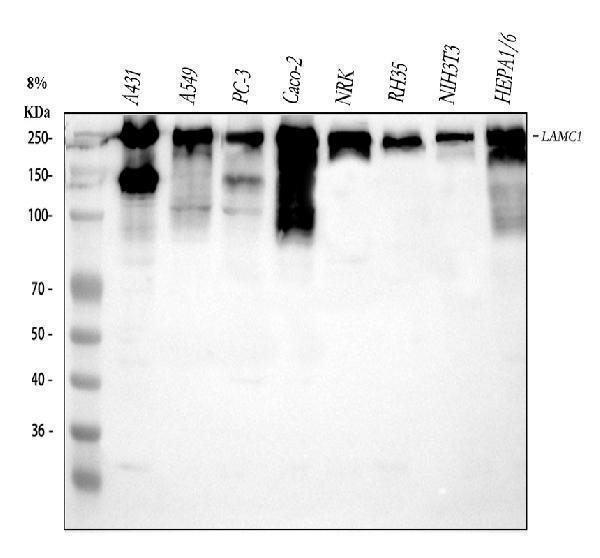
Click image to see more details
Western blot analysis of Laminin using anti-Laminin antibody (A03522).
Electrophoresis was performed on a 5-20% SDS-PAGE gel at 70V (Stacking gel) / 90V (Resolving gel) for 2-3 hours. The sample well of each lane was loaded with 30 ug of sample under reducing conditions.
Lane 1: human A431 whole cell lysates,
Lane 2: human A549 whole cell lysates,
Lane 3: human PC-3 whole cell lysates,
Lane 4: human CACO-2 whole cell lysates,
Lane 5: rat NRK whole cell lysates,
Lane 6: rat RH35 whole cell lysates,
Lane 7: mouse NIH/3T3 whole cell lysates,
Lane 8: mouse HEPA1-6 whole cell lysates.
After electrophoresis, proteins were transferred to a nitrocellulose membrane at 150 mA for 50-90 minutes. Blocked the membrane with 5% non-fat milk/TBS for 1.5 hour at RT. The membrane was incubated with rabbit anti-Laminin antigen affinity purified polyclonal antibody (Catalog # A03522) at 0.5 μg/mL overnight at 4°C, then washed with TBS-0.1%Tween 3 times with 5 minutes each and probed with a goat anti-rabbit IgG-HRP secondary antibody at a dilution of 1:5000 for 1.5 hour at RT. The signal is developed using an Enhanced Chemiluminescent detection (ECL) kit (Catalog # EK1002) with Tanon 5200 system. A specific band was detected for Laminin at approximately 150, 220-250 kDa. The expected band size for Laminin is at 177 kDa.

Click image to see more details
IHC analysis of Laminin using anti-Laminin antibody (A03522).
Laminin was detected in paraffin-embedded section of mouse heart tissue . Heat mediated antigen retrieval was performed in citrate buffer (pH6, epitope retrieval solution) for 20 mins. The tissue section was blocked with 10% goat serum. The tissue section was then incubated with 1μg/ml rabbit anti-Laminin Antibody (A03522) overnight at 4°C. Biotinylated goat anti-rabbit IgG was used as secondary antibody and incubated for 30 minutes at 37°C. The tissue section was developed using Strepavidin-Biotin-Complex (SABC)(Catalog # SA1022) with DAB as the chromogen.

Click image to see more details
IHC analysis of LAMC1/LAMC2/LAMC3 using anti-LAMC1/LAMC2/LAMC3 antibody (A03522).
LAMC1/LAMC2/LAMC3 was detected in a paraffin-embedded section of human kidney tissue. Heat mediated antigen retrieval was performed in EDTA buffer (pH 8.0, epitope retrieval solution). The tissue section was blocked with 10% goat serum. The tissue section was then incubated with 2 μg/ml rabbit anti-LAMC1/LAMC2/LAMC3 Antibody (A03522) overnight at 4°C. Peroxidase Conjugated Goat Anti-rabbit IgG was used as secondary antibody and incubated for 30 minutes at 37°C. The tissue section was developed using HRP Conjugated Rabbit IgG Super Vision Assay Kit (Catalog # SV0002) with DAB as the chromogen.

Click image to see more details
IHC analysis of Laminin using anti-Laminin antibody (A03522).
Laminin was detected in paraffin-embedded section of mouse kidney tissue . Heat mediated antigen retrieval was performed in citrate buffer (pH6, epitope retrieval solution) for 20 mins. The tissue section was blocked with 10% goat serum. The tissue section was then incubated with 1μg/ml rabbit anti-Laminin Antibody (A03522) overnight at 4°C. Biotinylated goat anti-rabbit IgG was used as secondary antibody and incubated for 30 minutes at 37°C. The tissue section was developed using Strepavidin-Biotin-Complex (SABC)(Catalog # SA1022) with DAB as the chromogen.

Click image to see more details
IHC analysis of Laminin using anti-Laminin antibody (A03522).
Laminin was detected in paraffin-embedded section of rat cardiac muscle tissue. Heat mediated antigen retrieval was performed in citrate buffer (pH6, epitope retrieval solution) for 20 mins. The tissue section was blocked with 10% goat serum. The tissue section was then incubated with 1μg/ml rabbit anti-Laminin Antibody (A03522) overnight at 4°C. Biotinylated goat anti-rabbit IgG was used as secondary antibody and incubated for 30 minutes at 37°C. The tissue section was developed using Strepavidin-Biotin-Complex (SABC)(Catalog # SA1022) with DAB as the chromogen.

Click image to see more details
IHC analysis of Laminin using anti-Laminin antibody (A03522).
Laminin was detected in paraffin-embedded section of rat kidney tissue . Heat mediated antigen retrieval was performed in citrate buffer (pH6, epitope retrieval solution) for 20 mins. The tissue section was blocked with 10% goat serum. The tissue section was then incubated with 1μg/ml rabbit anti-Laminin Antibody (A03522) overnight at 4°C. Biotinylated goat anti-rabbit IgG was used as secondary antibody and incubated for 30 minutes at 37°C. The tissue section was developed using Strepavidin-Biotin-Complex (SABC)(Catalog # SA1022) with DAB as the chromogen.

Click image to see more details
IF analysis of Laminin using anti-Laminin antibody (A03522).
Laminin was detected in a paraffin-embedded section of mouse skeletal muscle tissue. Heat mediated antigen retrieval was performed in EDTA buffer (pH 8.0, epitope retrieval solution). The tissue section was blocked with 10% goat serum. The tissue section was then incubated with 5 μg/mL rabbit anti-Laminin Antibody (A03522) overnight at 4°C. DyLight®488 Conjugated Goat Anti-Rabbit IgG (BA1127) was used as secondary antibody at 1:500 dilution and incubated for 30 minutes at 37°C. The section was counterstained with DAPI. Visualize using a fluorescence microscope and filter sets appropriate for the label used.

Click image to see more details
Transportation increased apoptosis and deceased cell viability, and BAM15 changed this trend adversely. a Immunofluorescence staining result showed transportation increased expression of Caspase-3 (green) in retinal cups of 30 days, but imposed little effects on expression of Laminin (red). The use of DMSO and BAM15 in transportation was able to alleviate the intensified expression of Caspase-3. b In retinal cups of 60 days, the addition of DMSO or BAM15 was able to reduce the elevated expression of Caspase-3 (green) in transportation. In addition, staining results indicated expression of Laminin (red) varied slightly in different groups. c Real-time PCR revealed dynamic expression changes of apoptotic-related genes NFκB, TNF-α and P53 in retinal tissue of 30 days. d Changes of NFκB, TNF-α, and P53 expression was assessed in retinal cups of 60 days by real-time PCR analysis. e Cell viability of retinal cups was assessed by CCK8 assay in transportation. Scale = 50 μm ( a ). Scale = 100 μm ( b )
Index in PubMed under a CC BY license. PMID: 30795805

Click image to see more details
IF analysis of Laminin using anti-Laminin antibody (A03522).
Laminin was detected in a paraffin-embedded section of mouse skeletal muscle tissue. Heat mediated antigen retrieval was performed in EDTA buffer (pH 8.0, epitope retrieval solution). The tissue section was blocked with 10% goat serum. The tissue section was then incubated with 5 μg/mL rabbit anti-Laminin Antibody (A03522) overnight at 4°C. DyLight®488 Conjugated Goat Anti-Rabbit IgG (BA1127) was used as secondary antibody at 1:500 dilution and incubated for 30 minutes at 37°C. The section was counterstained with DAPI. Visualize using a fluorescence microscope and filter sets appropriate for the label used.

Click image to see more details
IF analysis of Laminin using anti-Laminin antibody (A03522).
Laminin was detected in a paraffin-embedded section of rat skeletal muscle tissue. Heat mediated antigen retrieval was performed in EDTA buffer (pH 8.0, epitope retrieval solution). The tissue section was blocked with 10% goat serum. The tissue section was then incubated with 5 μg/mL rabbit anti-Laminin Antibody (A03522) overnight at 4°C. DyLight®488 Conjugated Goat Anti-Rabbit IgG (BA1127) was used as secondary antibody at 1:500 dilution and incubated for 30 minutes at 37°C. The section was counterstained with DAPI. Visualize using a fluorescence microscope and filter sets appropriate for the label used.
Specific Publications For Anti-Laminin/Lamc1/Lamc2/Lamc3 Antibody Picoband® (A03522)
Loading publications
Recommended Resources
Here are featured tools and databases that you might find useful.
- Boster's Pathways Library
- Protein Databases
- Bioscience Research Protocol Resources
- Data Processing & Analysis Software
- Photo Editing Software
- Scientific Literature Resources
- Research Paper Management Tools
- Molecular Biology Software
- Primer Design Tools
- Bioinformatics Tools
- Phylogenetic Tree Analysis
Customer Reviews
Have you used Anti-Laminin/Lamc1/Lamc2/Lamc3 Antibody Picoband®?
Share your experimental results or join a short interview to earn up to $1,000 in product credits or other rewards.
0 Reviews For Anti-Laminin/Lamc1/Lamc2/Lamc3 Antibody Picoband®
Customer Q&As
Have a question?
Find answers in Q&As, reviews.
Can't find your answer?
Submit your question
