Click image to see more details
-

-
-
-
-
+4
Product Info Summary
| SKU: | PB9262 |
|---|---|
| Size: | 100 μg/vial |
| Reactive Species: | Human, Mouse, Rat |
| Host: | Rabbit |
| Application: | Flow Cytometry, IF, IHC, ICC, WB |
Customers Who Bought This Also Bought

Product info
Product Name
Anti-MAD1/MAD1L1 Antibody Picoband®
SKU/Catalog Number
PB9262
Size
100 μg/vial
Form
Lyophilized
Description
Boster Bio Anti-MAD1/MAD1L1 Antibody Picoband® catalog # PB9262. Tested in Flow Cytometry, IF, IHC, ICC, WB applications. This antibody reacts with Human, Mouse, Rat. The brand Picoband indicates this is a premium antibody that guarantees superior quality, high affinity, and strong signals with minimal background in Western blot applications. Only our best-performing antibodies are designated as Picoband, ensuring unmatched performance.
Storage & Handling
Store at -20˚C for one year from date of receipt. After reconstitution, at 4˚C for one month. It can also be aliquotted and stored frozen at -20˚C for six months. Avoid repeated freeze-thaw cycles.
Cite This Product
Anti-MAD1/MAD1L1 Antibody Picoband® (Boster Biological Technology, Pleasanton CA, USA, Catalog # PB9262)
Host
Rabbit
Contents
Each vial contains antibody formulated with stabilizing components, 0.9 mg NaCl, 0.2 mg Na2HPO4, and 0.05 mg NaN3.
*This antibody is supplied in a stabilized formulation.
Compatibility with conjugation reactions depends on the chemistry of the conjugation method used.
For conjugation methods that are not compatible with the stabilizing components present in this formulation, a carrier-free antibody format is required.
Clonality
Polyclonal
Isotype
Rabbit IgG
Immunogen
E.coli-derived human MAD1 recombinant protein (Position: L362-A632). Human MAD1 shares 81% amino acid (aa) sequence identity with mouse MAD1.
Cross-reactivity
No cross-reactivity with other proteins
Reactive Species
PB9262 is reactive to MAD1L1 in Human, Mouse, Rat
Observed Molecular Weight
83 kDa
Calculated molecular weight
83.1 kDa
Background of MAD1L1
Mitotic spindle assembly checkpoint protein MAD1 is a protein that in humans is encoded by the MAD1L1 gene. It is mapped to 7p22.3. MAD1L1 is a component of the mitotic spindle-assembly checkpoint that prevents the onset of anaphase until all chromosome are properly aligned at the metaphase plate. MAD1L1 can function as a homodimer. It localizes to the centrosome during metaphase and to the spindle midzone and the midbody during anaphase and telophase. MAD1L1 may also play a role in cell cycle control and tumor suppression.
Antibody Validation
Boster validates all antibodies on WB, IHC, ICC, Immunofluorescence, and ELISA with known positive control and negative samples to ensure specificity and high affinity, including thorough antibody incubations.
Application & Images
Applications
PB9262 is guaranteed for Flow Cytometry, IF, IHC, ICC, WB Boster Guarantee
Assay Dilutions Recommendation
The recommendations below provide a starting point for assay optimization. The actual working concentration varies and should be decided by the user.
Western blot, 0.1-0.5μg/ml, Human, Mouse, Rat
Immunohistochemistry (Paraffin-embedded Section), 0.5-1μg/ml, Human, Mouse, Rat
Immunocytochemistry, 0.5-1μg/ml, Human
Immunocytochemistry/Immunofluorescence, 2μg/ml, Human
Flow Cytometry (Fixed), 1-3μg/1x106 cells, Human
Positive Control
WB: human Hela whole cell, human Raji whole cell, human Jurkat whole cell, human HepG2 whole cellhuman HL-60 whole cellhuman THP-1 whole cellhuman Caco-2 whole cell, rat brain tissue, rat kidney tissue, rat lung tissue, rat liver tissue, mouse brain tissue, mouse kidney tissue, mouse lung tissue, mouse liver tissue, mouse NIH3T3 whole cell
IHC: mouse intestine tissue, rat intestine tissue, human intestinal cancer tissue
ICC/IF: U20S cell
ICC: Hela cell
FCM: A431 cell
Validation Images & Assay Conditions
Click image to see more details
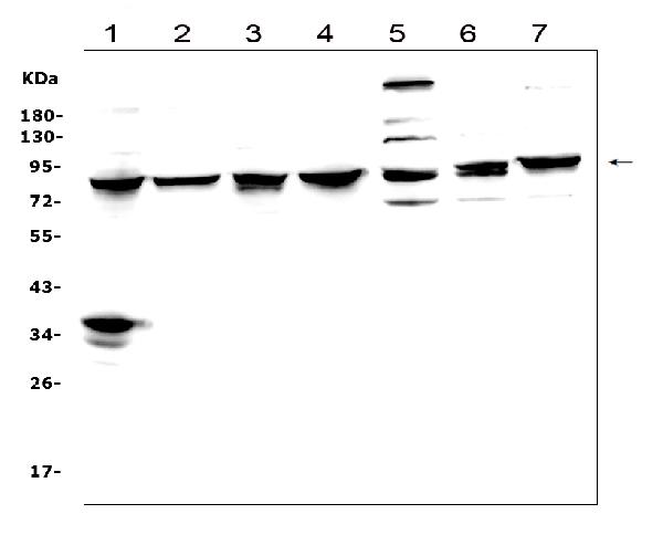
Western blot analysis of MAD1 using anti-MAD1 antibody (PB9262).
Electrophoresis was performed on a 5-20% SDS-PAGE gel at 70V (Stacking gel) / 90V (Resolving gel) for 2-3 hours. The sample well of each lane was loaded with 50ug of sample under reducing conditions.
Lane 1: human Hela whole cell lysates,
Lane 2: human Raji whole cell lysates,
Lane 3: human Jurkat whole cell lysates,
Lane 4: human HepG2 whole cell lysates.
Lane 5: human HL-60 whole cell lysates.
Lane 6: human THP-1 whole cell lysates.
Lane 7: human Caco-2 whole cell lysates.
After Electrophoresis, proteins were transferred to a Nitrocellulose membrane at 150mA for 50-90 minutes. Blocked the membrane with 5% Non-fat Milk/ TBS for 1.5 hour at RT. The membrane was incubated with rabbit anti-MAD1 antigen affinity purified polyclonal antibody (Catalog # PB9262) at 0.5 μg/mL overnight at 4°C, then washed with TBS-0.1%Tween 3 times with 5 minutes each and probed with a goat anti-rabbit IgG-HRP secondary antibody at a dilution of 1:10000 for 1.5 hour at RT. The signal is developed using an Enhanced Chemiluminescent detection (ECL) kit (Catalog # EK1002) with Tanon 5200 system. A specific band was detected for MAD1 at approximately 83KD. The expected band size for MAD1 is at 83KD.

Click image to see more details
Western blot analysis of MAD1 using anti-MAD1 antibody (PB9262).
Electrophoresis was performed on a 5-20% SDS-PAGE gel at 70V (Stacking gel) / 90V (Resolving gel) for 2-3 hours. The sample well of each lane was loaded with 50ug of sample under reducing conditions.
Lane 1: rat brain tissue lysates,
Lane 2: rat kidney tissue lysates,
Lane 3: rat lung tissue lysates,
Lane 4: rat liver tissue lysates,
Lane 5: mouse brain tissue lysates,
Lane 6: mouse kidney tissue lysates,
Lane 7: mouse lung tissue lysates,
Lane 8: mouse liver tissue lysates,
Lane 9: mouse NIH3T3 whole cell lysates.
After Electrophoresis, proteins were transferred to a Nitrocellulose membrane at 150mA for 50-90 minutes. Blocked the membrane with 5% Non-fat Milk/ TBS for 1.5 hour at RT. The membrane was incubated with rabbit anti-MAD1 antigen affinity purified polyclonal antibody (Catalog # PB9262) at 0.5 μg/mL overnight at 4°C, then washed with TBS-0.1%Tween 3 times with 5 minutes each and probed with a goat anti-rabbit IgG-HRP secondary antibody at a dilution of 1:10000 for 1.5 hour at RT. The signal is developed using an Enhanced Chemiluminescent detection (ECL) kit (Catalog # EK1002) with Tanon 5200 system. A specific band was detected for MAD1 at approximately 83KD. The expected band size for MAD1 is at 83KD.

Click image to see more details
IHC analysis of MAD1 using anti-MAD1 antibody (PB9262).
MAD1 was detected in paraffin-embedded section of mouse intestine tissues. Heat mediated antigen retrieval was performed in citrate buffer (pH6, epitope retrieval solution) for 20 mins. The tissue section was blocked with 10% goat serum. The tissue section was then incubated with 1μg/ml rabbit anti-MAD1 Antibody (PB9262) overnight at 4°C. Biotinylated goat anti-rabbit IgG was used as secondary antibody and incubated for 30 minutes at 37°C. The tissue section was developed using Strepavidin-Biotin-Complex (SABC)(Catalog # SA1022) with DAB as the chromogen.

Click image to see more details
IHC analysis of MAD1 using anti-MAD1 antibody (PB9262).
MAD1 was detected in paraffin-embedded section of rat intestine tissues. Heat mediated antigen retrieval was performed in citrate buffer (pH6, epitope retrieval solution) for 20 mins. The tissue section was blocked with 10% goat serum. The tissue section was then incubated with 1μg/ml rabbit anti-MAD1 Antibody (PB9262) overnight at 4°C. Biotinylated goat anti-rabbit IgG was used as secondary antibody and incubated for 30 minutes at 37°C. The tissue section was developed using Strepavidin-Biotin-Complex (SABC)(Catalog # SA1022) with DAB as the chromogen.

Click image to see more details
IHC analysis of MAD1 using anti-MAD1 antibody (PB9262).
MAD1 was detected in paraffin-embedded section of human intestinal cancer tissues. Heat mediated antigen retrieval was performed in citrate buffer (pH6, epitope retrieval solution) for 20 mins. The tissue section was blocked with 10% goat serum. The tissue section was then incubated with 1μg/ml rabbit anti-MAD1 Antibody (PB9262) overnight at 4°C. Biotinylated goat anti-rabbit IgG was used as secondary antibody and incubated for 30 minutes at 37°C. The tissue section was developed using Strepavidin-Biotin-Complex (SABC)(Catalog # SA1022) with DAB as the chromogen.

Click image to see more details
IHC analysis of MAD1 using anti-MAD1 antibody (PB9262).
MAD1 was detected in immunocytochemical section of Hela cell. Enzyme antigen retrieval was performed using IHC enzyme antigen retrieval reagent (AR0022) for 15 mins. The cells were blocked with 10% goat serum. And then incubated with 1μg/ml rabbit anti-MAD1 Antibody (PB9262) overnight at 4°C. Biotinylated goat anti-rabbit IgG was used as secondary antibody and incubated for 30 minutes at 37°C. The section was developed using Strepavidin-Biotin-Complex (SABC)(Catalog # SA1022) with DAB as the chromogen.

Click image to see more details
Flow Cytometry analysis of A431 cells using anti-MAD1 antibody (PB9262).
Overlay histogram showing A431 cells stained with PB9262 (Blue line). To facilitate intracellular staining, cells were fixed with 4% paraformaldehyde and permeabilized with permeabilization buffer. The cells were blocked with 10% normal goat serum. And then incubated with rabbit anti-MAD1 Antibody (PB9262,1μg/1x106 cells) for 30 min at 20°C. DyLight®488 conjugated goat anti-rabbit IgG (BA1127, 5-10μg/1x106 cells) was used as secondary antibody for 30 minutes at 20°C. Isotype control antibody (Green line) was rabbit IgG (1μg/1x106) used under the same conditions. Unlabelled sample without incubation with primary antibody and secondary antibody (Red line) was used as a blank control.

Click image to see more details
IF analysis of MAD1 using anti-MAD1 antibody (PB9262).
MAD1 was detected in immunocytochemical section of U20S cells. Enzyme antigen retrieval was performed using IHC enzyme antigen retrieval reagent (AR0022) for 15 mins. The cells were blocked with 10% goat serum. And then incubated with 2μg/mL rabbit anti-MAD1 Antibody (PB9262) overnight at 4°C. DyLight®488 Conjugated Goat Anti-Rabbit IgG (BA1127) was used as secondary antibody at 1:100 dilution and incubated for 30 minutes at 37°C. The section was counterstained with DAPI. Visualize using a fluorescence microscope and filter sets appropriate for the label used.
Specific Publications For Anti-MAD1/MAD1L1 Antibody Picoband® (PB9262)
Loading publications
Recommended Resources
Here are featured tools and databases that you might find useful.
- Boster's Pathways Library
- Protein Databases
- Bioscience Research Protocol Resources
- Data Processing & Analysis Software
- Photo Editing Software
- Scientific Literature Resources
- Research Paper Management Tools
- Molecular Biology Software
- Primer Design Tools
- Bioinformatics Tools
- Phylogenetic Tree Analysis
Customer Reviews
Have you used Anti-MAD1/MAD1L1 Antibody Picoband®?
Share your experimental results or join a short interview to earn up to $1,000 in product credits or other rewards.
0 Reviews For Anti-MAD1/MAD1L1 Antibody Picoband®
Customer Q&As
Have a question?
Find answers in Q&As, reviews.
Can't find your answer?
Submit your question
5 Customer Q&As for Anti-MAD1/MAD1L1 Antibody Picoband®
Question
See below the WB image, lot number and protocol we used for embryonic kidney using anti-MAD1/MAD1L1 antibody PB9262. Please let me know if you require anything else.
Verified Customer
Verified customer
Asked: 2020-02-10
Answer
Thank you very much for the data. Our lab team are working to resolve this as quickly as possible, and we appreciate your patience and understanding! You have provided everything we needed. Please let me know if there is anything you need in the meantime.
Boster Scientific Support
Answered: 2020-02-10
Question
We are currently using anti-MAD1/MAD1L1 antibody PB9262 for mouse tissue, and we are content with the IHC-F results. The species of reactivity given in the datasheet says human, mouse, rat. Is it true that the antibody can work on canine tissues as well?
Verified Customer
Verified customer
Asked: 2020-02-03
Answer
The anti-MAD1/MAD1L1 antibody (PB9262) has not been tested for cross reactivity specifically with canine tissues, though there is a good chance of cross reactivity. We have an innovator award program that if you test this antibody and show it works in canine you can get your next antibody for free. Please contact me if I can help you with anything.
Boster Scientific Support
Answered: 2020-02-03
Question
I see that the anti-MAD1/MAD1L1 antibody PB9262 works with ICC, what is the protocol used to produce the result images on the product page?
Verified Customer
Verified customer
Asked: 2019-12-03
Answer
You can find protocols for ICC on the "support/technical resources" section of our navigation menu. If you have any further questions, please send an email to support@bosterbio.com
Boster Scientific Support
Answered: 2019-12-03
Question
Would PB9262 anti-MAD1/MAD1L1 antibody work on parafin embedded sections? If so, which fixation method do you recommend we use (PFA, paraformaldehyde, other)?
Verified Customer
Verified customer
Asked: 2019-10-01
Answer
You can see on the product datasheet, PB9262 anti-MAD1/MAD1L1 antibody as been validated on ICC. It is best to use PFA for fixation because it has better tissue penetration ability. PFA needs to be prepared fresh before use. Long term stored PFA turns into formalin, as the PFA molecules congregate and become formalin.
Boster Scientific Support
Answered: 2019-10-01
Question
My question regarding product PB9262, anti-MAD1/MAD1L1 antibody. I was wondering if it would be possible to conjugate this antibody with biotin. I would need it to be without BSA or sodium azide. I am planning on using a buffer exchange of sodium azide with PBS only. Would there be problems for me to conjugate the antibody and store it in -20 degrees in small aliquots?
Verified Customer
Verified customer
Asked: 2019-07-19
Answer
We do not advise storing this antibody with PBS buffer only in -20 degrees. If you want to store it in -20 degrees it is best to add some cryoprotectant like glycerol. If you want carrier free PB9262 anti-MAD1/MAD1L1 antibody, we can provide it to you in a special formula with trehalose and/or glycerol. These molecules will not interfere with conjugation chemistry and provide a good level of protection for the antibody from degradation. Please be sure to specify this in your purchase order.
Boster Scientific Support
Answered: 2019-07-19
