Click image to see more details
-

-
-
-
-
+4
Product Info Summary
| SKU: | A00784-1 |
|---|---|
| Size: | 100 μg/vial |
| Reactive Species: | Human |
| Host: | Rabbit |
| Application: | ELISA, WB |
Customers Who Bought This Also Bought

Product info
Product Name
Anti-MADH7/SMAD7 Antibody Picoband®
SKU/Catalog Number
A00784-1
Size
100 μg/vial
Form
Lyophilized
Description
Boster Bio Anti-MADH7/SMAD7 Antibody Picoband® catalog # A00784-1. Tested in ELISA, WB applications. This antibody reacts with Human. The brand Picoband indicates this is a premium antibody that guarantees superior quality, high affinity, and strong signals with minimal background in Western blot applications. Only our best-performing antibodies are designated as Picoband, ensuring unmatched performance.
Storage & Handling
Store at -20˚C for one year from date of receipt. After reconstitution, at 4˚C for one month. It can also be aliquotted and stored frozen at -20˚C for six months. Avoid repeated freeze-thaw cycles.
Cite This Product
Anti-MADH7/SMAD7 Antibody Picoband® (Boster Biological Technology, Pleasanton CA, USA, Catalog # A00784-1)
Host
Rabbit
Contents
Each vial contains 4mg Trehalose, 0.9mg NaCl and 0.2mg Na2HPO4.
Clonality
Polyclonal
Isotype
Rabbit IgG
Immunogen
E.coli-derived human MADH7/SMAD7 recombinant protein (Position: R353-Q388).
Cross-reactivity
No cross-reactivity with other proteins.
Reactive Species
A00784-1 is reactive to SMAD7 in Human
Observed Molecular Weight
50 kDa
Calculated molecular weight
46.4 kDa
Background of SMAD7
Mothers against decapentaplegic homolog 7 or SMAD7 is a protein that in humans is encoded by the SMAD7 gene. The protein encoded by this gene is a nuclear protein that binds the E3 ubiquitin ligase SMURF2. Upon binding, this complex translocates to the cytoplasm, where it interacts with TGF-beta receptor type-1 (TGFBR1), leading to the degradation of both the encoded protein and TGFBR1. Expression of this gene is induced by TGFBR1. Variations in this gene are a cause of susceptibility to colorectal cancer type 3 (CRCS3). Several transcript variants encoding different isoforms have been found for this gene.
Antibody Validation
Boster validates all antibodies on WB, IHC, ICC, Immunofluorescence, and ELISA with known positive control and negative samples to ensure specificity and high affinity, including thorough antibody incubations.
Application & Images
Applications
A00784-1 is guaranteed for ELISA, WB Boster Guarantee
Recommend Dilution
| Application | Dilution | Species |
|---|---|---|
| Western blot | 0.25-0.5μg/ml | Human |
| ELISA | 0.1-0.5μg/ml | - |
Tested application
Suggested blocking solution with 5% non-fat milk or BSA; (*)Recommended protein loading: 20-40 µg per lane
Validation Images & Assay Conditions
Click image to see more details
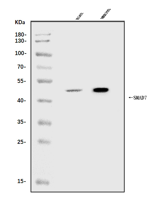
Western blot analysis of MADH7/SMAD7 using anti-MADH7/SMAD7 antibody (A00784-1).
Electrophoresis was performed on a 5-20% SDS-PAGE gel at 70V (Stacking gel) / 90V (Resolving gel) for 2-3 hours. The sample well of each lane was loaded with 30ug of sample under reducing conditions.
Lane 1: human K562 whole cell lysates,
Lane 2: human HEK293 whole cell lysates.
After Electrophoresis, proteins were transferred to a Nitrocellulose membrane at 150mA for 50-90 minutes. Blocked the membrane with 5% Non-fat Milk/ TBS for 1.5 hour at RT. The membrane was incubated with rabbit anti-MADH7/SMAD7 antigen affinity purified polyclonal antibody (Catalog # A00784-1) at 0.5 μg/mL overnight at 4°C, then washed with TBS-0.1%Tween 3 times with 5 minutes each and probed with a goat anti-rabbit IgG-HRP secondary antibody at a dilution of 1:5000 for 1.5 hour at RT. The signal is developed using an Enhanced Chemiluminescent detection (ECL) kit (Catalog # EK1002) with Tanon 5200 system. A specific band was detected for MADH7/SMAD7 at approximately 50KD. The expected band size for MADH7/SMAD7 is at 50KD.

Click image to see more details
Effects of CGA on the TGF-β1/miR-21/Smad7 signaling pathway in LX2 cells after TGF-β1 stimulation. (A–C) The mRNA levels were detected by real-time quantitative PCR. (D) The protein levels were assayed by western blotting. The data from three independent experiments are expressed as the means ± SD. ∗ P < 0.05 compared with the experimental group; ∗∗ P < 0.01 compared with experimental group; ## P < 0.01 compared with the normal group.
Index in PubMed under a CC BY license. PMID: 29311932

Click image to see more details
Verification of downstream signaling molecules in LX2 cells after miR-21 overexpression. (A) LX2 cells were transfected with lentivirus, and the expression of GFP was observed with a fluorescence microscope after 48 and 72 h. (B) The expression of miR-21, Smad7 and CTGF were measured by quantitative real-time PCR. (C) The protein expression was detected by western blotting. Data are shown means ± SD and significant differences were determined by one-way ANOVA. ## P < 0.01 for lentivirus-up group vs. normal group.
Index in PubMed under a CC BY license. PMID: 29311932

Click image to see more details
The illustration of CGA protecting against liver fibrosis in vitro and in vivo by regulating miR-21-regulated TGF-β1/Smad7 signaling pathway.
Index in PubMed under a CC BY license. PMID: 29311932

Click image to see more details
Effects of CGA on the TGF-β1/miR-21/Smad7 signaling pathway in LX2 cells after TGF-β1 overexpression. (A–C) The mRNA levels were detected by real-time quantitative PCR. (D) The protein levels were assayed by western blotting. The data from three independent experiments are expressed as the means ± SD. ∗ P < 0.05 for lentivirus-up/TGF-β1/CGA vs. lentivirus-up/TGF-β1; ∗∗ P < 0.01 for lentivirus-up/TGF-β1/CGA vs. lentivirus-up/TGF-β1; # P < 0.05 compared with the normal group; ## P < 0.01 compared with the normal group.
Index in PubMed under a CC BY license. PMID: 29311932

Click image to see more details
Verification of downstream signaling molecules in LX2 cells after miR-21 knockdown. (A) LX2 cells were transfected with lentivirus, and the expression of GFP was observed with a fluorescence microscope after 48 and 72 h. (B) The expression of miR-21, Smad7 and CTGF was measured by quantitative real-time PCR. (C) The protein expression was detected by western blotting. Data are shown as the means ± SD, and significant differences were determined by one-way ANOVA. ## P < 0.01 for lentivirus-down group vs. normal group.
Index in PubMed under a CC BY license. PMID: 29311932

Click image to see more details
Effects of CGA on the TGF-β1/miR-21/Smad7 signaling pathway in LX2 cells after TGF-β1 knockdown. (A–C) The mRNA levels were detected by real-time quantitative PCR. (D) The protein levels were assayed by western blotting. The data from three independent experiments are expressed as the means ± SD. ∗ P < 0.05 for lentivirus-down/TGF-β1/CGA vs. lentivirus-down/TGF-β1; ∗∗ P < 0.01 for lentivirus-down/TGF-β1/CGA vs. lentivirus-down/TGF-β1; ## P < 0.01 lentivirus-down/TGF-β1 vs. lentivirus-down.
Index in PubMed under a CC BY license. PMID: 29311932

Click image to see more details
Effect of CGA on the TGF-β1/miR-21/Smad7 signaling pathway in CCl4-induced rats. (A–C) The mRNA levels were measured by real-time quantitative PCR. (D) The protein levels were assayed by western blotting. The data from three independent experiments are expressed as the means ± SD. ∗ P < 0.05 compared with the experimental group; ∗∗ P < 0.01 compared with experimental group; # P < 0.05 compared with the normal group; ## P < 0.01 compared with the normal group.
Index in PubMed under a CC BY license. PMID: 29311932
Specific Publications For Anti-MADH7/SMAD7 Antibody Picoband® (A00784-1)
Loading publications
Recommended Resources
Here are featured tools and databases that you might find useful.
- Boster's Pathways Library
- Protein Databases
- Bioscience Research Protocol Resources
- Data Processing & Analysis Software
- Photo Editing Software
- Scientific Literature Resources
- Research Paper Management Tools
- Molecular Biology Software
- Primer Design Tools
- Bioinformatics Tools
- Phylogenetic Tree Analysis
Customer Reviews
Have you used Anti-MADH7/SMAD7 Antibody Picoband®?
Share your experimental results or join a short interview to earn up to $1,000 in product credits or other rewards.
0 Reviews For Anti-MADH7/SMAD7 Antibody Picoband®
Customer Q&As
Have a question?
Find answers in Q&As, reviews.
Can't find your answer?
Submit your question
