Click image to see more details
-

-
-
-
-
+4
Product Info Summary
| SKU: | A01201-4 |
|---|---|
| Size: | 100 μg/vial |
| Reactive Species: | Human, Mouse, Rat |
| Host: | Rabbit |
| Application: | ELISA, IHC, WB |
Customers Who Bought This Also Bought

Product info
Product Name
Anti-MAP2 Antibody Picoband®
SKU/Catalog Number
A01201-4
Size
100 μg/vial
Form
Lyophilized
Description
Boster Bio Anti-MAP2 Antibody Picoband® catalog # A01201-4 Tested in ELISA, IHC, WB applications. This antibody reacts with Human, Mouse, Rat. The brand Picoband indicates this is a premium antibody that guarantees superior quality, high affinity, and strong signals with minimal background in Western blot applications. Only our best-performing antibodies are designated as Picoband, ensuring unmatched performance.
Storage & Handling
At -20°C for one year from date of receipt. After reconstitution, at 4°C for one month. It can also be aliquotted and stored frozen at -20°C for six months. Avoid repeated freezing and thawing.
Cite This Product
Anti-MAP2 Antibody Picoband® (Boster Biological Technology, Pleasanton CA, USA, Catalog # A01201-4)
Host
Rabbit
Contents
Each vial contains 4 mg Trehalose, 0.9 mg NaCl, 0.2 mg Na2HPO4.
Clonality
Polyclonal
Isotype
Rabbit IgG
Immunogen
E.coli-derived human MAP2 recombinant protein (Position: A360-E1101).
Cross-reactivity
No cross-reactivity with other proteins.
Reactive Species
A01201-4 is reactive to MAP2 in Human, Mouse, Rat
Observed Molecular Weight
280 kDa
Calculated molecular weight
199.5 kDa
Background of MAP2
Microtubule-associated protein 2 is a protein that in humans is encoded by the MAP2 gene. This gene encodes a protein that belongs to the microtubule-associated protein family. The proteins of this family are thought to be involved in microtubule assembly, which is an essential step in neurogenesis. The products of similar genes in rat and mouse are neuron-specific cytoskeletal proteins that are enriched in dentrites, implicating a role in determining and stabilizing dentritic shape during neuron development. A number of alternatively spliced variants encoding distinct isoforms have been described.
Antibody Validation
Boster validates all antibodies on WB, IHC, ICC, Immunofluorescence, and ELISA with known positive control and negative samples to ensure specificity and high affinity, including thorough antibody incubations.
Application & Images
Applications
A01201-4 is guaranteed for ELISA, IHC, WB Boster Guarantee
Recommend Dilution
| Application | Dilution | Species |
|---|---|---|
| Western blot | 0.25-0.5 μg/ml | Human |
| Immunohistochemistry(Paraffin-embedded Section) | 2-5 μg/ml | Mouse, Rat |
| ELISA | 0.1-0.5 μg/ml | - |
Tested application
Suggested blocking solution with 5% non-fat milk or BSA; (*)Recommended protein loading: 20-40 µg per lane
Use TE buffer pH 9.0 for antigen retrieval; (*) citrate buffer pH 6.0 is an alternative.
Validation Images & Assay Conditions
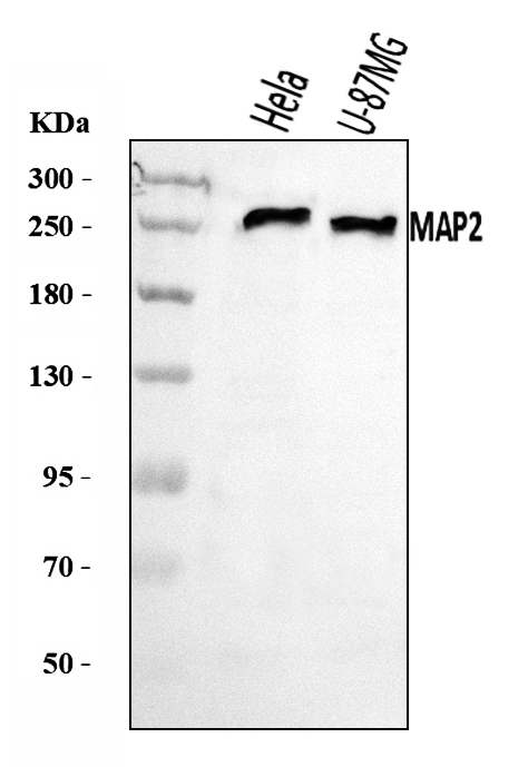
Click image to see more details
Western blot analysis of MAP2 using anti-MAP2 antibody (A01201-4).
Electrophoresis was performed on a 5-20% SDS-PAGE gel at 70V (Stacking gel) / 90V (Resolving gel) for 2-3 hours. The sample well of each lane was loaded with 30 ug of sample under reducing conditions.
Lane 1: human Hela whole cell lysates,
Lane 2: human U-87MG whole cell lysates.
After electrophoresis, proteins were transferred to a nitrocellulose membrane at 150 mA for 50-90 minutes. Blocked the membrane with 5% non-fat milk/TBS for 1.5 hour at RT. The membrane was incubated with rabbit anti-MAP2 antigen affinity purified polyclonal antibody (Catalog # A01201-4) at 0.5 μg/mL overnight at 4°C, then washed with TBS-0.1%Tween 3 times with 5 minutes each and probed with a goat anti-rabbit IgG-HRP secondary antibody at a dilution of 1:5000 for 1.5 hour at RT. The signal is developed using an Enhanced Chemiluminescent detection (ECL) kit (Catalog # EK1002) with Tanon 5200 system. A specific band was detected for MAP2 at approximately 280 kDa. The expected band size for MAP2 is at 200 kDa.

Click image to see more details
IHC analysis of MAP2 using anti-MAP2 antibody (A01201-4).
MAP2 was detected in a paraffin-embedded section of human brain tissue. Heat mediated antigen retrieval was performed in EDTA buffer (pH 8.0, epitope retrieval solution). The tissue section was blocked with 10% goat serum. The tissue section was then incubated with 2 μg/ml rabbit anti-MAP2 Antibody (A01201-4) overnight at 4°C. Peroxidase Conjugated Goat Anti-rabbit IgG was used as secondary antibody and incubated for 30 minutes at 37°C. The tissue section was developed using HRP Conjugated Rabbit IgG Super Vision Assay Kit (Catalog # SV0002) with DAB as the chromogen.

Click image to see more details
BSHX decoction decreased the damage of tissue and promoted axon regeneration after SCI. A Co-immunofluorescence images showed GFAP (red) and MAP2 (green) at day 14 after SCI. B Co-immunofluorescence images showed the axonal regeneration (GFAP, red; GAP43, green) in the lesion site at day after SCI. FS Fibrotic scar
Index in PubMed under a CC BY license. PMID: 35820953

Click image to see more details
Detection of the mature retinal markers in the proliferation-cultured E13.5 RPCs. After 4 day proliferation culture in vitro, E13.5 RPCs showed no obvious expression of the mature retinal markers Rhodopsin ( A-C ), Map2 ( D-F ), or GS ( G-I ). Bars were 20 μm.
Index in PubMed under a CC BY license. PMID: 19960071

Click image to see more details
Immunofluorescence detection in differentiated E13.5 and E17.5 RPCs. After 8 days of differentiation, the expression of Brn3b (panel A , B ), Map2 (panel C , D ), GFAP (panel E , F ), glutamine synthetase (panel G , H ), and Rhodopsin (panel I , J ) in E13.5 and E17.5 RPCs was investigated. K is the statistical ratio of positive cells in both RPCs. Note that the expression ratios of Brn3b and MAP2 in E13.5 RPCs were significantly higher than in E17.5 RPCs. In contrast, E17.5 RPCs expressed GFAP , glutamine synthetase ( GS ), and rhodopsin in higher percentages. The values were mean±standard deviation from three experiments. The symbols * and ** represent p<0.05 and p<0.01, respectively, versus E13.5 RPCs. Bars were 20 μm. The significance was evaluated by the Student t -test.
Index in PubMed under a CC BY license. PMID: 19960071

Click image to see more details
LMW-ASP upregulated the expression of MAP-2. (A) MAP-2 expression was detected by immunohistochemistry (400×, scale bar = 100 μm). (B) The IODs of MAP-2 (mean ± SD, n = 6). ### indicates p < 0.001 (sham versus model). ** indicates p < 0.01 (model versus LMW-ASP).
Index in PubMed under a CC BY license. PMID: 41019183

Click image to see more details
IHC analysis of MAP2 using anti-MAP2 antibody (A01201-4).
MAP2 was detected in a paraffin-embedded section of mouse brain tissue. Heat mediated antigen retrieval was performed in EDTA buffer (pH 8.0, epitope retrieval solution). The tissue section was blocked with 10% goat serum. The tissue section was then incubated with 2 μg/ml rabbit anti-MAP2 Antibody (A01201-4) overnight at 4°C. Biotinylated goat anti-rabbit IgG was used as secondary antibody and incubated for 30 minutes at 37°C. The tissue section was developed using Strepavidin-Biotin-Complex (SABC) (Catalog # SA1022) with DAB as the chromogen.

Click image to see more details
IHC analysis of MAP2 using anti-MAP2 antibody (A01201-4).
MAP2 was detected in a paraffin-embedded section of rat brain tissue. Heat mediated antigen retrieval was performed in EDTA buffer (pH 8.0, epitope retrieval solution). The tissue section was blocked with 10% goat serum. The tissue section was then incubated with 2 μg/ml rabbit anti-MAP2 Antibody (A01201-4) overnight at 4°C. Biotinylated goat anti-rabbit IgG was used as secondary antibody and incubated for 30 minutes at 37°C. The tissue section was developed using Strepavidin-Biotin-Complex (SABC) (Catalog # SA1022) with DAB as the chromogen.
Specific Publications For Anti-MAP2 Antibody Picoband® (A01201-4)
Loading publications
Recommended Resources
Here are featured tools and databases that you might find useful.
- Boster's Pathways Library
- Protein Databases
- Bioscience Research Protocol Resources
- Data Processing & Analysis Software
- Photo Editing Software
- Scientific Literature Resources
- Research Paper Management Tools
- Molecular Biology Software
- Primer Design Tools
- Bioinformatics Tools
- Phylogenetic Tree Analysis
Customer Reviews
Have you used Anti-MAP2 Antibody Picoband®?
Share your experimental results or join a short interview to earn up to $1,000 in product credits or other rewards.
0 Reviews For Anti-MAP2 Antibody Picoband®
Customer Q&As
Have a question?
Find answers in Q&As, reviews.
Can't find your answer?
Submit your question
