Click image to see more details
-

-
-
-
-
+2
Product Info Summary
| SKU: | MA1057 |
|---|---|
| Size: | 100 μg/vial |
| Reactive Species: | Human, Mouse, Rat |
| Host: | Mouse |
| Application: | IHC, WB |
Customers Who Bought This Also Bought

Product info
Product Name
Anti-MAP2 Antibody (Monoclonal, HM-2)
SKU/Catalog Number
MA1057
BM1243 is an alternative SKU for this antibody, used in previous lots.
Size
100 μg/vial
Form
Lyophilized
Description
Boster Bio Anti-MAP2 Antibody (Monoclonal, HM-2) catalog # MA1057. Tested in IHC, WB applications. This antibody reacts with Human, Mouse, Rat.
Storage & Handling
Store at -20˚C for one year from date of receipt. After reconstitution, at 4˚C for one month. It can also be aliquotted and stored frozen at -20˚C for six months. Avoid repeated freeze-thaw cycles.
Cite This Product
Anti-MAP2 Antibody (Monoclonal, HM-2) (Boster Biological Technology, Pleasanton CA, USA, Catalog # MA1057)
Host
Mouse
Contents
Mouse IgG in stabilizing components, 1.2% sodium acetate and 0.01mg NaN3.
Clonality
Monoclonal
Clone Number
HM-2
Isotype
Mouse IgG1
Immunogen
Rat brain microtubule-associated proteins (MAPs).
Cross-reactivity
No cross-reactivity with other proteins
Reactive Species
MA1057 is reactive to Map2 in Human, Mouse, Rat
Observed Molecular Weight
280 kDa
Calculated molecular weight
202.4 kDa
Background of Map2
MAP2, a 280-kD protein, is highly concentrated in neuronal somata and dendrites. Microtubule-associated protein 2 (MAP2)is a neurosteroid receptor. MAP2 gene contains 19 exons, and located in segment 2q34-q35. The transgenic MAP2c was present in dendrites but not in axons but transgenic MAP2c messenger RNA was limited to cell bodies.
Antibody Validation
Boster validates all antibodies on WB, IHC, ICC, Immunofluorescence, and ELISA with known positive control and negative samples to ensure specificity and high affinity, including thorough antibody incubations.
Application & Images
Applications
MA1057 is guaranteed for IHC, WB Boster Guarantee
Assay Dilutions Recommendation
The recommendations below provide a starting point for assay optimization. The actual working concentration varies and should be decided by the user.
Immunohistochemistry (Paraffin-embedded Section), 1-2μg/ml, Human, Mouse, Rat
Western blot, 0.5-2μg/ml, Mouse, rat
Positive Control
WB: rat brain tissue, mouse brain tissue
IHC: Rat Brain Tissue
Validation Images & Assay Conditions
Click image to see more details
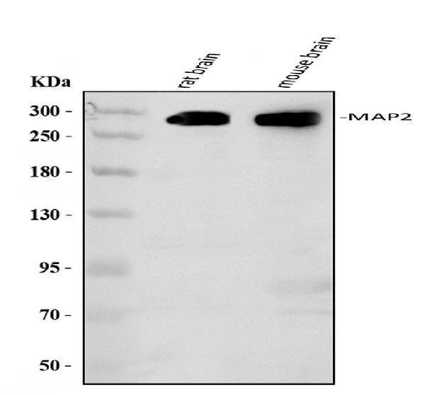
Western blot analysis of MAP2 using anti-MAP2 antibody (MA1057).
Electrophoresis was performed on a 5-20% SDS-PAGE gel at 70V (Stacking gel) / 90V (Resolving gel) for 2-3 hours. The sample well of each lane was loaded with 30 ug of sample under reducing conditions.
Lane 1: rat brain tissue lysates,
Lane 2: mouse brain tissue lysates.
After electrophoresis, proteins were transferred to a membrane. Then the membrane was incubated with rabbit anti-MAP2 antigen affinity purified monoclonal antibody (MA1057) at 1:500 overnight at 4°C, then washed with TBS-0.1%Tween 3 times with 5 minutes each and probed with a goat anti-mouse IgG-HRP secondary antibody at a dilution of 1:500 for 1.5 hour at RT. The signal is developed using an Enhanced Chemiluminescent detection (ECL) kit (Catalog # EK1002) with Tanon 5200 system. A specific band was detected for MAP2 at approximately 280 kDa. The expected band size for MAP2 is at 200 kDa.

Click image to see more details
Anti-MAP2 antibody (monoclonal), MA1057, IHC(P)
IHC(P): Rat Brain Tissue

Click image to see more details
Ptf1a reprograms human foreskin fibroblasts (HFF) into tripotent iNSCs. a , b Ectopic expression of Ptf1a in HFFs by lentiviruses induced the formation of neurospheres. c , d In the absence of doxycycline (Dox), Ptf1a-induced neurosphere cells were capable of forming neurospheres before passage 20 ( c ), but lost the capacity after passage 20 and became monolayered ( d ). e – i Ptf1a-induced human neural stem cells (hiNSCs) were highly immunoreactive for PTF1A, SOX2, PAX6, NESTIN, OLIG2, and FABP7. j – p Ptf1a-induced hiNSCs were capable of differentiating into neurons immunoreactive for TUJ1, MAP2, NEUN, TAU, or GABA, astrocytes labeled by GFAP, or oligodendrocytes marked by O1. q Neurons differentiated from hiNSCs were immunoreactive for both Tuj1 and synapsin. Cells in f – q were counterstained with nuclear DAPI. r Voltage-clamp recordings indicated fast activated and inactivated inward sodium currents as well as outward potassium currents on a differentiated neuron. s Current-clamp recordings revealed action potential responses of a differentiated neuron under current injection. t An action potential was induced after depolarization of the neuron. u Spontaneous postsynaptic currents recorded from a differentiated neuron. Scale bars, 80 μm ( a – d ) and 40 μm ( e – q )
Index in PubMed under a CC BY license. PMID: 30030434

Click image to see more details
In vitro differentiation potential of Ptf1a-reprogrammed miNSCs. a Schematic showing that miNSCs reprogrammed directly from MEFs by Ptf1a are able to differentiate into neurons, astrocytes, and oligodendrocytes under proper culture conditions. b miNSC10 cells underwent drastic morphological changes to form neuron-like cells when the culture medium was switched from NSC medium to neural differentiation medium. c miNSC10 cells could be differentiated into neurons immunoreactive for Tuj1, Map2, Dcx, NeuN, Tau, Peripherin, or GABA. They were also capable of differentiating into astrocytes (immunoreactive for GFAP) and oligodendrocytes (immunoreactive for O1, CNP, or MBP). d In vitro differentiated neurons were immunoreactive for both Tuj1 and synapsin. Cells in c and d were counterstained with nuclear DAPI. e Quantification of Map2 + neurons, GFAP+ astrocytes, and O1+ oligodendrocytes differentiated from miNSC10 cells under different differentiation conditions. f A merged micrograph showing a typical GFP-tagged neuron differentiated from miNSCs that was chosen for patch-clamp recording. g Voltage-clamp recordings indicated fast activated and inactivated inward sodium currents as well as outward potassium currents on a differentiated neuron. h Current-clamp recordings revealed action potential responses of the differentiated neuron under current injection. i Multiple action potentials were induced after depolarization of the neuron. j Spontaneous postsynaptic currents recorded from an in vitro differentiated neuron. Scale bars, 80 μm ( b ) and 40 μm ( c , d )
Index in PubMed under a CC BY license. PMID: 30030434

Click image to see more details
Granule cell morphology and synaptic integrity changes between anxiety-like and depression-like behaviors. (A,B) Representative Golgi-Cox staining images of dendritic spines and spine density in the hippocampal DG granule cells in CRS-treated rats, CFA-treated rats, and the controls. n = 10 for each group, bar = 10 μm. (C) Representative images of Golgi-Cox -stained granule cells in the DG area (top) and the reconstruction of its dendritic branches (bottom) from each group. Bar = 50 μm. (D) There were no significant effects of CRS and CFA injection on the dendritic length of the DG granule cells. (E) Sholl analysis of dendritic length in DG granule cells. CRS reduced dendrite intersection in the region 285–345 mm away from the soma compared with the control group. A repeated measures analysis of variance (ANOVA), * p < 0.05 vs. CON, n = 10 for each group; all graphs represent mean ± SEM. (F,G) Expression of SYP and MAP2 in the hippocampus was evaluated by Western blotting. Semi-quantitative analyses of SYP and MAP2 expression were performed. Note that the expression of SYP was significantly reduced in CRS-treated rats but was increased in CFA -treated rats. DG: dentate gyrus; CFA: complete Freund's adjuvant; CRS, chronic restraint stress; SYP, synaptophysin; MAP2, microtubule-associated protein 2. * p < 0.05, vs. control group (one-way ANOVA). All of the data are presented as mean ± SEM.
Index in PubMed under a CC BY license. PMID: 30740068

Click image to see more details
TUDCA promoted neuron regeneration along endogenous NSCs migration at day 7 after SCI. (A) Co-immunofluorescence showed endogenous NSCs (Nestin, green) and reactive astrocytes (GFAP, red) at the margin of the lesion site at day 7 after SCI. (B) Endogenous NSCs (Nestin, green) and neuron (NeuN, red) at the margin of the lesion site at day 7 after SCI. (C, D) Quantitative polymerase chain reaction (qPCR) showing the expression of Nestin, GFAP, NeuN, MAP2 and Oligo 2 at day 7 after SCI. All experiments were performed in triplicated and data were presented means ± SEM, n = 3 per group. *P < 0.05, **P < 0.01.
Index in PubMed under a CC BY license. PMID: 40276612
Specific Publications For Anti-MAP2 Antibody (Monoclonal, HM-2) (MA1057)
Loading publications
Recommended Resources
Here are featured tools and databases that you might find useful.
- Boster's Pathways Library
- Protein Databases
- Bioscience Research Protocol Resources
- Data Processing & Analysis Software
- Photo Editing Software
- Scientific Literature Resources
- Research Paper Management Tools
- Molecular Biology Software
- Primer Design Tools
- Bioinformatics Tools
- Phylogenetic Tree Analysis
Customer Reviews
Have you used Anti-MAP2 Antibody (Monoclonal, HM-2)?
Share your experimental results or join a short interview to earn up to $1,000 in product credits or other rewards.
0 Reviews For Anti-MAP2 Antibody (Monoclonal, HM-2)
Customer Q&As
Have a question?
Find answers in Q&As, reviews.
Can't find your answer?
Submit your question
17 Customer Q&As for Anti-MAP2 Antibody (Monoclonal, HM-2)
Question
Is there a BSA free version of anti-MAP2 antibody (Monoclonal, HM-2) MA1057 available?
Verified Customer
Verified customer
Asked: 2020-05-01
Answer
We appreciate your recent telephone inquiry. I can confirm that some lots of this anti-MAP2 antibody (Monoclonal, HM-2) MA1057 are BSA free. For now, these lots are available and we can make a BSA free formula for you free of charge. It will take 3 extra days to prepare. If you require this antibody BSA free again in future, please do not hesitate to contact me and I will be pleased to check which lots we have in stock that are BSA free.
Boster Scientific Support
Answered: 2020-05-01
Question
Would anti-MAP2 antibody (Monoclonal, HM-2) MA1057 work on primate WB with dorsolateral prefrontal cortex?
Verified Customer
Verified customer
Asked: 2020-04-06
Answer
Our lab technicians have not tested anti-MAP2 antibody (Monoclonal, HM-2) MA1057 on primate. You can run a BLAST between primate and the immunogen sequence of anti-MAP2 antibody (Monoclonal, HM-2) MA1057 to see if they may cross-react. If the sequence homology is close, then you can perform a pilot test. Keep in mind that since we have not validated primate samples, this use of the antibody is not covered by our guarantee. However we have an innovator award program that if you test this antibody and show it works in primate dorsolateral prefrontal cortex in WB, you can get your next antibody for free.
Boster Scientific Support
Answered: 2020-04-06
Question
We are currently using anti-MAP2 antibody (Monoclonal, HM-2) MA1057 for mouse tissue, and we are satisfied with the IHC results. The species of reactivity given in the datasheet says human, mouse, rat. Is it likely that the antibody can work on pig tissues as well?
Verified Customer
Verified customer
Asked: 2020-02-27
Answer
The anti-MAP2 antibody (Monoclonal, HM-2) (MA1057) has not been validated for cross reactivity specifically with pig tissues, but there is a good chance of cross reactivity. We have an innovator award program that if you test this antibody and show it works in pig you can get your next antibody for free. Please contact me if I can help you with anything.
Boster Scientific Support
Answered: 2020-02-27
Question
We have observed staining in mouse liver. Are there any suggestions? Is anti-MAP2 antibody (Monoclonal, HM-2) supposed to stain liver positively?
Verified Customer
Verified customer
Asked: 2019-12-26
Answer
From what I have seen in literature liver does express MAP2. From what I have seen in Uniprot.org, MAP2 is expressed in dorsolateral prefrontal cortex, brain, brain pancreas, cervix carcinoma, liver, among other tissues. Regarding which tissues have MAP2 expression, here are a few articles citing expression in various tissues:
Brain, Pubmed ID: 8294038
Brain, and Pancreas, Pubmed ID: 15489334
Cervix carcinoma, Pubmed ID: 17081983, 18220336
Liver, Pubmed ID: 24275569
Boster Scientific Support
Answered: 2019-12-26
Question
Our lab want to know about to test anti-MAP2 antibody (Monoclonal, HM-2) MA1057 on human brain pancreas for research purposes, then I may be interested in using anti-MAP2 antibody (Monoclonal, HM-2) MA1057 for diagnostic purposes as well. Is the antibody suitable for diagnostic purposes?
E. Taylor
Verified customer
Asked: 2019-12-20
Answer
The products we sell, including anti-MAP2 antibody (Monoclonal, HM-2) MA1057, are only intended for research use. They would not be suitable for use in diagnostic work. If you have the means to develop a product into diagnostic use, and are interested in collaborating with us and develop our product into an IVD product, please contact us for more discussions.
Boster Scientific Support
Answered: 2019-12-20
Question
We bought anti-MAP2 antibody (Monoclonal, HM-2) for WB on liver in the past. I am using mouse, and We are going to use the antibody for IHC next. I am looking for examining liver as well as dorsolateral prefrontal cortex in our next experiment. Could you please give me some suggestion on which antibody would work the best for IHC?
Verified Customer
Verified customer
Asked: 2019-11-21
Answer
I have checked the website and datasheets of our anti-MAP2 antibody (Monoclonal, HM-2) and it appears that MA1057 has been tested on mouse in both WB and IHC. Thus MA1057 should work for your application. Our Boster satisfaction guarantee will cover this product for IHC in mouse even if the specific tissue type has not been validated. We do have a comprehensive range of products for IHC detection and you can check out our website bosterbio.com to find out more information about them.
Boster Scientific Support
Answered: 2019-11-21
Question
I have a question about product MA1057, anti-MAP2 antibody (Monoclonal, HM-2). I was wondering if it would be possible to conjugate this antibody with biotin. I would need it to be without BSA or sodium azide. I am planning on using a buffer exchange of sodium azide with PBS only. Would there be problems for me to conjugate the antibody and store it in -20 degrees in small aliquots?
Verified Customer
Verified customer
Asked: 2019-08-07
Answer
We do not recommend storing this antibody with PBS buffer only in -20 degrees. If you want to store it in -20 degrees it is best to add some cryoprotectant like glycerol. If you want carrier free MA1057 anti-MAP2 antibody (Monoclonal, HM-2), we can provide it to you in a special formula with trehalose and/or glycerol. These molecules will not interfere with conjugation chemistry and provide a good level of protection for the antibody from degradation. Please be sure to specify this in your purchase order.
Boster Scientific Support
Answered: 2019-08-07
Question
I was wanting to use your anti-MAP2 antibody (Monoclonal, HM-2) for WB for human brain pancreas on frozen tissues, but I want to know if it has been tested for this particular application. Has this antibody been tested and is this antibody a good choice for human brain pancreas identification?
Verified Customer
Verified customer
Asked: 2019-07-15
Answer
As indicated on the product datasheet, MA1057 anti-MAP2 antibody (Monoclonal, HM-2) has been tested for IHC, WB on human, mouse, rat tissues. We have an innovator award program that if you test this antibody and show it works in human brain pancreas in IHC-frozen, you can get your next antibody for free.
Boster Scientific Support
Answered: 2019-07-15
Question
Is a blocking peptide available for product anti-MAP2 antibody (Monoclonal, HM-2) (MA1057)?
Verified Customer
Verified customer
Asked: 2019-06-04
Answer
We do provide the blocking peptide for product anti-MAP2 antibody (Monoclonal, HM-2) (MA1057). If you would like to place an order for it please contact support@bosterbio.com and make a special request.
Boster Scientific Support
Answered: 2019-06-04
Question
I appreciate helping with my inquiry over the phone. Here are the WB image, lot number and protocol we used for brain pancreas using anti-MAP2 antibody (Monoclonal, HM-2) MA1057. Let me know if you need anything else.
Verified Customer
Verified customer
Asked: 2018-10-02
Answer
We appreciate the data. You have provided everything we needed. Our lab team are working to resolve your inquiry as quickly as possible, and we appreciate your patience and understanding! Please let me know if there is anything you need in the meantime.
Boster Scientific Support
Answered: 2018-10-02
Question
I see that the anti-MAP2 antibody (Monoclonal, HM-2) MA1057 works with WB, what is the protocol used to produce the result images on the product page?
Verified Customer
Verified customer
Asked: 2018-05-16
Answer
You can find protocols for WB on the "support/technical resources" section of our navigation menu. If you have any further questions, please send an email to support@bosterbio.com
Boster Scientific Support
Answered: 2018-05-16
Question
See attached the WB image, lot number and protocol we used for brain pancreas using anti-MAP2 antibody (Monoclonal, HM-2) MA1057. Please let me know if you require anything else.
Verified Customer
Verified customer
Asked: 2017-11-23
Answer
Thank you very much for the data. Our lab team are working to resolve this as quickly as possible, and we appreciate your patience and understanding! You have provided everything we needed. Please let me know if there is anything you need in the meantime.
Boster Scientific Support
Answered: 2017-11-23
Question
Would anti-MAP2 antibody (Monoclonal, HM-2) MA1057 work for WB with brain pancreas?
C. Anderson
Verified customer
Asked: 2017-09-14
Answer
According to the expression profile of brain pancreas, MAP2 is highly expressed in brain pancreas. So, it is likely that anti-MAP2 antibody (Monoclonal, HM-2) MA1057 will work for WB with brain pancreas.
Boster Scientific Support
Answered: 2017-09-14
Question
I am looking for using your anti-MAP2 antibody (Monoclonal, HM-2) for microtubule cytoskeleton organization studies. Has this antibody been tested with western blotting on rat brain tissue? We would like to see some validation images before ordering.
J. Zhao
Verified customer
Asked: 2016-08-10
Answer
Thank you for your inquiry. This MA1057 anti-MAP2 antibody (Monoclonal, HM-2) is validated on rat brain tissue. It is guaranteed to work for IHC, WB in human, mouse, rat. Our Boster guarantee will cover your intended experiment even if the sample type has not been be directly tested.
Boster Scientific Support
Answered: 2016-08-10
Question
Is this MA1057 anti-MAP2 antibody (Monoclonal, HM-2) reactive to the isotypes of MAP2?
P. Taylor
Verified customer
Asked: 2015-10-07
Answer
The immunogen of MA1057 anti-MAP2 antibody (Monoclonal, HM-2) is Rat brain microtubule-associated proteins(MAPs). Could you tell me which isotype you are interested in so I can help see if the immunogen is part of this isotype?
Boster Scientific Support
Answered: 2015-10-07
Question
Would MA1057 anti-MAP2 antibody (Monoclonal, HM-2) work on parafin embedded sections? If so, which fixation method do you recommend we use (PFA, paraformaldehyde, other)?
A. Gonzalez
Verified customer
Asked: 2014-10-30
Answer
It shows on the product datasheet, MA1057 anti-MAP2 antibody (Monoclonal, HM-2) as been validated on WB. It is best to use PFA for fixation because it has better tissue penetration ability. PFA needs to be prepared fresh before use. Long term stored PFA turns into formalin, as the PFA molecules congregate and become formalin.
Boster Scientific Support
Answered: 2014-10-30
Question
We were content with the WB result of your anti-MAP2 antibody (Monoclonal, HM-2). However we have seen positive staining in brain cytoskeleton. cell using this antibody. Is that expected? Could you tell me where is MAP2 supposed to be expressed?
J. Edwards
Verified customer
Asked: 2013-08-16
Answer
According to literature, brain does express MAP2. Generally MAP2 expresses in cytoplasm, cytoskeleton. cell. Regarding which tissues have MAP2 expression, here are a few articles citing expression in various tissues:
Brain, Pubmed ID: 8294038
Brain, and Pancreas, Pubmed ID: 15489334
Cervix carcinoma, Pubmed ID: 17081983, 18220336
Liver, Pubmed ID: 24275569
Boster Scientific Support
Answered: 2013-08-16
