Click image to see more details
-

-
-
-
-
+2
Product Info Summary
| SKU: | A08547-1 |
|---|---|
| Size: | 100 μg/vial |
| Reactive Species: | Human, Mouse, Rat |
| Host: | Rabbit |
| Application: | IHC, WB |
Customers Who Bought This Also Bought

Product info
Product Name
Anti-MCUR1 Antibody Picoband®
SKU/Catalog Number
A08547-1
Size
100 μg/vial
Form
Lyophilized
Description
Boster Bio Anti-MCUR1 Antibody Picoband® catalog # A08547-1. Tested in IHC, WB applications. This antibody reacts with Human, Mouse, Rat. The brand Picoband indicates this is a premium antibody that guarantees superior quality, high affinity, and strong signals with minimal background in Western blot applications. Only our best-performing antibodies are designated as Picoband, ensuring unmatched performance.
Storage & Handling
Store at -20˚C for one year from date of receipt. After reconstitution, at 4˚C for one month. It can also be aliquotted and stored frozen at -20˚C for six months. Avoid repeated freeze-thaw cycles.
Cite This Product
Anti-MCUR1 Antibody Picoband® (Boster Biological Technology, Pleasanton CA, USA, Catalog # A08547-1)
Host
Rabbit
Contents
Each vial contains 4mg Trehalose, 0.9mg NaCl, 0.2mg Na2HPO4, 0.05mg NaN3.
Clonality
Polyclonal
Isotype
Rabbit IgG
Immunogen
A synthetic peptide corresponding to a sequence in the middle region of human MCUR1, which shares 79.4% and 82.3% amino acid (aa) sequence identity with mouse and rat MCUR1, respectively.
Cross-reactivity
No cross-reactivity with other proteins.
Reactive Species
A08547-1 is reactive to MCUR1 in Human, Mouse, Rat
Observed Molecular Weight
40 kDa
Calculated molecular weight
39.7 kDa
Background of MCUR1
MCUR1 is an inner mitochondrial membrane protein that has been shown to function as a component of the mitochondrial Ca (2+) uniporter, in the assembly of mitochondrial respiratory complex IV, and in mitochondrial permeability transition (MPT). The MCUR1 gene is mapped to chromosome 6p23 based on an alignment of the MCUR1 sequence with the genomic sequence.
Antibody Validation
Boster validates all antibodies on WB, IHC, ICC, Immunofluorescence, and ELISA with known positive control and negative samples to ensure specificity and high affinity, including thorough antibody incubations.
Application & Images
Applications
A08547-1 is guaranteed for IHC, WB Boster Guarantee
Recommend Dilution
| Application | Dilution | Species |
|---|---|---|
| Western blot | 0.1-0.5μg/ml | |
| Immunohistochemistry (Paraffin-embedded Section) | 0.5-1μg/ml |
Tested application
Suggested blocking solution with 5% non-fat milk or BSA; (*)Recommended protein loading: 20-40 µg per lane
Use TE buffer pH 9.0 for antigen retrieval; (*) citrate buffer pH 6.0 is an alternative.
Validation Images & Assay Conditions
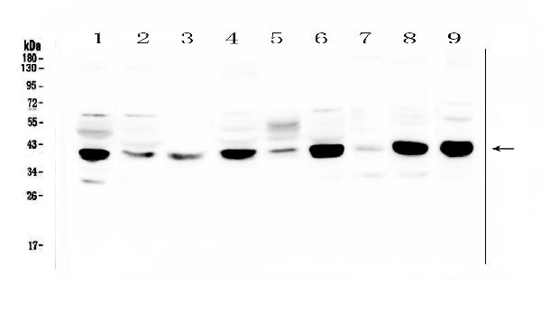
Click image to see more details
Western blot analysis of MCUR1 using anti-MCUR1 antibody (A08547-1).
Electrophoresis was performed on a 5-20% SDS-PAGE gel at 70V (Stacking gel) / 90V (Resolving gel) for 2-3 hours. The sample well of each lane was loaded with 50ug of sample under reducing conditions.
Lane 1: human Hela whole cell lysates.
Lane 2: human MDA-MB-231 whole cell lysates,
Lane 3: human HL-60 whole cell lysates,
Lane 4: human MDA-MB-453 whole cell lysates,
Lane 5: human A431 whole cell lysates,
Lane 6: human Caco-2 whole cell lysates,
Lane 7: rat spleen tissue lysates,
Lane 8: mouse lung tissue lysates,
Lane 9: mouse Ana-1 whole cell lysates,
After Electrophoresis, proteins were transferred to a Nitrocellulose membrane at 150mA for 50-90 minutes. Blocked the membrane with 5% Non-fat Milk/ TBS for 1.5 hour at RT. The membrane was incubated with rabbit anti-MCUR1 antigen affinity purified polyclonal antibody (Catalog # A08547-1) at 0.5 μg/mL overnight at 4°C, then washed with TBS-0.1%Tween 3 times with 5 minutes each and probed with a goat anti-rabbit IgG-HRP secondary antibody at a dilution of 1:10000 for 1.5 hour at RT. The signal is developed using an Enhanced Chemiluminescent detection (ECL) kit (Catalog # EK1002) with Tanon 5200 system. A specific band was detected for MCUR1 at approximately 40KD. The expected band size for MCUR1 is at 40KD.

Click image to see more details
IHC analysis of MCUR1 using anti-MCUR1 antibody (A08547-1).
MCUR1 was detected in paraffin-embedded section of human oesophagus squama cancer tissue. Heat mediated antigen retrieval was performed in citrate buffer (pH6, epitope retrieval solution) for 20 mins. The tissue section was blocked with 10% goat serum. The tissue section was then incubated with 1μg/ml rabbit anti-MCUR1 Antibody (A08547-1) overnight at 4°C. Biotinylated goat anti-rabbit IgG was used as secondary antibody and incubated for 30 minutes at 37°C. The tissue section was developed using Strepavidin-Biotin-Complex (SABC)(Catalog # SA1022) with DAB as the chromogen.

Click image to see more details
IHC analysis of MCUR1 using anti-MCUR1 antibody (A08547-1).
MCUR1 was detected in paraffin-embedded section of human ovary cancer tissue. Heat mediated antigen retrieval was performed in citrate buffer (pH6, epitope retrieval solution) for 20 mins. The tissue section was blocked with 10% goat serum. The tissue section was then incubated with 1μg/ml rabbit anti-MCUR1 Antibody (A08547-1) overnight at 4°C. Biotinylated goat anti-rabbit IgG was used as secondary antibody and incubated for 30 minutes at 37°C. The tissue section was developed using Strepavidin-Biotin-Complex (SABC)(Catalog # SA1022) with DAB as the chromogen.

Click image to see more details
IHC analysis of MCUR1 using anti-MCUR1 antibody (A08547-1).
MCUR1 was detected in paraffin-embedded section of human lung cancer tissue. Heat mediated antigen retrieval was performed in citrate buffer (pH6, epitope retrieval solution) for 20 mins. The tissue section was blocked with 10% goat serum. The tissue section was then incubated with 1μg/ml rabbit anti-MCUR1 Antibody (A08547-1) overnight at 4°C. Biotinylated goat anti-rabbit IgG was used as secondary antibody and incubated for 30 minutes at 37°C. The tissue section was developed using Strepavidin-Biotin-Complex (SABC)(Catalog # SA1022) with DAB as the chromogen.

Click image to see more details
IHC analysis of MCUR1 using anti-MCUR1 antibody (A08547-1).
MCUR1 was detected in paraffin-embedded section of human placenta tissue. Heat mediated antigen retrieval was performed in citrate buffer (pH6, epitope retrieval solution) for 20 mins. The tissue section was blocked with 10% goat serum. The tissue section was then incubated with 1μg/ml rabbit anti-MCUR1 Antibody (A08547-1) overnight at 4°C. Biotinylated goat anti-rabbit IgG was used as secondary antibody and incubated for 30 minutes at 37°C. The tissue section was developed using Strepavidin-Biotin-Complex (SABC)(Catalog # SA1022) with DAB as the chromogen.

Click image to see more details
IHC analysis of MCUR1 using anti-MCUR1 antibody (A08547-1).
MCUR1 was detected in paraffin-embedded section of human tonsil tissue. Heat mediated antigen retrieval was performed in citrate buffer (pH6, epitope retrieval solution) for 20 mins. The tissue section was blocked with 10% goat serum. The tissue section was then incubated with 1μg/ml rabbit anti-MCUR1 Antibody (A08547-1) overnight at 4°C. Biotinylated goat anti-rabbit IgG was used as secondary antibody and incubated for 30 minutes at 37°C. The tissue section was developed using Strepavidin-Biotin-Complex (SABC)(Catalog # SA1022) with DAB as the chromogen.
Specific Publications For Anti-MCUR1 Antibody Picoband® (A08547-1)
Loading publications
Recommended Resources
Here are featured tools and databases that you might find useful.
- Boster's Pathways Library
- Protein Databases
- Bioscience Research Protocol Resources
- Data Processing & Analysis Software
- Photo Editing Software
- Scientific Literature Resources
- Research Paper Management Tools
- Molecular Biology Software
- Primer Design Tools
- Bioinformatics Tools
- Phylogenetic Tree Analysis
Customer Reviews
Have you used Anti-MCUR1 Antibody Picoband®?
Share your experimental results or join a short interview to earn up to $1,000 in product credits or other rewards.
0 Reviews For Anti-MCUR1 Antibody Picoband®
Customer Q&As
Have a question?
Find answers in Q&As, reviews.
Can't find your answer?
Submit your question
7 Customer Q&As for Anti-MCUR1 Antibody Picoband®
Question
We are currently using anti-MCUR1 antibody A08547-1 for rat tissue, and we are happy with the WB results. The species of reactivity given in the datasheet says human, mouse, rat. Is it possible that the antibody can work on monkey tissues as well?
Verified Customer
Verified customer
Asked: 2019-12-20
Answer
The anti-MCUR1 antibody (A08547-1) has not been validated for cross reactivity specifically with monkey tissues, but there is a good chance of cross reactivity. We have an innovator award program that if you test this antibody and show it works in monkey you can get your next antibody for free. Please contact me if I can help you with anything.
Boster Scientific Support
Answered: 2019-12-20
Question
Will anti-MCUR1 antibody A08547-1 work for IHC-P with lung ovary?
N. Johnson
Verified customer
Asked: 2019-09-16
Answer
According to the expression profile of lung ovary, MCUR1 is highly expressed in lung ovary. So, it is likely that anti-MCUR1 antibody A08547-1 will work for IHC-P with lung ovary.
Boster Scientific Support
Answered: 2019-09-16
Question
Would anti-MCUR1 antibody A08547-1 work on feline WB with adipose tissue?
Verified Customer
Verified customer
Asked: 2019-08-29
Answer
Our lab technicians have not tested anti-MCUR1 antibody A08547-1 on feline. You can run a BLAST between feline and the immunogen sequence of anti-MCUR1 antibody A08547-1 to see if they may cross-react. If the sequence homology is close, then you can perform a pilot test. Keep in mind that since we have not validated feline samples, this use of the antibody is not covered by our guarantee. However we have an innovator award program that if you test this antibody and show it works in feline adipose tissue in WB, you can get your next antibody for free.
Boster Scientific Support
Answered: 2019-08-29
Question
Thanks for helping with my inquiry over the phone. Here are the WB image, lot number and protocol we used for lung ovary using anti-MCUR1 antibody A08547-1. Let me know if you need anything else.
Verified Customer
Verified customer
Asked: 2019-04-25
Answer
I appreciate the data. You have provided everything we needed. Our lab team are working to resolve your inquiry as quickly as possible, and we appreciate your patience and understanding! Please let me know if there is anything you need in the meantime.
Boster Scientific Support
Answered: 2019-04-25
Question
See attached the WB image, lot number and protocol we used for lung ovary using anti-MCUR1 antibody A08547-1. Please let me know if you require anything else.
Verified Customer
Verified customer
Asked: 2018-09-03
Answer
Thank you very much for the data. Our lab team are working to resolve this as quickly as possible, and we appreciate your patience and understanding! You have provided everything we needed. Please let me know if there is anything you need in the meantime.
Boster Scientific Support
Answered: 2018-09-03
Question
I was wanting to use your anti-MCUR1 antibody for IHC-P for human lung ovary on frozen tissues, but I want to know if it has been validated for this particular application. Has this antibody been validated and is this antibody a good choice for human lung ovary identification?
C. Mangal
Verified customer
Asked: 2014-11-13
Answer
You can see on the product datasheet, A08547-1 anti-MCUR1 antibody has been tested for IHC-P, WB on human, mouse, rat tissues. We have an innovator award program that if you test this antibody and show it works in human lung ovary in IHC-frozen, you can get your next antibody for free.
Boster Scientific Support
Answered: 2014-11-13
Question
We want to test anti-MCUR1 antibody A08547-1 on human lung ovary for research purposes, then I may be interested in using anti-MCUR1 antibody A08547-1 for diagnostic purposes as well. Is the antibody suitable for diagnostic purposes?
J. Carter
Verified customer
Asked: 2013-12-12
Answer
The products we sell, including anti-MCUR1 antibody A08547-1, are only intended for research use. They would not be suitable for use in diagnostic work. If you have the means to develop a product into diagnostic use, and are interested in collaborating with us and develop our product into an IVD product, please contact us for more discussions.
Boster Scientific Support
Answered: 2013-12-12
