Click image to see more details
-

-
-
-
-
+4
Product Info Summary
| SKU: | A01065 |
|---|---|
| Size: | 100 μg/vial |
| Reactive Species: | Human, Mouse, Rat |
| Host: | Rabbit |
| Application: | Flow Cytometry, IHC, WB |
Customers Who Bought This Also Bought

Product info
Product Name
Anti-Methylmalonyl Coenzyme A mutase Antibody Picoband®
SKU/Catalog Number
A01065
Size
100 μg/vial
Form
Lyophilized
Description
Boster Bio Anti-Methylmalonyl Coenzyme A mutase Antibody Picoband® catalog # A01065. Tested in Flow Cytometry, IHC, WB applications. This antibody reacts with Human, Mouse, Rat. The brand Picoband indicates this is a premium antibody that guarantees superior quality, high affinity, and strong signals with minimal background in Western blot applications. Only our best-performing antibodies are designated as Picoband, ensuring unmatched performance.
Storage & Handling
Store at -20˚C for one year from date of receipt. After reconstitution, at 4˚C for one month. It can also be aliquotted and stored frozen at -20˚C for six months. Avoid repeated freeze-thaw cycles.
Cite This Product
Anti-Methylmalonyl Coenzyme A mutase Antibody Picoband® (Boster Biological Technology, Pleasanton CA, USA, Catalog # A01065)
Host
Rabbit
Contents
Each vial contains 4mg Trehalose, 0.9mg NaCl, 0.2mg Na2HPO4, 0.01mg NaN3.
Clonality
Polyclonal
Isotype
Rabbit IgG
Immunogen
A synthetic peptide corresponding to a sequence at the N-terminus of human MUT, different from the related mouse sequence by one amino acid.
Cross-reactivity
No cross-reactivity with other proteins
Reactive Species
A01065 is reactive to MUT in Human, Mouse, Rat
Observed Molecular Weight
83 kDa
Calculated molecular weight
83.1 kDa
Background of MUT
Methylmalonyl-CoA mutase (MUT) is a mitochondrial enzyme that catalyzes the isomerization of methylmalonyl-CoA to succinyl-CoA. This gene is mapped to 6p12.3. MUT requires a vitamin B12-derived prosthetic group, adenosylcobalamin (commonly referred to as AdoCbl), to function. And the product of this enzyme, succinyl-CoA, is a key molecule of the TCA cycle.
Antibody Validation
Boster validates all antibodies on WB, IHC, ICC, Immunofluorescence, and ELISA with known positive control and negative samples to ensure specificity and high affinity, including thorough antibody incubations.
Application & Images
Applications
A01065 is guaranteed for Flow Cytometry, IHC, WB Boster Guarantee
Assay Dilutions Recommendation
The recommendations below provide a starting point for assay optimization. The actual working concentration varies and should be decided by the user.
Western blot, 0.1-0.5μg/ml, Human, Mouse, Rat
Immunohistochemistry (Paraffin-embedded Section), 2-5μg/ml, Human
Flow Cytometry (Fixed), 1-3μg/1x106 cells, Human
Positive Control
WB: human Hela whole cell, human PC-3 whole cell, human A431 whole cell, human A564 whole cell, rat liver tissue, rat PC-12 whole cell, mouse liver tissue, mouse SP2/0 whole cell
IHC: human bladder urothelial carcinoma tissue, human colorectal adenocarcinoma tissue, human esophageal squamous carcinoma tissue, human liver cancer tissue, human lung cancer tissue
FCM: SiHa cell, HepG2 cell
Validation Images & Assay Conditions
Click image to see more details
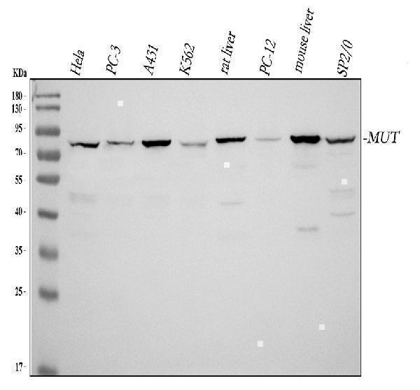
Western blot analysis of MUT using anti-MUT antibody (A01065).
Electrophoresis was performed on a 10% SDS-PAGE gel at 80V (Stacking gel) / 120V (Resolving gel) for 2 hours. The sample well of each lane was loaded with 30 ug of sample under reducing conditions.
lane 1: human Hela whole cell lysates,
lane 2: human PC-3 whole cell lysates,
lane 3: human A431 whole cell lysates,
lane 4: human A564 whole cell lysates,
lane 5: rat liver tissue lysates,
lane 6: rat PC-12 whole cell lysates,
lane 7: mouse liver tissue lysates,
lane 8: mouse SP2/0 whole cell lysates.
After Electrophoresis, proteins were transferred to a Nitrocellulose membrane at 150mA for 50-90 minutes. Blocked the membrane with 5% Non-fat Milk/ TBS for 1.5 hour at RT. The membrane was incubated with rabbit anti-MUT antigen affinity purified polyclonal antibody (Catalog # A01065) at 0.5 μg/mL overnight at 4°C, then washed with TBS-0.1%Tween 3 times with 5 minutes each and probed with a goat anti-rabbit IgG-HRP secondary antibody at a dilution of 1:10000 for 1.5 hour at RT. The signal is developed using an ECL Plus Western Blotting Substrate (Catalog # AR1196-200) with Tanon 5200 system. A specific band was detected for MUT at approximately 83 kDa. The expected band size for MUT is at 83 kDa.

Click image to see more details
IHC analysis of MUT using anti-MUT antibody (A01065).
MUT was detected in a paraffin-embedded section of human bladder urothelial carcinoma tissue. Heat mediated antigen retrieval was performed in EDTA buffer (pH 8.0, epitope retrieval solution). The tissue section was blocked with 10% goat serum. The tissue section was then incubated with 2 μg/ml rabbit anti-MUT Antibody (A01065) overnight at 4°C. Peroxidase Conjugated Goat Anti-rabbit IgG was used as secondary antibody and incubated for 30 minutes at 37°C. The tissue section was developed using HRP Conjugated Rabbit IgG Super Vision Assay Kit (Catalog # SV0002) with DAB as the chromogen.

Click image to see more details
IHC analysis of MUT using anti-MUT antibody (A01065).
MUT was detected in a paraffin-embedded section of human colorectal adenocarcinoma tissue. Heat mediated antigen retrieval was performed in EDTA buffer (pH 8.0, epitope retrieval solution). The tissue section was blocked with 10% goat serum. The tissue section was then incubated with 2 μg/ml rabbit anti-MUT Antibody (A01065) overnight at 4°C. Peroxidase Conjugated Goat Anti-rabbit IgG was used as secondary antibody and incubated for 30 minutes at 37°C. The tissue section was developed using HRP Conjugated Rabbit IgG Super Vision Assay Kit (Catalog # SV0002) with DAB as the chromogen.

Click image to see more details
IHC analysis of MUT using anti-MUT antibody (A01065).
MUT was detected in a paraffin-embedded section of human esophageal squamous carcinoma tissue. Heat mediated antigen retrieval was performed in EDTA buffer (pH 8.0, epitope retrieval solution). The tissue section was blocked with 10% goat serum. The tissue section was then incubated with 2 μg/ml rabbit anti-MUT Antibody (A01065) overnight at 4°C. Peroxidase Conjugated Goat Anti-rabbit IgG was used as secondary antibody and incubated for 30 minutes at 37°C. The tissue section was developed using HRP Conjugated Rabbit IgG Super Vision Assay Kit (Catalog # SV0002) with DAB as the chromogen.

Click image to see more details
IHC analysis of MUT using anti-MUT antibody (A01065).
MUT was detected in a paraffin-embedded section of human liver cancer tissue. Heat mediated antigen retrieval was performed in EDTA buffer (pH 8.0, epitope retrieval solution). The tissue section was blocked with 10% goat serum. The tissue section was then incubated with 2 μg/ml rabbit anti-MUT Antibody (A01065) overnight at 4°C. Peroxidase Conjugated Goat Anti-rabbit IgG was used as secondary antibody and incubated for 30 minutes at 37°C. The tissue section was developed using HRP Conjugated Rabbit IgG Super Vision Assay Kit (Catalog # SV0002) with DAB as the chromogen.

Click image to see more details
IHC analysis of MUT using anti-MUT antibody (A01065).
MUT was detected in a paraffin-embedded section of human lung cancer tissue. Heat mediated antigen retrieval was performed in EDTA buffer (pH 8.0, epitope retrieval solution). The tissue section was blocked with 10% goat serum. The tissue section was then incubated with 2 μg/ml rabbit anti-MUT Antibody (A01065) overnight at 4°C. Peroxidase Conjugated Goat Anti-rabbit IgG was used as secondary antibody and incubated for 30 minutes at 37°C. The tissue section was developed using HRP Conjugated Rabbit IgG Super Vision Assay Kit (Catalog # SV0002) with DAB as the chromogen.

Click image to see more details
Flow Cytometry analysis of SiHa cells using anti-MUT antibody (A01065).
Overlay histogram showing SiHa cells stained with A01065 (Blue line). To facilitate intracellular staining, cells were fixed with 4% paraformaldehyde and permeabilized with permeabilization buffer. The cells were blocked with 10% normal goat serum. And then incubated with rabbit anti-MUT Antibody (A01065, 1 μg/1x106 cells) for 30 min at 20°C. DyLight®488 conjugated goat anti-rabbit IgG (BA1127, 5-10 μg/1x106 cells) was used as secondary antibody for 30 minutes at 20°C. Isotype control antibody (Green line) was rabbit IgG (1 μg/1x106) used under the same conditions. Unlabelled sample without incubation with primary antibody and secondary antibody (Red line) was used as a blank control.

Click image to see more details
Flow Cytometry analysis of HepG2 cells using anti-MUT antibody (A01065).
Overlay histogram showing HepG2 cells stained with A01065 (Blue line). To facilitate intracellular staining, cells were fixed with 4% paraformaldehyde and permeabilized with permeabilization buffer. The cells were blocked with 10% normal goat serum. And then incubated with rabbit anti-MUT Antibody (A01065, 1 μg/1x106 cells) for 30 min at 20°C. DyLight®488 conjugated goat anti-rabbit IgG (BA1127, 5-10 μg/1x106 cells) was used as secondary antibody for 30 minutes at 20°C. Isotype control antibody (Green line) was rabbit IgG (1 μg/1x106) used under the same conditions. Unlabelled sample without incubation with primary antibody and secondary antibody (Red line) was used as a blank control.
Specific Publications For Anti-Methylmalonyl Coenzyme A mutase Antibody Picoband® (A01065)
Loading publications
Recommended Resources
Here are featured tools and databases that you might find useful.
- Boster's Pathways Library
- Protein Databases
- Bioscience Research Protocol Resources
- Data Processing & Analysis Software
- Photo Editing Software
- Scientific Literature Resources
- Research Paper Management Tools
- Molecular Biology Software
- Primer Design Tools
- Bioinformatics Tools
- Phylogenetic Tree Analysis
Customer Reviews
Have you used Anti-Methylmalonyl Coenzyme A mutase Antibody Picoband®?
Share your experimental results or join a short interview to earn up to $1,000 in product credits or other rewards.
0 Reviews For Anti-Methylmalonyl Coenzyme A mutase Antibody Picoband®
Customer Q&As
Have a question?
Find answers in Q&As, reviews.
Can't find your answer?
Submit your question
4 Customer Q&As for Anti-Methylmalonyl Coenzyme A mutase Antibody Picoband®
Question
Can A01065 be used for flow cytometry? Do you have the tested data?
Verified customer
Asked: 2021-11-11
Answer
The Anti-Methylmalonyl Coenzyme A Mutase Antibody Picoband™ (A01065) can be used for flow cytometry and here is the tested data: https://www.bosterbio.com/anti-mut-picoband-trade-antibody-a01065-boster.html
Boster Scientific Support
Answered: 2021-11-16
Question
We are currently using anti-Methylmalonyl Coenzyme A mutase antibody A01065 for rat tissue, and we are satisfied with the WB results. The species of reactivity given in the datasheet says human, mouse, rat. Is it possible that the antibody can work on dog tissues as well?
Verified Customer
Verified customer
Asked: 2019-09-18
Answer
The anti-Methylmalonyl Coenzyme A mutase antibody (A01065) has not been tested for cross reactivity specifically with dog tissues, but there is a good chance of cross reactivity. We have an innovator award program that if you test this antibody and show it works in dog you can get your next antibody for free. Please contact me if I can help you with anything.
Boster Scientific Support
Answered: 2019-09-18
Question
Would A01065 anti-Methylmalonyl Coenzyme A mutase antibody work on parafin embedded sections? If so, which fixation method do you recommend we use (PFA, paraformaldehyde, other)?
Verified Customer
Verified customer
Asked: 2018-01-08
Answer
You can see on the product datasheet, A01065 anti-Methylmalonyl Coenzyme A mutase antibody as been tested on WB. It is best to use PFA for fixation because it has better tissue penetration ability. PFA needs to be prepared fresh before use. Long term stored PFA turns into formalin, as the PFA molecules congregate and become formalin.
Boster Scientific Support
Answered: 2018-01-08
Question
Is a blocking peptide available for product anti-Methylmalonyl Coenzyme A mutase antibody (A01065)?
Verified Customer
Verified customer
Asked: 2017-10-24
Answer
We do provide the blocking peptide for product anti-Methylmalonyl Coenzyme A mutase antibody (A01065). If you would like to place an order for it please contact support@bosterbio.com and make a special request.
Boster Scientific Support
Answered: 2017-10-24
