Click image to see more details
-

-
-
-
-
+5
Product Info Summary
| SKU: | A01577-3 |
|---|---|
| Size: | 100 μg/vial |
| Reactive Species: | Human, Mouse, Rat |
| Host: | Rabbit |
| Application: | ELISA, Flow Cytometry, IF, IHC, ICC, WB |
Customers Who Bought This Also Bought

Product info
Product Name
Anti-N Cadherin/CDH2 Antibody Picoband®
SKU/Catalog Number
A01577-3
Size
100 μg/vial
Form
Lyophilized
Description
Boster Bio Anti-N Cadherin/CDH2 Antibody Picoband® catalog # A01577-3. Tested in ELISA, Flow Cytometry, IF, IHC, ICC, WB applications. This antibody reacts with Human, Mouse, Rat. The brand Picoband indicates this is a premium antibody that guarantees superior quality, high affinity, and strong signals with minimal background in Western blot applications. Only our best-performing antibodies are designated as Picoband, ensuring unmatched performance.
Storage & Handling
Store at -20˚C for one year from date of receipt. After reconstitution, at 4˚C for one month. It can also be aliquotted and stored frozen at -20˚C for six months. Avoid repeated freeze-thaw cycles.
Cite This Product
Anti-N Cadherin/CDH2 Antibody Picoband® (Boster Biological Technology, Pleasanton CA, USA, Catalog # A01577-3)
Host
Rabbit
Contents
Each vial contains 4mg Trehalose, 0.9mg NaCl and 0.2mg Na2HPO4.
Clonality
Polyclonal
Isotype
Rabbit IgG
Immunogen
E.coli-derived human N Cadherin/CDH2 recombinant protein (Position: E170-E266).
Cross-reactivity
No cross-reactivity with other proteins.
Reactive Species
A01577-3 is reactive to CDH2 in Human, Mouse, Rat
Observed Molecular Weight
150 kDa
Calculated molecular weight
99.8 kDa
Background of CDH2
N-cadherin (NCAD) is a member of the cadherin cell-cell adhesion receptor family that includes P-, E-, and R-cadherin and liver cell adhesion molecule (L-CAM). N-Cadherin,, also known as Cadherin-2, encodes a 907-amino acid protein that includes a 159-amino acid signal sequence. Human and mouse nucleotide sequences are 96% identical. Mouse Ncad gene consists of 16 exons dispersed over more than 200 kb of genomic DNA. Human N-cadherin gene contains 16 exons and its sequence is highly similar to both the mouse NCAD gene (including the large first and second introns) and other cadherin genes. N-cadherin is mapped to 18q11.2. Cadherin regulates dendritic spine morphogenesis.
Antibody Validation
Boster validates all antibodies on WB, IHC, ICC, Immunofluorescence, and ELISA with known positive control and negative samples to ensure specificity and high affinity, including thorough antibody incubations.
Application & Images
Applications
A01577-3 is guaranteed for ELISA, Flow Cytometry, IF, IHC, ICC, WB Boster Guarantee
Assay Dilutions Recommendation
The recommendations below provide a starting point for assay optimization. The actual working concentration varies and should be decided by the user.
Western blot, 0.25-0.5μg/ml, Human, Mouse, Rat
Immunohistochemistry (Paraffin-embedded Section), 2-5μg/ml, Human
Immunocytochemistry/Immunofluorescence, 5 μg/ml, Human
Immunofluorescence, 5 μg/ml, Human
Flow Cytometry (Fixed), 1-3μg/1x106 cells, Human
ELISA, 0.1-0.5μg/ml, -
Positive Control
WB: human A549 whole cell, human HEK293 whole cell, rat liver tissue, mouse liver tissue
IHC: human liver cancer tissue, human lung cancer tissue, human pancreatic cancer tissue
IF: human liver cancer tissue
ICC/IF: Hela cell
FCM: HEPG2 cell
Validation Images & Assay Conditions
Click image to see more details
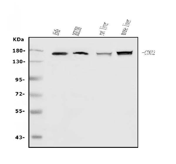
Western blot analysis of N Cadherin/CDH2 using anti-N Cadherin/CDH2 antibody (A01577-3).
Electrophoresis was performed on a 5-20% SDS-PAGE gel at 70V (Stacking gel) / 90V (Resolving gel) for 2-3 hours. The sample well of each lane was loaded with 50ug of sample under reducing conditions.
Lane 1: human A549 whole cell lysates,
Lane 2: human HEK293 whole cell lysates,
Lane 3: rat liver tissue lysates,
Lane 4: mouse liver tissue lysates.
After Electrophoresis, proteins were transferred to a Nitrocellulose membrane at 150mA for 50-90 minutes. Blocked the membrane with 5% Non-fat Milk/ TBS for 1.5 hour at RT. The membrane was incubated with rabbit anti-N Cadherin/CDH2 antigen affinity purified polyclonal antibody (Catalog # A01577-3) at 0.5 μg/mL overnight at 4°C, then washed with TBS-0.1%Tween 3 times with 5 minutes each and probed with a goat anti-rabbit IgG-HRP secondary antibody at a dilution of 1:5000 for 1.5 hour at RT. The signal is developed using an Enhanced Chemiluminescent detection (ECL) kit (Catalog # EK1002) with Tanon 5200 system. A specific band was detected for N Cadherin/CDH2 at approximately 150KD. The expected band size for N Cadherin/CDH2 is at 150KD.

Click image to see more details
IHC analysis of N Cadherin/CDH2 using anti-N Cadherin/CDH2 antibody (A01577-3).
N Cadherin/CDH2 was detected in paraffin-embedded section of human liver cancer tissue. Heat mediated antigen retrieval was performed in EDTA buffer (pH8.0, epitope retrieval solution). The tissue section was blocked with 10% goat serum. The tissue section was then incubated with 2μg/ml rabbit anti-N Cadherin/CDH2 Antibody (A01577-3) overnight at 4°C. Biotinylated goat anti-rabbit IgG was used as secondary antibody and incubated for 30 minutes at 37°C. The tissue section was developed using Strepavidin-Biotin-Complex (SABC) (Catalog # SA1022) with DAB as the chromogen.

Click image to see more details
IHC analysis of N Cadherin/CDH2 using anti-N Cadherin/CDH2 antibody (A01577-3).
N Cadherin/CDH2 was detected in paraffin-embedded section of human lung cancer tissue. Heat mediated antigen retrieval was performed in EDTA buffer (pH8.0, epitope retrieval solution). The tissue section was blocked with 10% goat serum. The tissue section was then incubated with 2μg/ml rabbit anti-N Cadherin/CDH2 Antibody (A01577-3) overnight at 4°C. Biotinylated goat anti-rabbit IgG was used as secondary antibody and incubated for 30 minutes at 37°C. The tissue section was developed using Strepavidin-Biotin-Complex (SABC) (Catalog # SA1022) with DAB as the chromogen.

Click image to see more details
IHC analysis of N Cadherin/CDH2 using anti-N Cadherin/CDH2 antibody (A01577-3).
N Cadherin/CDH2 was detected in paraffin-embedded section of human pancreatic cancer tissue. Heat mediated antigen retrieval was performed in EDTA buffer (pH8.0, epitope retrieval solution). The tissue section was blocked with 10% goat serum. The tissue section was then incubated with 2μg/ml rabbit anti-N Cadherin/CDH2 Antibody (A01577-3) overnight at 4°C. Biotinylated goat anti-rabbit IgG was used as secondary antibody and incubated for 30 minutes at 37°C. The tissue section was developed using Strepavidin-Biotin-Complex (SABC) (Catalog # SA1022) with DAB as the chromogen.

Click image to see more details
IF analysis of N Cadherin/CDH2 using anti-N Cadherin/CDH2 antibody (A01577-3).
N Cadherin/CDH2 was detected in a paraffin-embedded section of human liver cancer tissue. Heat mediated antigen retrieval was performed in EDTA buffer (pH 8.0, epitope retrieval solution). The tissue section was blocked with 10% goat serum. The tissue section was then incubated with 5 μg/mL rabbit anti-N Cadherin/CDH2 Antibody (A01577-3) overnight at 4°C. Biotin conjugated goat anti-rabbit IgG (BA1003) was used as secondary antibody and incubated for 30 minutes at 37°C. The tissue section was developed using DyLight®488 Conjugated Avidin (BA1128). The section was counterstained with DAPI. Visualize using a fluorescence microscope and filter sets appropriate for the label used.

Click image to see more details
IF analysis of N Cadherin/CDH2 using anti-N Cadherin/CDH2 antibody (A01577-3) and anti-Tubulin Alpha antibody (M03989-3).
N Cadherin/CDH2 was detected in immunocytochemical section of Hela cell. Enzyme antigen retrieval was performed using IHC enzyme antigen retrieval reagent (AR0022) for 15 mins. The cells were blocked with 10% goat serum. And then incubated with 5 μg/mL rabbit anti-N Cadherin/CDH2 Antibody (A01577-3) and mouse anti-Tubulin Alpha antibody (M03989-3) overnight at 4°C. DyLight®488 Conjugated Goat Anti-Rabbit IgG (BA1127) and Cy3 Conjugated Goat Anti-Mouse IgG (BA1031) were used as secondary antibody at 1:500 dilution and incubated for 30 minutes at 37°C. Visualize using a fluorescence microscope and filter sets appropriate for the label used.

Click image to see more details
Flow Cytometry analysis of HEPG2 cells using anti-N Cadherin/CDH2 antibody (A01577-3).
Overlay histogram showing HEPG2 cells stained with A01577-3 (Blue line). The cells were fixed with 4% paraformaldehyde and blocked with 10% normal goat serum. And then incubated with rabbit anti-N Cadherin/CDH2 Antibody (A01577-3, 1μg/1x106 cells) for 30 min at 20°C. DyLight®488 conjugated goat anti-rabbit IgG (BA1127, 5-10μg/1x106 cells) was used as secondary antibody for 30 minutes at 20°C. Isotype control antibody (Green line) was rabbit IgG (1μg/1x106) used under the same conditions. Unlabelled sample without incubation with primary antibody and secondary antibody (Red line) was used as a blank control.

Click image to see more details
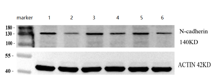
Western blot analysis of N Cadherin/CDH2 using anti-N Cadherin/CDH2 antibody (A01577-3).
Electrophoresis was performed on a 5-20% SDS-PAGE gel at 70V (Stacking gel) / 90V (Resolving gel) for 2-3 hours. The sample well of each lane was loaded with 50ug of sample under reducing conditions.
Lane 1: human cervical cancer tissue lysates,
Lane 2: human cervical cancer adjacent tissue lysates,
Lane 3: human cervical cancer tissue lysates,
Lane 4: human cervical cancer adjacent tissue lysates.
Lane 5: human cervical cancer tissue lysates,
Lane 6: human cervical cancer adjacent tissue lysates.
After Electrophoresis, proteins were transferred to a Nitrocellulose membrane at 150mA for 50-90 minutes. Blocked the membrane with 5% Non-fat Milk/ TBS for 1.5 hour at RT. The membrane was incubated with rabbit anti-N Cadherin/CDH2 antigen affinity purified polyclonal antibody (Catalog # A01577-3) at 1:1000 overnight at 4°C, then washed with TBS-0.1%Tween 3 times with 5 minutes each and probed with a goat anti-rabbit IgG-HRP secondary antibody at a dilution of 1:5000 for 1.5 hour at RT. The signal is developed using an Enhanced Chemiluminescent detection (ECL) kit (Catalog # EK1002) with ChemiDoc MP system. A specific band was detected for N Cadherin/CDH2 at approximately 140KD. The expected band size for N Cadherin/CDH2 is at 150KD.

Click image to see more details
Silibinin inhibited the growth and promoted the apoptosis of LA795 cells. (A) . Migration of LA795 cells in the presence of 0, 3, 10, 30, 100, and 300 μM silibinin assessed by wound healing assay ( n = 4). (B) . Statistical results of the percentage of wound area in (A) ( n = 4) (C) . Statistical results of LA795 cells viability incubated with different concentrations of silibinin ( n = 6). (D) . Expression of β -catenin, E-cadherin, N-cadherin, and vimentin of LA795 cells after treated with 200 μM silibinin for 24 h ( n = 3). (E) . Representative real-time images of LA795 cells incubated with different concentrations of silibinin ( n = 6). (F) . Representative images of apoptotic cells after 200 μM silibinin treatment for 24 h detected by annexin V apoptosis assay ( n = 3). (G) . Expression of cleaved-caspase 3 and cleaved-caspase 9 of LA795 cells after 200 μM silibinin treatment for 24 h ( n = 3).
Index in PubMed under a CC BY license. PMID: 33935737
Specific Publications For Anti-N Cadherin/CDH2 Antibody Picoband® (A01577-3)
Loading publications
Recommended Resources
Here are featured tools and databases that you might find useful.
- Boster's Pathways Library
- Protein Databases
- Bioscience Research Protocol Resources
- Data Processing & Analysis Software
- Photo Editing Software
- Scientific Literature Resources
- Research Paper Management Tools
- Molecular Biology Software
- Primer Design Tools
- Bioinformatics Tools
- Phylogenetic Tree Analysis
Customer Reviews
Have you used Anti-N Cadherin/CDH2 Antibody Picoband®?
Share your experimental results or join a short interview to earn up to $1,000 in product credits or other rewards.
1 Reviews For Anti-N Cadherin/CDH2 Antibody Picoband®
N Cadherin/CDH2 Antibody Picoband® (A01577-3) shows clear, specific bands in human cervical cancer and adjacent tissues by WB, with higher levels in cancer tissues, demonstrating excellent antibody performance.
Excellent
| SKU | A01577-3 |
|---|---|
| Application | Western Blot |
| Sample | human cervical cancer and adjacent tissues |
| Sample Processing Description | Three samples each of human cervical cancer and adjacent tissues were collected, and total protein was extracted. |
| Other Reagents | RIPA lysis buffer, Protease inhibitor, Running buffer, Transfer buffer, Blocking buffer |
| Primary Antibody | N Cadherin/CDH2 Antibody Picoband® |
| Primary Incubation | 1:1000, overnight at 4 ℃ |
| Secondary Antibody | HRP Conjugated AffiniPure Goat Anti-Rabbit IgG (H+L) (BA1054) |
| Secondary Incubation | 1:10000, 1 h in RT |
| Detection | Substrate: ECL substrate, Image system: ChemiDoc MP |
| Results Summary | The purpose of this study was to evaluate the expression differences of N-cadherin in human cervical cancer and adjacent tissues. Results from three samples showed that N-cadherin levels in adjacent tissues were significantly lower than in cancer tissues. |
Xiaoyuan Qu, Shandong First Medical University
Verified customer
Submitted 2026-03-26
Customer Q&As
Have a question?
Find answers in Q&As, reviews.
Can't find your answer?
Submit your question
