Click image to see more details
Product Info Summary
| SKU: | PA1058-1 |
|---|---|
| Size: | 100μg/vial |
| Reactive Species: | Human, Mouse, Rat |
| Host: | Rabbit |
| Application: | IHC, WB |
Customers Who Bought This Also Bought

Product info
Product Name
Anti-NMDAR2A/GRIN2A Antibody Picoband®
SKU/Catalog Number
PA1058-1
Size
100μg/vial
Form
Lyophilized
Description
Boster Bio Anti-NMDAR2A/GRIN2A Antibody catalog # PA1058-1. Tested in IHC, WB applications. This antibody reacts with Human, Mouse, Rat. The brand Picoband indicates this is a premium antibody that guarantees superior quality, high affinity, and strong signals with minimal background in Western blot applications. Only our best-performing antibodies are designated as Picoband, ensuring unmatched performance.
Storage & Handling
Store at -20˚C for one year from date of receipt. After reconstitution, at 4˚C for one month. It can also be aliquotted and stored frozen at -20˚C for six months. Avoid repeated freeze-thaw cycles.
Cite This Product
Anti-NMDAR2A/GRIN2A Antibody Picoband® (Boster Biological Technology, Pleasanton CA, USA, Catalog # PA1058-1)
Host
Rabbit
Contents
Each vial contains 4 mg Trehalose, 0.9 mg NaCl and 0.2 mg Na2HPO4.
Clonality
Polyclonal
Immunogen
A synthetic peptide corresponding to a sequence at the C-terminus of human NMDAR2A, different from the related mouse sequence by three amino acids, and from the related rat sequence by four amino acids.
Cross-reactivity
No cross reactivity with other proteins
Reactive Species
PA1058-1 is reactive to GRIN2A in Human, Mouse, Rat
Observed Molecular Weight
180 kDa
Calculated molecular weight
165.3 kDa
Background of GRIN2A
GRIN2A is also known as N-methyl-D-aspartate receptor channel, subunit epsilon-1 (NMDAR2A). This gene encodes a member of the glutamate-gated ion channel protein family. The encoded protein is an N-methyl-D-aspartate (NMDA) receptor subunit. NMDA receptors are both ligand-gated and voltage-dependent, and are involved in long-term potentiation, an activity-dependent increase in the efficiency of synaptic transmission thought to underlie certain kinds of memory and learning. These receptors are permeable to calcium ions, and activation results in a calcium influx into post-synaptic cells, which results in the activation of several signaling cascades. Disruption of this gene is associated with focal epilepsy and speech disorder with or without mental retardation. Alternative splicing results in multiple transcript variants.
Antibody Validation
Boster validates all antibodies on WB, IHC, ICC, Immunofluorescence, and ELISA with known positive control and negative samples to ensure specificity and high affinity, including thorough antibody incubations.
Application & Images
Applications
PA1058-1 is guaranteed for IHC, WB Boster Guarantee
Recommend Dilution
| Application | Dilution | Species |
|---|---|---|
| Western blot | 0.1-0.5μg/ml | Mouse, Rat |
| Immunohistochemistry (Paraffin-embedded Section) | 2-5μg/ml | Rat |
Tested application
Suggested blocking solution with 5% non-fat milk or BSA; (*)Recommended protein loading: 20-40 µg per lane
Use TE buffer pH 9.0 for antigen retrieval; (*) citrate buffer pH 6.0 is an alternative.
Validation Images & Assay Conditions
Click image to see more details
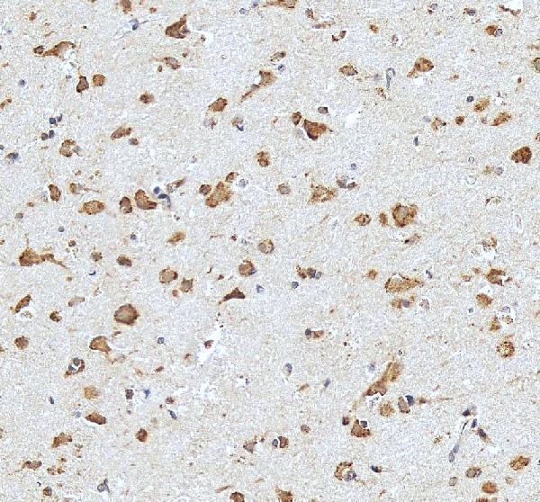
IHC analysis of NMDAR2A/GRIN2A using anti-NMDAR2A/GRIN2A antibody (PA1058-1).
NMDAR2A/GRIN2A was detected in a paraffin-embedded section of human brain tissue. Heat mediated antigen retrieval was performed in EDTA buffer (pH 8.0, epitope retrieval solution). The tissue section was blocked with 10% goat serum. The tissue section was then incubated with 2 μg/ml rabbit anti-NMDAR2A/GRIN2A Antibody (PA1058-1) overnight at 4°C. Peroxidase Conjugated Goat Anti-rabbit IgG was used as secondary antibody and incubated for 30 minutes at 37°C. The tissue section was developed using HRP Conjugated Rabbit IgG Super Vision Assay Kit (Catalog # SV0002) with DAB as the chromogen.

Click image to see more details
IHC analysis of NMDAR2A/GRIN2A using anti-NMDAR2A/GRIN2A antibody (PA1058-1).
NMDAR2A/GRIN2A was detected in a paraffin-embedded section of human heart tissue. Heat mediated antigen retrieval was performed in EDTA buffer (pH 8.0, epitope retrieval solution). The tissue section was blocked with 10% goat serum. The tissue section was then incubated with 2 μg/ml rabbit anti-NMDAR2A/GRIN2A Antibody (PA1058-1) overnight at 4°C. Peroxidase Conjugated Goat Anti-rabbit IgG was used as secondary antibody and incubated for 30 minutes at 37°C. The tissue section was developed using HRP Conjugated Rabbit IgG Super Vision Assay Kit (Catalog # SV0002) with DAB as the chromogen.

Click image to see more details
Western blot analysis of GRIN2A using anti-GRIN2A antibody (PA1058-1).
Electrophoresis was performed on a 5-20% SDS-PAGE gel at 70V (Stacking gel) / 90V (Resolving gel) for 2-3 hours. The sample well of each lane was loaded with 30 ug of sample under reducing conditions.
Lane 1: rat brain tissue lysates,
Lane 2: mouse brain tissue lysates.
After electrophoresis, proteins were transferred to a nitrocellulose membrane at 150 mA for 50-90 minutes. Blocked the membrane with 5% non-fat milk/TBS for 1.5 hour at RT. The membrane was incubated with rabbit anti-GRIN2A antigen affinity purified polyclonal antibody (Catalog # PA1058-1) at 0.5 μg/mL overnight at 4°C, then washed with TBS-0.1%Tween 3 times with 5 minutes each and probed with a goat anti-rabbit IgG-HRP secondary antibody at a dilution of 1:5000 for 1.5 hour at RT. The signal is developed using an Enhanced Chemiluminescent detection (ECL) kit (Catalog # EK1002) with Tanon 5200 system. A specific band was detected for GRIN2A at approximately 180 kDa. The expected band size for GRIN2A is at 165 kDa.

Click image to see more details
IHC analysis of GRIN2A using anti-GRIN2A antibody (PA1058-1).
GRIN2A was detected in a paraffin-embedded section of rat brain tissue. Heat mediated antigen retrieval was performed in EDTA buffer (pH 8.0, epitope retrieval solution). The tissue section was blocked with 10% goat serum. The tissue section was then incubated with 2 μg/ml rabbit anti-GRIN2A Antibody (PA1058-1) overnight at 4°C. Peroxidase Conjugated Goat Anti-rabbit IgG was used as secondary antibody and incubated for 30 minutes at 37°C. The tissue section was developed using HRP Conjugated Rabbit IgG Super Vision Assay Kit (Catalog # SV0002) with DAB as the chromogen.
Specific Publications For Anti-NMDAR2A/GRIN2A Antibody Picoband® (PA1058-1)
Loading publications
Recommended Resources
Here are featured tools and databases that you might find useful.
- Boster's Pathways Library
- Protein Databases
- Bioscience Research Protocol Resources
- Data Processing & Analysis Software
- Photo Editing Software
- Scientific Literature Resources
- Research Paper Management Tools
- Molecular Biology Software
- Primer Design Tools
- Bioinformatics Tools
- Phylogenetic Tree Analysis
Customer Reviews
Have you used Anti-NMDAR2A/GRIN2A Antibody Picoband®?
Share your experimental results or join a short interview to earn up to $1,000 in product credits or other rewards.
0 Reviews For Anti-NMDAR2A/GRIN2A Antibody Picoband®
Customer Q&As
Have a question?
Find answers in Q&As, reviews.
Can't find your answer?
Submit your question
6 Customer Q&As for Anti-NMDAR2A/GRIN2A Antibody Picoband®
Question
Would anti-NMDAR2A/GRIN2A antibody PA1058-1 work for IHC with cerebellum?
Verified Customer
Verified customer
Asked: 2019-12-18
Answer
According to the expression profile of cerebellum, GRIN2A is highly expressed in cerebellum. So, it is likely that anti-NMDAR2A/GRIN2A antibody PA1058-1 will work for IHC with cerebellum.
Boster Scientific Support
Answered: 2019-12-18
Question
Our lab want to know about to test anti-NMDAR2A/GRIN2A antibody PA1058-1 on mouse cerebellum for research purposes, then I may be interested in using anti-NMDAR2A/GRIN2A antibody PA1058-1 for diagnostic purposes as well. Is the antibody suitable for diagnostic purposes?
Verified Customer
Verified customer
Asked: 2019-07-02
Answer
The products we sell, including anti-NMDAR2A/GRIN2A antibody PA1058-1, are only intended for research use. They would not be suitable for use in diagnostic work. If you have the means to develop a product into diagnostic use, and are interested in collaborating with us and develop our product into an IVD product, please contact us for more discussions.
Boster Scientific Support
Answered: 2019-07-02
Question
Is this PA1058-1 anti-NMDAR2A/GRIN2A antibody reactive to the isotypes of GRIN2A?
Verified Customer
Verified customer
Asked: 2019-05-27
Answer
The immunogen of PA1058-1 anti-NMDAR2A/GRIN2A antibody is A synthetic peptide corresponding to a sequence at the C-terminus of human NMDAR2A(1360-1376aa, DHTSDNPFLHSHRDDQR), different from the related mouse sequence by three amino acids, and from the related rat sequence by four amino acids. Could you tell me which isotype you are interested in so I can help see if the immunogen is part of this isotype?
Boster Scientific Support
Answered: 2019-05-27
Question
Do you have a BSA free version of anti-NMDAR2A/GRIN2A antibody PA1058-1 available?
Verified Customer
Verified customer
Asked: 2019-01-15
Answer
Thanks for your recent telephone inquiry. I can confirm that some lots of this anti-NMDAR2A/GRIN2A antibody PA1058-1 are BSA free. For now, these lots are available and we can make a BSA free formula for you free of charge. It will take 3 extra days to prepare. If you require this antibody BSA free again in future, please do not hesitate to contact me and I will be pleased to check which lots we have in stock that are BSA free.
Boster Scientific Support
Answered: 2019-01-15
Question
I see that the anti-NMDAR2A/GRIN2A antibody PA1058-1 works with IHC, what is the protocol used to produce the result images on the product page?
P. Baker
Verified customer
Asked: 2015-06-01
Answer
You can find protocols for IHC on the "support/technical resources" section of our navigation menu. If you have any further questions, please send an email to support@bosterbio.com
Boster Scientific Support
Answered: 2015-06-01
Question
I was wanting to use your anti-NMDAR2A/GRIN2A antibody for IHC for mouse cerebellum on frozen tissues, but I want to know if it has been validated for this particular application. Has this antibody been validated and is this antibody a good choice for mouse cerebellum identification?
D. Roberts
Verified customer
Asked: 2015-02-24
Answer
It shows on the product datasheet, PA1058-1 anti-NMDAR2A/GRIN2A antibody has been validated for IHC, WB on human, mouse, rat tissues. We have an innovator award program that if you test this antibody and show it works in mouse cerebellum in IHC-frozen, you can get your next antibody for free.
Boster Scientific Support
Answered: 2015-02-24
