Click image to see more details
Product Info Summary
| SKU: | PA1666 |
|---|---|
| Size: | 100 μg/vial |
| Reactive Species: | Mouse, Rat |
| Host: | Rabbit |
| Application: | IHC, WB |
Customers Who Bought This Also Bought

Product info
Product Name
Anti-NADPH oxidase 1 NOX1 Antibody Picoband®
SKU/Catalog Number
PA1666
BA3720 is an alternative SKU for this antibody, used in previous lots.
Size
100 μg/vial
Form
Lyophilized
Description
Boster Bio Anti-NADPH oxidase 1 NOX1 Antibody catalog # PA1666. Tested in IHC, WB applications. This antibody reacts with Mouse, Rat. The brand Picoband indicates this is a premium antibody that guarantees superior quality, high affinity, and strong signals with minimal background in Western blot applications. Only our best-performing antibodies are designated as Picoband, ensuring unmatched performance.
Storage & Handling
Store at -20˚C for one year from date of receipt. After reconstitution, at 4˚C for one month. It can also be aliquotted and stored frozen at -20˚C for six months. Avoid repeated freeze-thaw cycles.
Cite This Product
Anti-NADPH oxidase 1 NOX1 Antibody Picoband® (Boster Biological Technology, Pleasanton CA, USA, Catalog # PA1666)
Host
Rabbit
Contents
Each vial contains 4 mg Trehalose, 0.9 mg NaCl and 0.2 mg Na2HPO4.
Clonality
Polyclonal
Isotype
Rabbit IgG
Immunogen
A synthetic peptide corresponding to a sequence in the middle region of rat NOX1, different from the related mouse sequence by one amino acid.
Cross-reactivity
No cross-reactivity with other proteins
Reactive Species
PA1666 is reactive to Nox1 in Mouse, Rat
Observed Molecular Weight
65 kDa
Calculated molecular weight
65.2 kDa
Background of Nox1
NOX1 (NADPH OXIDASE 1), also known as NOH1, MOX1 or GP91-2, is an enzyme that in humans is encoded by the NOX1 gene. It is also a homolog of the catalytic subunit of the superoxide-generating NADPH oxidase of phagocytes, gp91phox. The NOX1 gene is mapped to Xq22.1. NOX1 was expressed in colon, prostate, uterus, and vascular smooth muscle, but not in peripheral blood leukocytes. The deduced 564-amino acid NOX1 protein, which is 58% identical to CYBB, contains 6 membrane-spanning regions, conserved flavin and pyridine nucleotide-binding sites, and histidines possibly involved in heme ligation. Overexpression of MOX1 in NIH 3T3 cells increased superoxide generation and cell growth. Cells expressing MOX1 had a transformed appearance, showed anchorage-independent growth, and produced tumors in athymic mice. Disruption of either Nox1 or Nox2 significantly delayed progression of motor neuron disease in these mice. However, 50% survival rates were enhanced significantly more by Nox2 deletion than Nox1 deletion.
Antibody Validation
Boster validates all antibodies on WB, IHC, ICC, Immunofluorescence, and ELISA with known positive control and negative samples to ensure specificity and high affinity, including thorough antibody incubations.
Application & Images
Applications
PA1666 is guaranteed for IHC, WB Boster Guarantee
Recommend Dilution
| Application | Dilution | Species |
|---|---|---|
| Western blot | 0.1-0.5μg/ml | Mouse, Rat |
| Immunohistochemistry (Paraffin-embedded Section) | 2-5μg/ml | Rat |
Tested application
Suggested blocking solution with 5% non-fat milk or BSA; (*)Recommended protein loading: 20-40 µg per lane
Use TE buffer pH 9.0 for antigen retrieval; (*) citrate buffer pH 6.0 is an alternative.
Validation Images & Assay Conditions
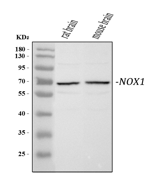
Click image to see more details
Western blot analysis of NOX1 using anti-NOX1 antibody (PA1666).
Electrophoresis was performed on a 5-20% SDS-PAGE gel at 70V (Stacking gel) / 90V (Resolving gel) for 2-3 hours. The sample well of each lane was loaded with 30 ug of sample under reducing conditions.
Lane 1: rat brain tissue lysates,
Lane 2: mouse brain tissue lysates.
After electrophoresis, proteins were transferred to a nitrocellulose membrane at 150 mA for 50-90 minutes. Blocked the membrane with 5% non-fat milk/TBS for 1.5 hour at RT. The membrane was incubated with rabbit anti-NOX1 antigen affinity purified polyclonal antibody (Catalog # PA1666) at 0.5 μg/mL overnight at 4°C, then washed with TBS-0.1%Tween 3 times with 5 minutes each and probed with a goat anti-rabbit IgG-HRP secondary antibody at a dilution of 1:5000 for 1.5 hour at RT. The signal is developed using an Enhanced Chemiluminescent detection (ECL) kit (Catalog # EK1002) with Tanon 5200 system. A specific band was detected for NOX1 at approximately 65 kDa. The expected band size for NOX1 is at 65 kDa.

Click image to see more details
Effect of GDM on the ROS level in offspring. Heart tissues were isolated from male offspring. ( A ) ROS levels in the left ventricle (LV) tissues isolated from both control (□) and STZ-treated (■) groups were measured using in vitro ROS/RNS assay kit. ( B ) NOX1, 2, and 4 protein abundances in the LV tissues isolated from both control (□) and STZ-treated (■) groups were determined by Western blot analysis. Their protein densities were normalized to internal control (GAPDH). ( C ) After NAC pretreatment, ROS levels in the LV tissues isolated from both control (□) and STZ-treated (■) groups were measured using in vitro ROS/RNS assay kit. Data are means ± SEM (n=4 animals/group). *P < 0.05 vs. control, as determined by Student's t-test.
Index in PubMed under a CC BY license. PMID: 31223283

Click image to see more details
IHC analysis of NOX1 using anti-NOX1 antibody (PA1666).
NOX1 was detected in a paraffin-embedded section of rat colon tissue. Heat mediated antigen retrieval was performed in EDTA buffer (pH 8.0, epitope retrieval solution). The tissue section was blocked with 10% goat serum. The tissue section was then incubated with 2 μg/ml rabbit anti-NOX1 Antibody (PA1666) overnight at 4°C. Peroxidase Conjugated Goat Anti-rabbit IgG was used as secondary antibody and incubated for 30 minutes at 37°C. The tissue section was developed using HRP Conjugated Rabbit IgG Super Vision Assay Kit (Catalog # SV0002) with DAB as the chromogen.
Specific Publications For Anti-NADPH oxidase 1 NOX1 Antibody Picoband® (PA1666)
Loading publications
Recommended Resources
Here are featured tools and databases that you might find useful.
- Boster's Pathways Library
- Protein Databases
- Bioscience Research Protocol Resources
- Data Processing & Analysis Software
- Photo Editing Software
- Scientific Literature Resources
- Research Paper Management Tools
- Molecular Biology Software
- Primer Design Tools
- Bioinformatics Tools
- Phylogenetic Tree Analysis
Customer Reviews
Have you used Anti-NADPH oxidase 1 NOX1 Antibody Picoband®?
Share your experimental results or join a short interview to earn up to $1,000 in product credits or other rewards.
0 Reviews For Anti-NADPH oxidase 1 NOX1 Antibody Picoband®
Customer Q&As
Have a question?
Find answers in Q&As, reviews.
Can't find your answer?
Submit your question
