Click image to see more details
Product Info Summary
| SKU: | A00804-3 |
|---|---|
| Size: | 100 µg/vial |
| Reactive Species: | Mouse, Rat |
| Host: | Rabbit |
| Application: | ELISA, WB |
Customers Who Bought This Also Bought

Product info
Product Name
Anti-Osm Antibody Picoband®
SKU/Catalog Number
A00804-3
Size
100 µg/vial
Form
Lyophilized
Description
Boster Bio Anti-Osm Antibody Picoband® catalog # A00804-3. Tested in WB, ELISA applications. This antibody reacts with Mouse, Rat. The brand Picoband indicates this is a premium antibody that guarantees superior quality, high affinity, and strong signals with minimal background in Western blot applications. Only our best-performing antibodies are designated as Picoband, ensuring unmatched performance.
Storage & Handling
At -20°C for one year from date of receipt. After reconstitution, at 4°C for one month. It can also be aliquotted and stored frozen at -20°C for six months. Avoid repeated freezing and thawing.
Cite This Product
Anti-Osm Antibody Picoband® (Boster Biological Technology, Pleasanton CA, USA, Catalog # A00804-3)
Host
Rabbit
Contents
Each vial contains 4 mg Trehalose, 0.9 mg NaCl, 0.2 mg Na2HPO4.
Clonality
Polyclonal
Isotype
IgG
Immunogen
E.coli-derived rat Osm recombinant protein (Position: E55-D158). Rat Osm shares 46% and 71.2% amino acid (aa) sequence identity with human and mouse Osm, respectively.
Cross-reactivity
No cross reactivity with other proteins.
Reactive Species
A00804-3 is reactive to Osm in Mouse, Rat
Observed Molecular Weight
27 kDa
Calculated molecular weight
27.1 kDa
Background of Osm
OSM (ONCOSTATIN M) is a member of a cytokine family that includes leukemia-inhibitory factor, granulocyte colony-stimulating factor, and interleukin 6. This gene encodes a growth regulator which inhibits the proliferation of a number of tumor cell lines. It regulates cytokine production, including IL-6, G-CSF and GM-CSF from endothelial cells. OSM is mapped on 22q12.2. OSM has the ability to inhibit the growth of human A375 melanoma cells but not normal human fibroblasts. Treatment with recombinant OSM leads to the inhibition of proliferation and changes in cellular morphology of a number of tumor cell lines derived from a wide variety of tissue types. OSM also has the ability to inhibit the proliferation of murine M1 myeloid leukemic cells and can induce their differentiation into macrophage-like cells, a function shared by LIF, CSF3, and IL6. The direction of gene transcription was telomeric to centromeric, with the OSM gene located upstream of the LIF gene.
Antibody Validation
Boster validates all antibodies on WB, IHC, ICC, Immunofluorescence, and ELISA with known positive control and negative samples to ensure specificity and high affinity, including thorough antibody incubations.
Application & Images
Applications
A00804-3 is guaranteed for ELISA, WB Boster Guarantee
Recommend Dilution
| Application | Dilution | Species |
|---|---|---|
| Western blot | 0.25-0.5 μg/ml | Mouse, Rat |
| ELISA | 0.1-0.5 μg/ml | Rat |
Tested application
Suggested blocking solution with 5% non-fat milk or BSA; (*)Recommended protein loading: 20-40 µg per lane
Validation Images & Assay Conditions
Click image to see more details
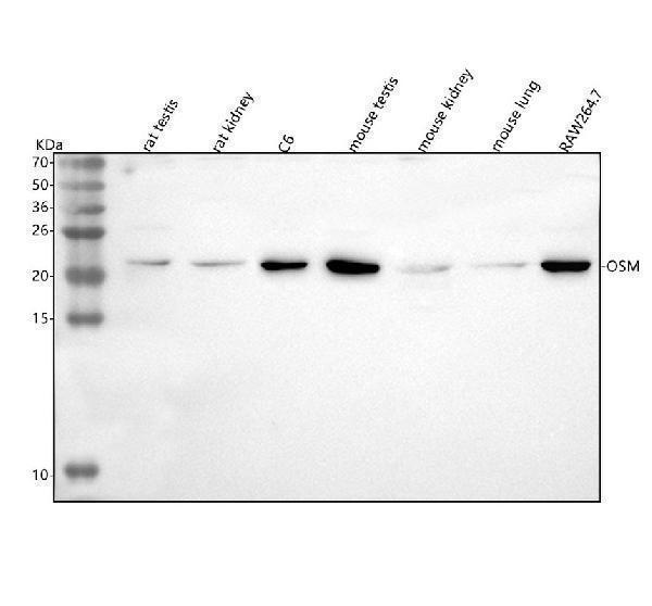
Western blot analysis of Osm using anti-Osm antibody (A00804-3).
Electrophoresis was performed on a 5-20% SDS-PAGE gel at 70V (Stacking gel) / 90V (Resolving gel) for 2-3 hours. The sample well of each lane was loaded with 30 ug of sample under reducing conditions.
Lane 1: rat testis tissue lysates,
Lane 2: rat kidney tissue lysates,
Lane 3: rat C6 whole cell lysates,
Lane 4: mouse tetis tissue lysates,
Lane 5: mouse kidney tissue lysates,
Lane 6: mouse lung tissue lysates,
Lane 7: mouse RAW264.7 whole cell lysates.
After electrophoresis, proteins were transferred to a nitrocellulose membrane at 150 mA for 50-90 minutes. Blocked the membrane with 5% non-fat milk/TBS for 1.5 hour at RT. The membrane was incubated with rabbit anti-Osm antigen affinity purified polyclonal antibody (Catalog # A00804-3) at 0.5 μg/mL overnight at 4°C, then washed with TBS-0.1%Tween 3 times with 5 minutes each and probed with a goat anti-rabbit IgG-HRP secondary antibody at a dilution of 1:5000 for 1.5 hour at RT. The signal is developed using an Enhanced Chemiluminescent detection (ECL) kit (Catalog # EK1002) with Tanon 5200 system. A specific band was detected for Osm at approximately 27 kDa. The expected band size for Osm is at 27 kDa.
Specific Publications For Anti-Osm Antibody Picoband® (A00804-3)
Loading publications
Recommended Resources
Here are featured tools and databases that you might find useful.
- Boster's Pathways Library
- Protein Databases
- Bioscience Research Protocol Resources
- Data Processing & Analysis Software
- Photo Editing Software
- Scientific Literature Resources
- Research Paper Management Tools
- Molecular Biology Software
- Primer Design Tools
- Bioinformatics Tools
- Phylogenetic Tree Analysis
Customer Reviews
Have you used Anti-Osm Antibody Picoband®?
Share your experimental results or join a short interview to earn up to $1,000 in product credits or other rewards.
0 Reviews For Anti-Osm Antibody Picoband®
Customer Q&As
Have a question?
Find answers in Q&As, reviews.
Can't find your answer?
Submit your question
