Click image to see more details
-

-
-
-
-
+3
Product Info Summary
| SKU: | M00145-3 |
|---|---|
| Size: | 100 μl |
| Reactive Species: | Human |
| Host: | Rabbit |
| Application: | IP, IF, IHC, ICC, WB |
Customers Who Bought This Also Bought

Product info
Product Name
Anti-p21 CDKN1A Rabbit Monoclonal Antibody
SKU/Catalog Number
M00145-3
BM3990 is an alternative SKU for this antibody, used in previous lots.
Size
100 μl
Form
Liquid
Description
Boster Bio Anti-p21 CDKN1A Rabbit Monoclonal Antibody catalog # M00145-3. Tested in WB, IHC, ICC/IF, IP applications. This antibody reacts with Human.
Storage & Handling
Store at -20°C for one year. For short term storage and frequent use, store at 4°C for up to one month. Avoid repeated freeze-thaw cycles.
Cite This Product
Anti-p21 CDKN1A Rabbit Monoclonal Antibody (Boster Biological Technology, Pleasanton CA, USA, Catalog # M00145-3)
Host
Rabbit
Contents
Rabbit IgG in stabilizing components, phosphate buffered saline, pH 7.4, 150mM NaCl, 0.02% sodium azide and 50% glycerol.
*This antibody is supplied in a stabilized formulation.
Compatibility with conjugation reactions depends on the chemistry of the conjugation method used.
For conjugation methods that are not compatible with the stabilizing components present in this formulation, a carrier-free antibody format is required.
Clonality
Monoclonal
Clone Number
ACG-3
Isotype
Rabbit IgG
Immunogen
A synthesized peptide derived from human p21
Reactive Species
M00145-3 is reactive to CDKN1A in Human
Observed Molecular Weight
21 kDa
Calculated molecular weight
18.1 kDa
Antibody Validation
Boster validates all antibodies on WB, IHC, ICC, Immunofluorescence, and ELISA with known positive control and negative samples to ensure specificity and high affinity, including thorough antibody incubations.
Application & Images
Applications
M00145-3 is guaranteed for IP, IF, IHC, ICC, WB Boster Guarantee
Recommend Dilution
WB 1:500-2000
IHC 1:50-200
ICC/IF 1:50-200
IP 1:50
Tested application
Use TE buffer pH 9.0 for antigen retrieval; (*) citrate buffer pH 6.0 is an alternative.
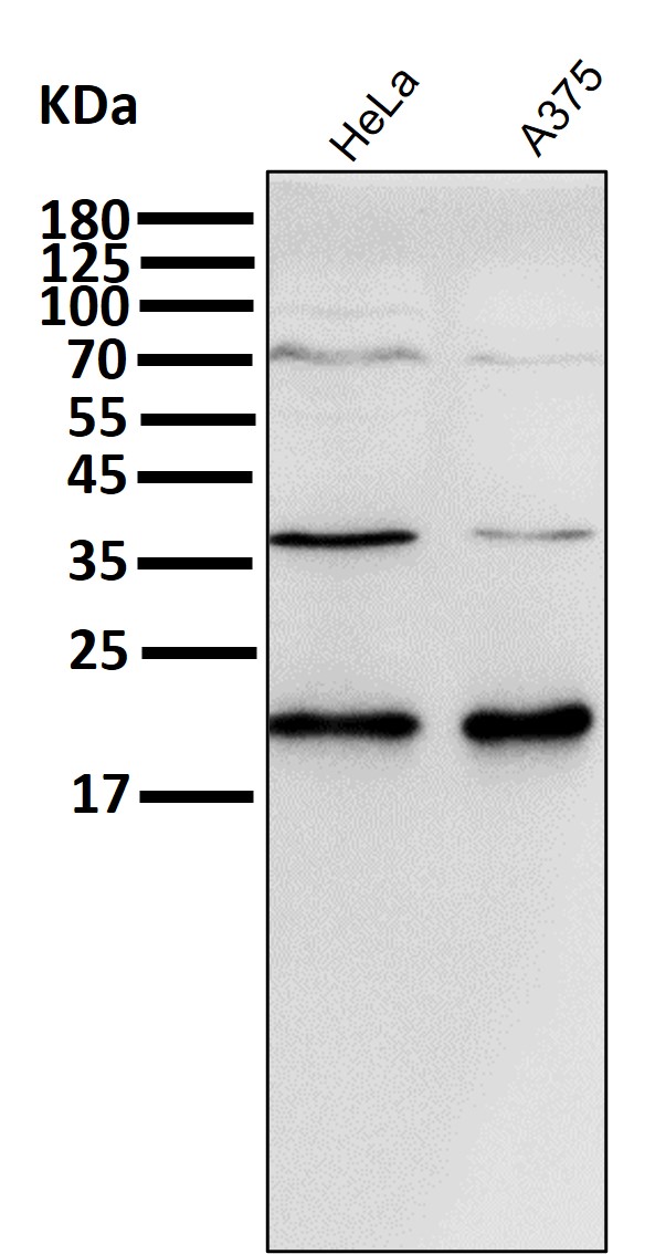
Validation Images & Assay Conditions
Click image to see more details
All lanes use the Antibody at 1:2K dilution for 1 hour at room temperature.

Click image to see more details
Immunohistochemical analysis of paraffin-embedded human colon, using p21 Antibody.

Click image to see more details
Exploration of mechanism underlying PSMC2-induced regulation of prostate cancer. a The expression levels of Akt, p-Akt, CDK6, Cyclin D1 and P21 detected by western blotting in PC-3 cells of shCtrl and shPSMC2 groups. b The expression levels of Akt, p-Akt, CDK6, Cyclin D1 and P21 detected by western blotting in PC-3 cells of Control and PSMC2 overexpression groups with or without treatment of MK-2206 (500 nM). c MTT assay was performed to assess the cell proliferation rate of PC-3 cells of Control and PSMC2 overexpression groups with or without treatment of MK-2206 (500 nM). Results were presented as mean ± SD. * P < 0.05, ** P < 0.01, *** P < 0.001
Index in PubMed under a CC BY license. PMID: 33902600

Click image to see more details
Short-term DMOG treatment reduces MSC senescence by activating HIF-1α and decreasing apoptosis. ( A , B ) Representative images of SA-β-gal staining showing the proportion of senescent cells in H₂O₂-treated MSCs before and after DMOG treatment. Scale bar = 500 μm. ( C ) Western blot analysis of p53 and p21 protein expression during oxidative stress-induced senescence. ( D , E ) SA-β-gal staining in replicative senescence (P15) MSCs, demonstrating the effect of DMOG in reducing senescence markers. Scale bar = 500 μm. ( F ) Western blot analysis of p53 and p21 protein levels in P5 (young) and P15 (senescent) MSCs. ( G ) Expression levels of senescence-associated genes (IL6, CXCL1, and MMP3) in both senescence models as assessed by qRT-PCR. ( H ) Western blotting and qPCR analysis showing increased HIF-1α protein and mRNA levels in both senescence models after DMOG treatment. ( I ) Calcein/PI live-dead staining revealed an increase in the proportion of live cells following DMOG treatment in both senescence models. ( J , K ) Flow cytometric analysis of apoptosis. Error bars represent the mean ± SD of three independent experiments. Statistical significance was set as p < 0.05. Full-length blots are presented in Supplementary Materials - WB Raw Data Full size image
Index in PubMed under a CC BY license. PMID: 40457488

Click image to see more details
Immunofluorescent analysis using the Antibody at 1:500 dilution.

Click image to see more details
Immunofluorescent analysis of MCF7 cells, using p21 Antibody .

Click image to see more details
Western blot analysis of p21 in (1) MCF-7 cell lysate; (2) LnCaP cell lysate.
Specific Publications For Anti-p21 CDKN1A Rabbit Monoclonal Antibody (M00145-3)
Loading publications
Recommended Resources
Here are featured tools and databases that you might find useful.
- Boster's Pathways Library
- Protein Databases
- Bioscience Research Protocol Resources
- Data Processing & Analysis Software
- Photo Editing Software
- Scientific Literature Resources
- Research Paper Management Tools
- Molecular Biology Software
- Primer Design Tools
- Bioinformatics Tools
- Phylogenetic Tree Analysis
Customer Reviews
Have you used Anti-p21 CDKN1A Rabbit Monoclonal Antibody?
Share your experimental results or join a short interview to earn up to $1,000 in product credits or other rewards.
0 Reviews For Anti-p21 CDKN1A Rabbit Monoclonal Antibody
Customer Q&As
Have a question?
Find answers in Q&As, reviews.
Can't find your answer?
Submit your question
1 Customer Q&As for Anti-p21 CDKN1A Rabbit Monoclonal Antibody
Question
We are currently using anti-p21 Rabbit Monoclonal antibody M00145-3 for human tissue, and we are well pleased with the IHC results. The species of reactivity given in the datasheet says human. Is it likely that the antibody can work on canine tissues as well?
Verified Customer
Verified customer
Asked: 2019-07-24
Answer
The anti-p21 Rabbit Monoclonal antibody (M00145-3) has not been validated for cross reactivity specifically with canine tissues, though there is a good chance of cross reactivity. We have an innovator award program that if you test this antibody and show it works in canine you can get your next antibody for free. Please contact me if I can help you with anything.
Boster Scientific Support
Answered: 2019-07-24
