Click image to see more details
-

-
-
-
-
+5
Product Info Summary
| SKU: | A00761-1 |
|---|---|
| Size: | 100 μg/vial |
| Reactive Species: | Human, Mouse, Rat |
| Host: | Rabbit |
| Application: | Flow Cytometry, IHC, WB |
Customers Who Bought This Also Bought

Product info
Product Name
Anti-PAH Antibody Picoband®
SKU/Catalog Number
A00761-1
Size
100 μg/vial
Form
Lyophilized
Description
Boster Bio Anti-PAH Antibody Picoband® catalog # A00761-1. Tested in Flow Cytometry(Intracellular), IHC, WB applications. This antibody reacts with Human, Mouse, Rat. The brand Picoband indicates this is a premium antibody that guarantees superior quality, high affinity, and strong signals with minimal background in Western blot applications. Only our best-performing antibodies are designated as Picoband, ensuring unmatched performance.
Storage & Handling
Store at -20˚C for one year from date of receipt. After reconstitution, at 4˚C for one month. It can also be aliquotted and stored frozen at -20˚C for six months. Avoid repeated freeze-thaw cycles.
Cite This Product
Anti-PAH Antibody Picoband® (Boster Biological Technology, Pleasanton CA, USA, Catalog # A00761-1)
Host
Rabbit
Contents
Each vial contains 4 mg Trehalose, 0.9 mg NaCl and 0.2 mg Na2HPO4.
Clonality
Polyclonal
Isotype
Rabbit IgG
Immunogen
E. coli-derived human PAH recombinant protein (Position: R71-H208). Human PAH shares 89.1% and 88.4% amino acid (aa) sequence identity with mouse and rat PAH, respectively.
Cross-reactivity
No cross-reactivity with other proteins.
Reactive Species
A00761-1 is reactive to PAH in Human, Mouse, Rat
Observed Molecular Weight
45 kDa
Calculated molecular weight
51.9 kDa
Background of PAH
Phenylalanine hydroxylase (PAH) is an enzyme that catalyzes the hydroxylation of the aromatic side-chain of phenylalanine to generate tyrosine. It is one of three members of the biopterin-dependent aromatic amino acid hydroxylases, a class of monooxygenase that uses tetrahydrobiopterin (BH4, a pteridine cofactor) and a non-heme iron for catalysis. Deficiency of this enzyme activity results in the autosomal recessive disorder phenylketonuria.
Antibody Validation
Boster validates all antibodies on WB, IHC, ICC, Immunofluorescence, and ELISA with known positive control and negative samples to ensure specificity and high affinity, including thorough antibody incubations.
Application & Images
Applications
A00761-1 is guaranteed for Flow Cytometry, IHC, WB Boster Guarantee
Recommend Dilution
| Application | Dilution | Species |
|---|---|---|
| Western blot | 0.1-0.5μg/ml | Human, Mouse, Rat, |
| Immunohistochemistry (Paraffin-embedded Section) | 2-5μg/ml | Human, Mouse, Rat |
| Flow Cytometry (Fixed) | 1-3 μg/1x106 cells | Human |
Tested application
Suggested blocking solution with 5% non-fat milk or BSA; (*)Recommended protein loading: 20-40 µg per lane
Use TE buffer pH 9.0 for antigen retrieval; (*) citrate buffer pH 6.0 is an alternative.
Validation Images & Assay Conditions
Click image to see more details
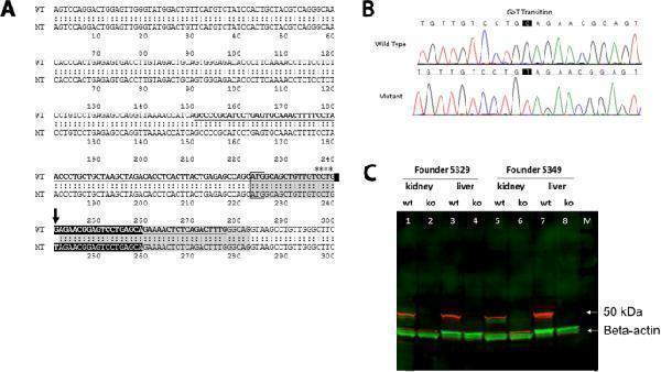
Creation of a Pah -KO mouse model. ( A ) The mouse genomic locus on chromosome 10 and the murine targeted allele after CRISPR gene editing are shown. Exon 1 contains the translation initiation codon. NCBI GeneID: 18478; RefSeq transcript: NM_008777. Exon 1 sequence is shadowed gray, the start codon (ATG) is framed, gRNA is shown as white letters on a black background, the asterisks and arrow indicate the position of PAM and G>T transition. The donor oligo for CRISPR targeting is in bold and underlined. ( B ) Sequencing genomic DNA samples of the founder 5349. The point mutation of G to T is indicated in the black boxes. ( C ) Western blot analyses to detect PAH protein in liver or kidney of wildtype (wt) or Hom (ko) mice. Solubilized proteins (60 µg) from mouse liver or kidney were fractionated and immune-blotted with an anti-PAH antibody. The PAH protein (red; expected size 50 kDa) is evident in wildtype mice (lanes 1, 3, 5 and 7) but is absent in Hom mice (2, 4, 6 and 8); lanes 1, 2, 5, 6) are from kidney; lanes 3, 4, 7, 8 are from liver. Equal loading is indicated by probing with antibody to beta-actin (green). M, molecular weight marker lane (bands not visible in this image).
Index in PubMed under a CC BY license. PMID: 33790381

Click image to see more details
Characterization of Hom and Het Pah -KO mice for 6 months. ( A ) The Hom mice showed lower bodyweights than the Het mice throughout the study ( B ) Summary of blood Phe at various timepoints is shown (n = 7/group) ( C ) Summary of blood Tyr at various timepoints is shown (n = 7/group). The graphical data are represented as mean ± SD (Phe and Tyr levels in Hom vs Het mice p < 0.0001) (n = 7 mice/group). ( D ) Western blot analysis of liver homogenate using anti-PAH antibody is shown. Note the lack of PAH protein in the Hom mice whereas Het mice showed a corresponding band (~ 50 kDa) for mouse PAH protein. Each lane contained 135 µg protein. Beta actin was used as an internal control. Full-length blots/gels are presented in Supplementary Fig. 2. ( E ) Summary of % PAH positive hepatocytes in the Hom and Het mice are shown. The graphical data are represented as mean ± SD (** p = 0.0012). ( F ) Representative liver and kidney PAH-IHC images show lack of IHC signal in a Hom mouse whereas liver from a Het mouse exhibit strong IHC signal in hepatocytes, mostly in the centrilobular region. Renal cortical epithelial cells from a Hom mouse show no IHC signal as compared to Het mouse, where most proximal convoluted tubular epithelium exhibit strong signal.
Index in PubMed under a CC BY license. PMID: 33790381

Click image to see more details
Analysis of Phe, Tyr and neurotransmitters in brains of Hom and Het Pah -KO mice at 6 months. Scatter plot charts show brain Phe, Tyr, serotonin, dopamine, L-DOPA, DOPAC, NE, 5-HIAA, and HVA. n = 6 Hom, 7 Het. Bars indicate group mean and the error bars indicate standard deviation (* p = 0.01, ** p = 0.003—0.007, *** p = 0.0001, **** p < 0.0001).
Index in PubMed under a CC BY license. PMID: 33790381

Click image to see more details
Hair coat color in Hom- Pah KO mice. The difference in hair coat color is grossly visible ( A ). Hair coat of the Hom mouse is light brown whereas it is dark brown for the Het mouse. ( B ) Hair shafts from the Hom mouse contain less melanin pigment when compared to the Het mouse. H&E stain 20× ( C ) Summary of hair melanin scores is shown in the scatter plot graph. n = 7 Hom, 7 Het. Bars indicate group median score and the error bars indicate range (*** p = 0.0006).
Index in PubMed under a CC BY license. PMID: 33790381

Click image to see more details
Western blot analysis of PAH using anti-PAH antibody (A00761-1).
Electrophoresis was performed on a 5-20% SDS-PAGE gel at 70V (Stacking gel) / 90V (Resolving gel) for 2-3 hours. The sample well of each lane was loaded with 30 ug of sample under reducing conditions.
Lane 1: human HepG2 whole cell lysates,
Lane 2: human CACO-2 whole cell lysates,
Lane 3: rat kidney tissue lysates,
Lane 4: rat liver tissue lysates,
Lane 5: mouse kidney tissue lysates,
Lane 6: mouse liver tissue lysates.
After electrophoresis, proteins were transferred to a nitrocellulose membrane at 150 mA for 50-90 minutes. Blocked the membrane with 5% non-fat milk/TBS for 1.5 hour at RT. The membrane was incubated with rabbit anti-PAH antigen affinity purified polyclonal antibody (Catalog # A00761-1) at 0.5 μg/mL overnight at 4°C, then washed with TBS-0.1%Tween 3 times with 5 minutes each and probed with a goat anti-rabbit IgG-HRP secondary antibody at a dilution of 1:5000 for 1.5 hour at RT. The signal is developed using an Enhanced Chemiluminescent detection (ECL) kit (Catalog # EK1002) with Tanon 5200 system. A specific band was detected for PAH at approximately 45 kDa. The expected band size for PAH is at 52 kDa.

Click image to see more details
IHC analysis of PAH using anti-PAH antibody (A00761-1).
PAH was detected in a paraffin-embedded section of human liver cancer tissue. Heat mediated antigen retrieval was performed in EDTA buffer (pH 8.0, epitope retrieval solution). The tissue section was blocked with 10% goat serum. The tissue section was then incubated with 2 μg/ml rabbit anti-PAH Antibody (A00761-1) overnight at 4°C. Peroxidase Conjugated Goat Anti-rabbit IgG was used as secondary antibody and incubated for 30 minutes at 37°C. The tissue section was developed using HRP Conjugated Rabbit IgG Super Vision Assay Kit (Catalog # SV0002) with DAB as the chromogen.

Click image to see more details
IHC analysis of PAH using anti-PAH antibody (A00761-1).
PAH was detected in a paraffin-embedded section of mouse kidney tissue. Heat mediated antigen retrieval was performed in EDTA buffer (pH 8.0, epitope retrieval solution). The tissue section was blocked with 10% goat serum. The tissue section was then incubated with 2 μg/ml rabbit anti-PAH Antibody (A00761-1) overnight at 4°C. Peroxidase Conjugated Goat Anti-rabbit IgG was used as secondary antibody and incubated for 30 minutes at 37°C. The tissue section was developed using HRP Conjugated Rabbit IgG Super Vision Assay Kit (Catalog # SV0002) with DAB as the chromogen.

Click image to see more details
IHC analysis of PAH using anti-PAH antibody (A00761-1).
PAH was detected in a paraffin-embedded section of rat kidney tissue. Heat mediated antigen retrieval was performed in EDTA buffer (pH 8.0, epitope retrieval solution). The tissue section was blocked with 10% goat serum. The tissue section was then incubated with 2 μg/ml rabbit anti-PAH Antibody (A00761-1) overnight at 4°C. Peroxidase Conjugated Goat Anti-rabbit IgG was used as secondary antibody and incubated for 30 minutes at 37°C. The tissue section was developed using HRP Conjugated Rabbit IgG Super Vision Assay Kit (Catalog # SV0002) with DAB as the chromogen.

Click image to see more details
Flow Cytometry analysis of HepG2 cells using anti-PAH antibody (A00761-1).
Overlay histogram showing HepG2 cells stained with A00761-1 (Blue line). To facilitate intracellular staining, cells were fixed with 4% paraformaldehyde and permeabilized with permeabilization buffer. The cells were blocked with 10% normal goat serum. And then incubated with rabbit anti-PAH Antibody (A00761-1, 1 μg/1x106 cells) for 30 min at 20°C. DyLight®488 conjugated goat anti-rabbit IgG (BA1127, 5-10 μg/1x106 cells) was used as secondary antibody for 30 minutes at 20°C. Isotype control antibody (Green line) was rabbit IgG (1 μg/1x106) used under the same conditions. Unlabelled sample (Red line) was also used as a control.
Specific Publications For Anti-PAH Antibody Picoband® (A00761-1)
Loading publications
Recommended Resources
Here are featured tools and databases that you might find useful.
- Boster's Pathways Library
- Protein Databases
- Bioscience Research Protocol Resources
- Data Processing & Analysis Software
- Photo Editing Software
- Scientific Literature Resources
- Research Paper Management Tools
- Molecular Biology Software
- Primer Design Tools
- Bioinformatics Tools
- Phylogenetic Tree Analysis
Customer Reviews
Have you used Anti-PAH Antibody Picoband®?
Share your experimental results or join a short interview to earn up to $1,000 in product credits or other rewards.
0 Reviews For Anti-PAH Antibody Picoband®
Customer Q&As
Have a question?
Find answers in Q&As, reviews.
Can't find your answer?
Submit your question
5 Customer Q&As for Anti-PAH Antibody Picoband®
Question
Will anti-PAH antibody A00761-1 work for WB with liver?
Verified Customer
Verified customer
Asked: 2020-05-05
Answer
According to the expression profile of liver, PAH is highly expressed in liver. So, it is likely that anti-PAH antibody A00761-1 will work for WB with liver.
Boster Scientific Support
Answered: 2020-05-05
Question
I appreciate helping with my inquiry over the phone. Here are the WB image, lot number and protocol we used for liver using anti-PAH antibody A00761-1. Let me know if you need anything else.
Verified Customer
Verified customer
Asked: 2020-03-27
Answer
Thank you for the data. You have provided everything we needed. Our lab team are working to resolve your inquiry as quickly as possible, and we appreciate your patience and understanding! Please let me know if there is anything you need in the meantime.
Boster Scientific Support
Answered: 2020-03-27
Question
We are currently using anti-PAH antibody A00761-1 for rat tissue, and we are content with the IF results. The species of reactivity given in the datasheet says human, mouse, rat. Is it true that the antibody can work on zebrafish tissues as well?
Verified Customer
Verified customer
Asked: 2020-01-13
Answer
The anti-PAH antibody (A00761-1) has not been validated for cross reactivity specifically with zebrafish tissues, though there is a good chance of cross reactivity. We have an innovator award program that if you test this antibody and show it works in zebrafish you can get your next antibody for free. Please contact me if I can help you with anything.
Boster Scientific Support
Answered: 2020-01-13
Question
Will anti-PAH antibody A00761-1 work on horse IHC-F with nephron tubule?
Verified Customer
Verified customer
Asked: 2019-02-06
Answer
Our lab technicians have not validated anti-PAH antibody A00761-1 on horse. You can run a BLAST between horse and the immunogen sequence of anti-PAH antibody A00761-1 to see if they may cross-react. If the sequence homology is close, then you can perform a pilot test. Keep in mind that since we have not validated horse samples, this use of the antibody is not covered by our guarantee. However we have an innovator award program that if you test this antibody and show it works in horse nephron tubule in IHC-F, you can get your next antibody for free.
Boster Scientific Support
Answered: 2019-02-06
Question
Is this A00761-1 anti-PAH antibody reactive to the isotypes of PAH?
L. Huang
Verified customer
Asked: 2014-05-15
Answer
The immunogen of A00761-1 anti-PAH antibody is E. coli-derived human PAH recombinant protein (Position: R71-H208). Human PAH shares 89.1% and 88.4% amino acid (aa) sequence identity with mouse and rat PAH, respectively. Could you tell me which isotype you are interested in so I can help see if the immunogen is part of this isotype?
Boster Scientific Support
Answered: 2014-05-15
