Click image to see more details
Product Info Summary
| SKU: | PA1563 |
|---|---|
| Size: | 100 μg/vial |
| Reactive Species: | Human, Mouse, Rat |
| Host: | Rabbit |
| Application: | WB |
Customers Who Bought This Also Bought

Product info
Product Name
Anti-PAK1 Antibody Picoband®
SKU/Catalog Number
PA1563
BA2954 is an alternative SKU for this antibody, used in previous lots.
Size
100 μg/vial
Form
Lyophilized
Description
Boster Bio Anti-PAK1 Antibody catalog # PA1563. Tested in WB applications. This antibody reacts with Human, Mouse, Rat. The brand Picoband indicates this is a premium antibody that guarantees superior quality, high affinity, and strong signals with minimal background in Western blot applications. Only our best-performing antibodies are designated as Picoband, ensuring unmatched performance.
Storage & Handling
Store at -20˚C for one year from date of receipt. After reconstitution, at 4˚C for one month. It can also be aliquotted and stored frozen at -20˚C for six months. Avoid repeated freeze-thaw cycles.
Cite This Product
Anti-PAK1 Antibody Picoband® (Boster Biological Technology, Pleasanton CA, USA, Catalog # PA1563)
Host
Rabbit
Contents
Each vial contains 4 mg Trehalose, 0.9 mg NaCl and 0.2 mg Na2HPO4.
Clonality
Polyclonal
Isotype
Rabbit IgG
Immunogen
A synthetic peptide corresponding to a sequence at the N-terminus of human PAK1, different from the related mouse and rat sequences by on amino acid.
Cross-reactivity
No cross-reactivity with other proteins
Reactive Species
PA1563 is reactive to PAK1 in Human, Mouse, Rat
Observed Molecular Weight
68 kDa
Calculated molecular weight
60.6 kDa
Background of PAK1
Serine/threonine-protein kinase PAK 1 is an enzyme that in humans is encoded by the PAK1 gene. PAK proteins are critical effectors that link RhoGTPases to cytoskeleton reorganization and nuclear signaling. PAK proteins, a family of serine/threonine p21-activated kinases, include PAK1, PAK2, PAK3 and PAK4. These proteins serve as targets for the small GTP binding proteins Cdc42 and Rac and have been implicated in a wide range of biological activities. PAK1 regulates cell motility and morphology. Alternative transcripts of this gene have been found, but their full-length natures have not yet been determined. The PAK1 gene is mapped to 11q13-q14 by inclusion within a mapped clone.
Antibody Validation
Boster validates all antibodies on WB, IHC, ICC, Immunofluorescence, and ELISA with known positive control and negative samples to ensure specificity and high affinity, including thorough antibody incubations.
Application & Images
Applications
PA1563 is guaranteed for WB Boster Guarantee
Recommend Dilution
| Application | Dilution | Species |
|---|---|---|
| Western blot | 0.1-0.5μg/ml | Human, Mouse, Rat |
Tested application
Suggested blocking solution with 5% non-fat milk or BSA; (*)Recommended protein loading: 20-40 µg per lane
Validation Images & Assay Conditions
Click image to see more details
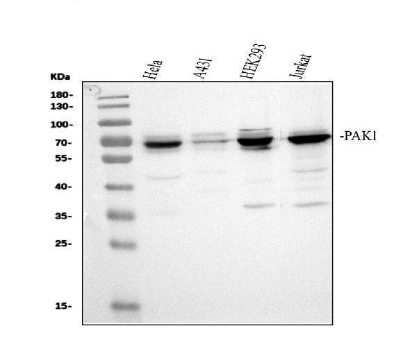
Western blot analysis of PAK1 using anti-PAK1 antibody (PA1563).
Electrophoresis was performed on a 10% SDS-PAGE gel at 80V (Stacking gel) / 120V (Resolving gel) for 2 hours. The sample well of each lane was loaded with 30 ug of sample under reducing conditions.
Lane 1: human Hela whole cell lysates,
Lane 2: human A431 whole cell lysates,
Lane 3: human HEK293 whole cell lysates,
Lane 4: human Jurkat whole cell lysates.
After electrophoresis, proteins were transferred to a nitrocellulose membrane at 150 mA for 50-90 minutes. Blocked the membrane with 5% non-fat milk/TBS for 1.5 hour at RT. The membrane was incubated with rabbit anti-PAK1 antigen affinity purified polyclonal antibody (PA1563) at 0.5 μg/mL overnight at 4°C, then washed with TBS-0.1%Tween 3 times with 5 minutes each and probed with a goat anti-rabbit IgG-HRP secondary antibody (Catalog # BA1054) at a dilution of 1:5000 for 1.5 hour at RT. The signal is developed using an ECL Plus Western Blotting Substrate (Catalog # AR1196-200) with Tanon 5200 system. A specific band was detected for PAK1 at approximately 68 kDa. The expected band size for Bcl-XS/BCL2L1 is at 61 kDa.

Click image to see more details
Western blot analysis of PAK1 using anti-PAK1 antibody (PA1563).
Electrophoresis was performed on a 10% SDS-PAGE gel at 80V (Stacking gel) / 120V (Resolving gel) for 2 hours. The sample well of each lane was loaded with 30 ug of sample under reducing conditions.
Lane 1: rat brain tissue lysates,
Lane 2: rat lung tissue lysates,
Lane 3: rat stomach tissue lysates,
Lane 4: rat PC-12 whole cell lysates,
Lane 5: mouse brain tissue lysates,
Lane 6: mouse lung tissue lysates,
Lane 7: mouse stomach tissue lysates,
Lane 8: mouse NIH/3T3 whole cell lysates.
After electrophoresis, proteins were transferred to a nitrocellulose membrane at 150 mA for 50-90 minutes. Blocked the membrane with 5% non-fat milk/TBS for 1.5 hour at RT. The membrane was incubated with rabbit anti-PAK1 antigen affinity purified polyclonal antibody (PA1563) at 0.5 μg/mL overnight at 4°C, then washed with TBS-0.1%Tween 3 times with 5 minutes each and probed with a goat anti-rabbit IgG-HRP secondary antibody (Catalog # BA1054) at a dilution of 1:5000 for 1.5 hour at RT. The signal is developed using an ECL Plus Western Blotting Substrate (Catalog # AR1196-200) with Tanon 5200 system. A specific band was detected for PAK1 at approximately 68 kDa. The expected band size for Bcl-XS/BCL2L1 is at 61 kDa.
Specific Publications For Anti-PAK1 Antibody Picoband® (PA1563)
Loading publications
Recommended Resources
Here are featured tools and databases that you might find useful.
- Boster's Pathways Library
- Protein Databases
- Bioscience Research Protocol Resources
- Data Processing & Analysis Software
- Photo Editing Software
- Scientific Literature Resources
- Research Paper Management Tools
- Molecular Biology Software
- Primer Design Tools
- Bioinformatics Tools
- Phylogenetic Tree Analysis
Customer Reviews
Have you used Anti-PAK1 Antibody Picoband®?
Share your experimental results or join a short interview to earn up to $1,000 in product credits or other rewards.
0 Reviews For Anti-PAK1 Antibody Picoband®
Customer Q&As
Have a question?
Find answers in Q&As, reviews.
Can't find your answer?
Submit your question
4 Customer Q&As for Anti-PAK1 Antibody Picoband®
Question
We have seen staining in human cervix carcinoma. Any tips? Is anti-PAK1 antibody supposed to stain cervix carcinoma positively?
Verified Customer
Verified customer
Asked: 2020-04-27
Answer
According to literature cervix carcinoma does express PAK1. According to Uniprot.org, PAK1 is expressed in middle temporal gyrus, placenta, cervix carcinoma, leukemic t-cell, cervix carcinoma erythroleukemia, liver, brain, among other tissues. Regarding which tissues have PAK1 expression, here are a few articles citing expression in various tissues:
Brain, Pubmed ID: 23503467
Cervix carcinoma, Pubmed ID: 16964243, 18669648
Cervix carcinoma, and Erythroleukemia, Pubmed ID: 23186163
Leukemic T-cell, Pubmed ID: 19690332
Liver, Pubmed ID: 24275569
Placenta, Pubmed ID: 8805275
Boster Scientific Support
Answered: 2020-04-27
Question
We are currently using anti-PAK1 antibody PA1563 for mouse tissue, and we are content with the WB results. The species of reactivity given in the datasheet says human, mouse, rat. Is it possible that the antibody can work on dog tissues as well?
Verified Customer
Verified customer
Asked: 2019-08-15
Answer
The anti-PAK1 antibody (PA1563) has not been tested for cross reactivity specifically with dog tissues, though there is a good chance of cross reactivity. We have an innovator award program that if you test this antibody and show it works in dog you can get your next antibody for free. Please contact me if I can help you with anything.
Boster Scientific Support
Answered: 2019-08-15
Question
I would like using your anti-PAK1 antibody for positive regulation of vascular smooth muscle cell proliferation studies. Has this antibody been tested with western blotting on tissue lysate? We would like to see some validation images before ordering.
Verified Customer
Verified customer
Asked: 2019-07-24
Answer
Thanks for your inquiry. This PA1563 anti-PAK1 antibody is tested on rat spleen tissue, testis tissue, tissue lysate, sw620 cell lysate, cem cell lysate, ht1080 cell lysate. It is guaranteed to work for WB in human, mouse, rat. Our Boster guarantee will cover your intended experiment even if the sample type has not been be directly tested.
Boster Scientific Support
Answered: 2019-07-24
Question
My boss were well pleased with the WB result of your anti-PAK1 antibody. However we have seen positive staining in placenta cytoplasm using this antibody. Is that expected? Could you tell me where is PAK1 supposed to be expressed?
Verified Customer
Verified customer
Asked: 2019-02-28
Answer
According to literature, placenta does express PAK1. Generally PAK1 expresses in cytoplasm. Regarding which tissues have PAK1 expression, here are a few articles citing expression in various tissues:
Brain, Pubmed ID: 23503467
Cervix carcinoma, Pubmed ID: 16964243, 18669648
Cervix carcinoma, and Erythroleukemia, Pubmed ID: 23186163
Leukemic T-cell, Pubmed ID: 19690332
Liver, Pubmed ID: 24275569
Placenta, Pubmed ID: 8805275
Boster Scientific Support
Answered: 2019-02-28
