Click image to see more details
Product Info Summary
| SKU: | A00943-4 |
|---|---|
| Size: | 100 µg/vial |
| Reactive Species: | Human, Mouse, Rat |
| Host: | Rabbit |
| Application: | ELISA, IF, ICC, WB |
Customers Who Bought This Also Bought

Product info
Product Name
Anti-PAX8 Antibody Picoband®
SKU/Catalog Number
A00943-4
Size
100 µg/vial
Form
Lyophilized
Description
Boster Bio Anti-PAX8 Antibody Picoband® catalog # A00943-4. Tested in WB, ICC/IF, ELISA applications. This antibody reacts with Human, Mouse, Rat. The brand Picoband indicates this is a premium antibody that guarantees superior quality, high affinity, and strong signals with minimal background in Western blot applications. Only our best-performing antibodies are designated as Picoband, ensuring unmatched performance.
Storage & Handling
At -20°C for one year from date of receipt. After reconstitution, at 4°C for one month. It can also be aliquotted and stored frozen at -20°C for six months. Avoid repeated freezing and thawing.
Cite This Product
Anti-PAX8 Antibody Picoband® (Boster Biological Technology, Pleasanton CA, USA, Catalog # A00943-4)
Host
Rabbit
Contents
Each vial contains 4 mg Trehalose, 0.9 mg NaCl, 0.2 mg Na2HPO4.
Clonality
Polyclonal
Isotype
IgG
Immunogen
E.coli-derived human PAX8 recombinant protein (Position: Q137-L450). Human PAX8 shares 95.3% and 95% amino acid (aa) sequence identity with mouse and rat PAX8, respectively.
Cross-reactivity
No cross reactivity with other proteins.
Reactive Species
A00943-4 is reactive to PAX8 in Human, Mouse, Rat
Observed Molecular Weight
48 kDa
Calculated molecular weight
48.2 kDa
Background of PAX8
Paired box gene 8, also known as PAX8, is a protein which in humans is encoded by the PAX8 gene. This gene encodes a member of the paired box (PAX) family of transcription factors. Members of this gene family typically encode proteins that contain a paired box domain, an octapeptide, and a paired-type homeodomain. This nuclear protein is involved in thyroid follicular cell development and expression of thyroid-specific genes. Mutations in this gene have been associated with thyroid dysgenesis, thyroid follicular carcinomas and atypical follicular thyroid adenomas. Alternatively spliced transcript variants encoding different isoforms have been described.
Antibody Validation
Boster validates all antibodies on WB, IHC, ICC, Immunofluorescence, and ELISA with known positive control and negative samples to ensure specificity and high affinity, including thorough antibody incubations.
Application & Images
Applications
A00943-4 is guaranteed for ELISA, IF, ICC, WB Boster Guarantee
Assay Dilutions Recommendation
The recommendations below provide a starting point for assay optimization. The actual working concentration varies and should be decided by the user.
Western blot, 0.25-0.5 μg/ml, Human, Mouse, Rat
Immunocytochemistry/Immunofluorescence, 5 μg/ml, Human
ELISA, 0.1-0.5 μg/ml, -
Positive Control
WB: human SW579 whole cell, human SK-OV-3 whole cell, rat kidney tissue, rat NRK whole cell, mouse NIH/3T3 whole cell
ICC/IF: HELA cell
Validation Images & Assay Conditions
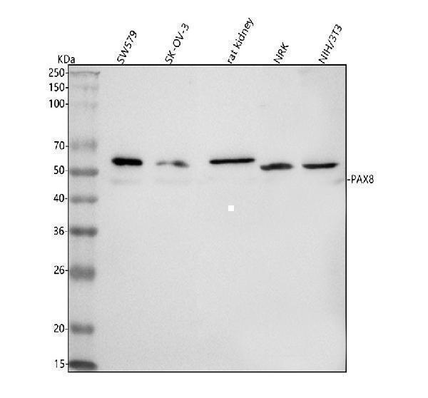
Click image to see more details
Western blot analysis of PAX8 using anti-PAX8 antibody (A00943-4).
Electrophoresis was performed on a 5-20% SDS-PAGE gel at 70V (Stacking gel) / 90V (Resolving gel) for 2-3 hours. The sample well of each lane was loaded with 30 ug of sample under reducing conditions.
Lane 1: human SW579 whole cell lysates,
Lane 2: human SK-OV-3 whole cell lysates,
Lane 3: rat kidney tissue lysates,
Lane 4: rat NRK whole cell lysates,
Lane 5: mouse NIH/3T3 whole cell lysates.
After electrophoresis, proteins were transferred to a nitrocellulose membrane at 150 mA for 50-90 minutes. Blocked the membrane with 5% non-fat milk/TBS for 1.5 hour at RT. The membrane was incubated with rabbit anti-PAX8 antigen affinity purified polyclonal antibody (Catalog # A00943-4) at 0.5 μg/mL overnight at 4°C, then washed with TBS-0.1%Tween 3 times with 5 minutes each and probed with a goat anti-rabbit IgG-HRP secondary antibody at a dilution of 1:5000 for 1.5 hour at RT. The signal is developed using an Enhanced Chemiluminescent detection (ECL) kit (Catalog # EK1002) with Tanon 5200 system. A specific band was detected for PAX8 at approximately 48 kDa. The expected band size for PAX8 is at 48 kDa.

Click image to see more details
IF analysis of PAX8 using anti-PAX8 antibody (A00943-4) and anti-Beta Tubulin antibody (M01857-3).
PAX8 was detected in immunocytochemical section of HELA cell. Enzyme antigen retrieval was performed using IHC enzyme antigen retrieval reagent (AR0022) for 15 mins. The cells were blocked with 10% goat serum. And then incubated with 5 μg/mL rabbit anti-PAX8 Antibody (A00943-4) and mouse anti-Beta Tubulin antibody (M01857-3) overnight at 4°C. DyLight®488 Conjugated Goat Anti-Rabbit IgG (BA1127) and Cy3 Conjugated Goat Anti-Mouse IgG (BA1031) were used as secondary antibody at 1:500 dilution and incubated for 30 minutes at 37°C. Visualize using a fluorescence microscope and filter sets appropriate for the label used.
Specific Publications For Anti-PAX8 Antibody Picoband® (A00943-4)
Loading publications
Recommended Resources
Here are featured tools and databases that you might find useful.
- Boster's Pathways Library
- Protein Databases
- Bioscience Research Protocol Resources
- Data Processing & Analysis Software
- Photo Editing Software
- Scientific Literature Resources
- Research Paper Management Tools
- Molecular Biology Software
- Primer Design Tools
- Bioinformatics Tools
- Phylogenetic Tree Analysis
Customer Reviews
Have you used Anti-PAX8 Antibody Picoband®?
Share your experimental results or join a short interview to earn up to $1,000 in product credits or other rewards.
0 Reviews For Anti-PAX8 Antibody Picoband®
Customer Q&As
Have a question?
Find answers in Q&As, reviews.
Can't find your answer?
Submit your question
