Click image to see more details
Product Info Summary
| SKU: | A00085-2 |
|---|---|
| Size: | 100 μg/vial |
| Reactive Species: | Mouse, Rat |
| Host: | Rabbit |
| Application: | ELISA, WB |
Customers Who Bought This Also Bought

Product info
Product Name
Anti-PCSK9 Antibody Picoband®
SKU/Catalog Number
A00085-2
Size
100 μg/vial
Form
Lyophilized
Description
Boster Bio Anti-PCSK9 Antibody Picoband® catalog # A00085-2. Tested in ELISA, WB applications. This antibody reacts with Mouse, Rat. The brand Picoband indicates this is a premium antibody that guarantees superior quality, high affinity, and strong signals with minimal background in Western blot applications. Only our best-performing antibodies are designated as Picoband, ensuring unmatched performance.
Storage & Handling
Store at -20˚C for one year from date of receipt. After reconstitution, at 4˚C for one month. It can also be aliquotted and stored frozen at -20˚C for six months. Avoid repeated freeze-thaw cycles.
Cite This Product
Anti-PCSK9 Antibody Picoband® (Boster Biological Technology, Pleasanton CA, USA, Catalog # A00085-2)
Host
Rabbit
Contents
Each vial contains 4mg Trehalose, 0.9mg NaCl and 0.2mg Na2HPO4.
Clonality
Polyclonal
Isotype
Rabbit IgG
Immunogen
E.coli-derived mouse PCSK9 recombinant protein (Position: Q182-H560).
Cross-reactivity
No cross-reactivity with other proteins.
Reactive Species
A00085-2 is reactive to Pcsk9 in Mouse, Rat
Observed Molecular Weight
74 kDa
Calculated molecular weight
74.8 kDa
Background of Pcsk9
Proprotein convertase subtilisin/kexin type 9, also known as PCSK9, is an enzyme that in humans is encoded by the PCSK9 gene. This gene encodes a proprotein convertase belonging to the proteinase K subfamily of the secretory subtilase family. By genomic sequence analysis, PCSK9 was mapped to chromosome 1p32. This gene is a crucial player in the regulation of plasma cholesterol homeostasis. It may prevent the recycling of LDLR from endosomes to the cell surface or direct it to lysosomes for degradation. PCSK9 can induce ubiquitination of LDLR leading to its subsequent degradation. This gene is involved in the disposal of non-acetylated intermediates of BACE1 in the early secretory pathway.
Antibody Validation
Boster validates all antibodies on WB, IHC, ICC, Immunofluorescence, and ELISA with known positive control and negative samples to ensure specificity and high affinity, including thorough antibody incubations.
Application & Images
Applications
A00085-2 is guaranteed for ELISA, WB Boster Guarantee
Assay Dilutions Recommendation
The recommendations below provide a starting point for assay optimization. The actual working concentration varies and should be decided by the user.
Western blot, 0.25-0.5μg/ml, Mouse, Rat
ELISA, 0.1-0.5μg/ml, -
Positive Control
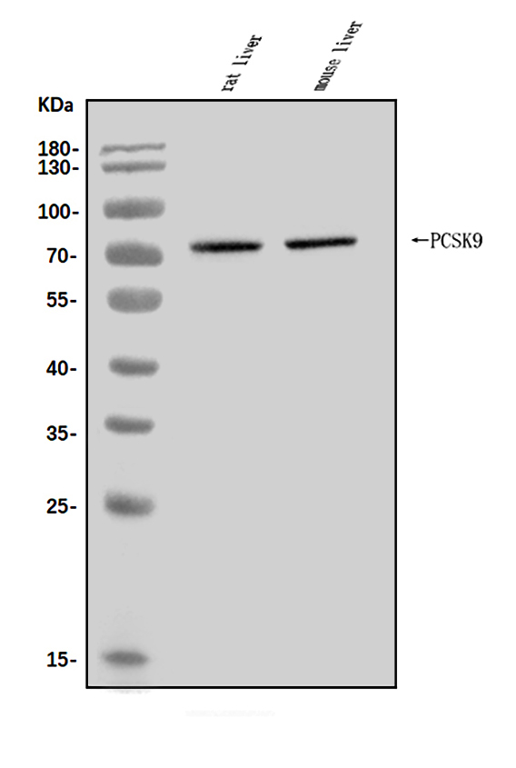
WB: rat liver tissue, mouse liver tissue
WB: mouse liver tissue
Validation Images & Assay Conditions
Click image to see more details
Western blot analysis of PCSK9/Pcsk9 using anti-PCSK9/Pcsk9 antibody (A00085-2).
Electrophoresis was performed on a 5-20% SDS-PAGE gel at 70V (Stacking gel) / 90V (Resolving gel) for 2-3 hours. The sample well of each lane was loaded with 30ug of sample under reducing conditions.
Lane 1: rat liver tissue lysates,
Lane 2: mouse liver tissue lysates.
After Electrophoresis, proteins were transferred to a Nitrocellulose membrane at 150mA for 50-90 minutes. Blocked the membrane with 5% Non-fat Milk/ TBS for 1.5 hour at RT. The membrane was incubated with rabbit anti-PCSK9/Pcsk9 antigen affinity purified polyclonal antibody (Catalog # A00085-2) at 0.5 μg/mL overnight at 4°C, then washed with TBS-0.1%Tween 3 times with 5 minutes each and probed with a goat anti-rabbit IgG-HRP secondary antibody at a dilution of 1:5000 for 1.5 hour at RT. The signal is developed using an Enhanced Chemiluminescent detection (ECL) kit (Catalog # EK1002) with Tanon 5200 system. A specific band was detected for PCSK9/Pcsk9 at approximately 74KD. The expected band size for PCSK9/Pcsk9 is at 74KD.

Click image to see more details
Western blot analysis of PCSK9 using anti-PCSK9 antibody (A00085-2).
Electrophoresis was performed on a 10% SDS-PAGE gel at 80V (Stacking gel) / 120V (Resolving gel) for 2 hours. The sample well of each lane was loaded with 30 μg of sample under reducing conditions.
Lane 1: mouse liver tissue lysates.
After electrophoresis, proteins were transferred to a nitrocellulose membrane at 150 mA for 50-90 minutes. Blocked the membrane with 5% non-fat milk/TBS for 1.5 hour at RT. The membrane was incubated with rabbit anti-PCSK9 antigen affinity purified polyclonal antibody (A00085-2) at 0.5 μg/mL overnight at 4°C, then washed with TBS-0.1%Tween-20 3 times with 5 minutes each and probed with a goat anti-rabbit IgG-HRP secondary antibody (Catalog # BA1054) at a dilution of 1:5000 for 1.5 hour at RT. The signal is developed using an ECL Plus Western Blotting Substrate (Catalog # AR1196-200) with Tanon 5200 system. A specific band was detected for PCSK9 at approximately 75 kDa. The expected band size for PCSK9 is at 75 kDa.
Specific Publications For Anti-PCSK9 Antibody Picoband® (A00085-2)
Loading publications
Recommended Resources
Here are featured tools and databases that you might find useful.
- Boster's Pathways Library
- Protein Databases
- Bioscience Research Protocol Resources
- Data Processing & Analysis Software
- Photo Editing Software
- Scientific Literature Resources
- Research Paper Management Tools
- Molecular Biology Software
- Primer Design Tools
- Bioinformatics Tools
- Phylogenetic Tree Analysis
Customer Reviews
Have you used Anti-PCSK9 Antibody Picoband®?
Share your experimental results or join a short interview to earn up to $1,000 in product credits or other rewards.
0 Reviews For Anti-PCSK9 Antibody Picoband®
Customer Q&As
Have a question?
Find answers in Q&As, reviews.
Can't find your answer?
Submit your question
