Click image to see more details
Product Info Summary
| SKU: | PB9994 |
|---|---|
| Size: | 100 μg/vial |
| Reactive Species: | Mouse, Rat |
| Host: | Rabbit |
| Application: | ELISA, WB |
Customers Who Bought This Also Bought

Product info
Product Name
Anti-PD-L1/CD274 Antibody Picoband®
SKU/Catalog Number
PB9994
PB1059 is an alternative SKU for this antibody, used in previous lots.
Size
100 μg/vial
Form
Lyophilized
Description
Boster Bio Anti-PD-L1/CD274 Antibody Picoband® catalog # PB9994. Tested in ELISA, WB applications. This antibody reacts with Mouse, Rat. The brand Picoband indicates this is a premium antibody that guarantees superior quality, high affinity, and strong signals with minimal background in Western blot applications. Only our best-performing antibodies are designated as Picoband, ensuring unmatched performance.
Storage & Handling
Store at -20˚C for one year from date of receipt. After reconstitution, at 4˚C for one month. It can also be aliquotted and stored frozen at -20˚C for six months. Avoid repeated freeze-thaw cycles.
Cite This Product
Anti-PD-L1/CD274 Antibody Picoband® (Boster Biological Technology, Pleasanton CA, USA, Catalog # PB9994)
Host
Rabbit
Contents
Each vial contains antibody formulated with stabilizing components, 0.9 mg NaCl, 0.2 mg Na2HPO4, and 0.05 mg NaN3.
*This antibody is supplied in a stabilized formulation.
Compatibility with conjugation reactions depends on the chemistry of the conjugation method used.
For conjugation methods that are not compatible with the stabilizing components present in this formulation, a carrier-free antibody format is required.
Clonality
Polyclonal
Isotype
Rabbit IgG
Immunogen
E. coli-derived mouse PD-L1 recombinant protein (Position: F19-T238). Mouse PD-L1 shares 78.6% amino acid (aa) sequence identity with human PD-L1.
Cross-reactivity
No cross-reactivity with other proteins.
Reactive Species
PB9994 is reactive to Cd274 in Mouse, Rat
Observed Molecular Weight
39 kDa
Calculated molecular weight
32.8 kDa
Background of Cd274
Programmed death-ligand 1 (PD-L1), also known as CD274 or B7-H1, is a protein that in humans is encoded by the CD274 gene. It is mapped to 9p24.1. PD-L1 is a 40kDa type 1 transmembrane protein that has been speculated to play a major role in suppressing the immune system during particular events such as pregnancy, tissue allografts, autoimmune disease and other disease states such as hepatitis. It has been concluded that upregulation of PD-L1 on tumor MDCs downregulates T-cell immunity and that PD-L1 blockade may represent an approach for cancer immunotherapy. Additionally, PD-L1 can provide positive costimulatory signals for innate and adaptive immunity and for protection against intracellular bacterial infection. What’s more, it has been found that PD1/PDL1 pathway may be a good target for restoring antitumor immunity in ovarian cancer.
Antibody Validation
Boster validates all antibodies on WB, IHC, ICC, Immunofluorescence, and ELISA with known positive control and negative samples to ensure specificity and high affinity, including thorough antibody incubations.
Application & Images
Applications
PB9994 is guaranteed for ELISA, WB Boster Guarantee
Assay Dilutions Recommendation
The recommendations below provide a starting point for assay optimization. The actual working concentration varies and should be decided by the user.
ELISA, 0.1-0.5μg/ml, -
Western blot, 0.1-0.5μg/ml, Mouse, Rat
Positive Control
WB: mouse brain tissue
Validation Images & Assay Conditions
Click image to see more details
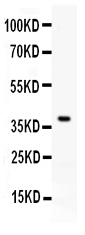
Western blot analysis of PD-L1 using anti-PD-L1 antibody (PB9994).
Electrophoresis was performed on a 5-20% SDS-PAGE gel at 70V (Stacking gel) / 90V (Resolving gel) for 2-3 hours. The sample well of each lane was loaded with 30 ug of sample under reducing conditions.
Lane 1: mouse brain tissue lysates.
After electrophoresis, proteins were transferred to a nitrocellulose membrane at 150 mA for 50-90 minutes. Blocked the membrane with 5% non-fat milk/TBS for 1.5 hour at RT. The membrane was incubated with rabbit anti-PD-L1 antigen affinity purified polyclonal antibody (Catalog # PB9994) at 0.5 μg/mL overnight at 4°C, then washed with TBS-0.1%Tween 3 times with 5 minutes each and probed with a goat anti-rabbit IgG-HRP secondary antibody at a dilution of 1:5000 for 1.5 hour at RT. The signal is developed using an Enhanced Chemiluminescent detection (ECL) kit (Catalog # EK1002) with Tanon 5200 system. A specific band was detected for PD-L1 at approximately 39 kDa. The expected band size for PD-L1 is at 33 kDa.
Specific Publications For Anti-PD-L1/CD274 Antibody Picoband® (PB9994)
Loading publications
Recommended Resources
Here are featured tools and databases that you might find useful.
- Boster's Pathways Library
- Protein Databases
- Bioscience Research Protocol Resources
- Data Processing & Analysis Software
- Photo Editing Software
- Scientific Literature Resources
- Research Paper Management Tools
- Molecular Biology Software
- Primer Design Tools
- Bioinformatics Tools
- Phylogenetic Tree Analysis
Customer Reviews
Have you used Anti-PD-L1/CD274 Antibody Picoband®?
Share your experimental results or join a short interview to earn up to $1,000 in product credits or other rewards.
0 Reviews For Anti-PD-L1/CD274 Antibody Picoband®
Customer Q&As
Have a question?
Find answers in Q&As, reviews.
Can't find your answer?
Submit your question
6 Customer Q&As for Anti-PD-L1/CD274 Antibody Picoband®
Question
We have been able to see staining in mouse chorionic villus. Any tips? Is anti-PD-L1/CD274 antibody supposed to stain chorionic villus positively?
Verified Customer
Verified customer
Asked: 2019-11-29
Answer
From literature chorionic villus does express CD274. From Uniprot.org, CD274 is expressed in chorionic villus, placenta, gastric carcinoma, lung, liver, among other tissues. Regarding which tissues have CD274 expression, here are a few articles citing expression in various tissues:
Liver, Pubmed ID: 19159218
Lung, Pubmed ID: 15489334
Placenta, Pubmed ID: 11015443, 14702039
Boster Scientific Support
Answered: 2019-11-29
Question
We are currently using anti-PD-L1/CD274 antibody PB9994 for mouse tissue, and we are satisfied with the WB results. The species of reactivity given in the datasheet says mouse. Is it true that the antibody can work on bovine tissues as well?
Verified Customer
Verified customer
Asked: 2019-06-13
Answer
The anti-PD-L1/CD274 antibody (PB9994) has not been tested for cross reactivity specifically with bovine tissues, though there is a good chance of cross reactivity. We have an innovator award program that if you test this antibody and show it works in bovine you can get your next antibody for free. Please contact me if I can help you with anything.
Boster Scientific Support
Answered: 2019-06-13
Question
We are currently using anti-PD-L1/CD274 antibody PB9994 for mouse tissue, and we are satisfied with the WB results. The species of reactivity given in the datasheet says mouse. Is it true that the antibody can work on bovine tissues as well?
Verified Customer
Verified customer
Asked: 2018-07-31
Answer
The anti-PD-L1/CD274 antibody (PB9994) has not been tested for cross reactivity specifically with bovine tissues, though there is a good chance of cross reactivity. We have an innovator award program that if you test this antibody and show it works in bovine you can get your next antibody for free. Please contact me if I can help you with anything.
Boster Scientific Support
Answered: 2018-07-31
Question
We purchased anti-PD-L1/CD274 antibody for ELISA on gastric carcinoma in the past. I am using mouse, and We want to use the antibody for WB next. I am interested in examining gastric carcinoma as well as lung in our next experiment. Could give a recommendation on which antibody would work the best for WB?
Verified Customer
Verified customer
Asked: 2018-07-04
Answer
I viewed the website and datasheets of our anti-PD-L1/CD274 antibody and it seems that PB9994 has been validated on mouse in both ELISA and WB. Thus PB9994 should work for your application. Our Boster satisfaction guarantee will cover this product for WB in mouse even if the specific tissue type has not been validated. We do have a comprehensive range of products for WB detection and you can check out our website bosterbio.com to find out more information about them.
Boster Scientific Support
Answered: 2018-07-04
Question
Our team were well pleased with the WB result of your anti-PD-L1/CD274 antibody. However we have observed positive staining in liver cell membrane using this antibody. Is that expected? Could you tell me where is CD274 supposed to be expressed?
P. Mitchell
Verified customer
Asked: 2014-05-26
Answer
From literature, liver does express CD274. Generally CD274 expresses in cell membrane. Regarding which tissues have CD274 expression, here are a few articles citing expression in various tissues:
Liver, Pubmed ID: 19159218
Lung, Pubmed ID: 15489334
Placenta, Pubmed ID: 11015443, 14702039
Boster Scientific Support
Answered: 2014-05-26
Question
I am looking for using your anti-PD-L1/CD274 antibody for immune response studies. Has this antibody been tested with western blotting on mouse brain? We would like to see some validation images before ordering.
C. Mitchell
Verified customer
Asked: 2013-03-14
Answer
We appreciate your inquiry. This PB9994 anti-PD-L1/CD274 antibody is validated on mouse brain. It is guaranteed to work for ELISA, WB in mouse. Our Boster guarantee will cover your intended experiment even if the sample type has not been be directly tested.
Boster Scientific Support
Answered: 2013-03-14
