Click image to see more details
-

-
-
-
-
+5
Product Info Summary
| SKU: | A01906-4 |
|---|---|
| Size: | 100 μl/vial |
| Reactive Species: | Human, Mouse, Rat |
| Host: | Rabbit |
| Application: | ELISA, IP, IF, IHC, ICC, WB |
Customers Who Bought This Also Bought

Product info
Product Name
Anti-PDH E1 Alpha/PDHA1 Antibody
SKU/Catalog Number
A01906-4
Size
100 μl/vial
Form
Liquid
Description
Boster Bio Anti-PDH E1 Alpha/PDHA1 Antibody catalog # A01906-4. Tested in WB, IHC, ICC, IF, IP, ELISA applications. This antibody reacts with Human, Mouse, Rat.
Storage & Handling
12 months from date of receipt,-20℃ as supplied. 6 months 2 to 8℃ after reconstitution. Avoid repeated freezing and thawing.
Cite This Product
Anti-PDH E1 Alpha/PDHA1 Antibody (Boster Biological Technology, Pleasanton CA, USA, Catalog # A01906-4)
Host
Rabbit
Contents
500 μg/ml antibody with PBS, 0.02% NaN3, 1 mg stabilizing protein and 50% glycerol
*This antibody is supplied in a stabilized formulation.
Compatibility with conjugation reactions depends on the chemistry of the conjugation method used.
For conjugation methods that are not compatible with the stabilizing components present in this formulation, a carrier-free antibody format is required.
Clonality
Polyclonal
Immunogen
E.coli-derived human PDH E1 Alpha/PDHA1 recombinant protein (Position: R45-S390).
Reactive Species
A01906-4 is reactive to PDHA1 in Human, Mouse, Rat
Observed Molecular Weight
40 kDa
Calculated molecular weight
43.3 kDa
Background of PDHA1
The pyruvate dehydrogenase (PDH) complex is a nuclear-encoded mitochondrial multienzyme complex that catalyzes the overall conversion of pyruvate to acetyl-CoA and CO(2), and provides the primary link between glycolysis and the tricarboxylic acid (TCA) cycle. The PDH complex is composed of multiple copies of three enzymatic components: pyruvate dehydrogenase (E1), dihydrolipoamide acetyltransferase (E2) and lipoamide dehydrogenase (E3). The E1 enzyme is a heterotetramer of two alpha and two beta subunits. This gene encodes the E1 alpha 1 subunit containing the E1 active site, and plays a key role in the function of the PDH complex. Mutations in this gene are associated with pyruvate dehydrogenase E1-alpha deficiency and X-linked Leigh syndrome. Alternatively spliced transcript variants encoding different isoforms have been found for this gene.
Antibody Validation
Boster validates all antibodies on WB, IHC, ICC, Immunofluorescence, and ELISA with known positive control and negative samples to ensure specificity and high affinity, including thorough antibody incubations.
Application & Images
Applications
A01906-4 is guaranteed for ELISA, IP, IF, IHC, ICC, WB Boster Guarantee
Recommend Dilution
| Application | Dilution | Species |
|---|---|---|
| Western blot | 1:500-2000 | |
| Immunohistochemistry | 1:50-400 | |
| Immunocytochemistry/Immunofluorescence | 1:50-400 | |
| Immunoprecipitation | 1:50 | |
| ELISA | 1:100-1000 |
Tested application
Suggested blocking solution with 5% non-fat milk or BSA; (*)Recommended protein loading: 20-40 µg per lane
Use TE buffer pH 9.0 for antigen retrieval; (*) citrate buffer pH 6.0 is an alternative.
Validation Images & Assay Conditions
Click image to see more details
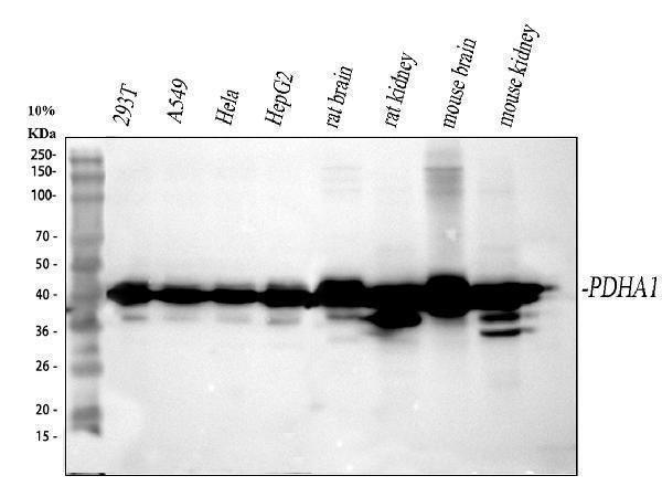
Western blot analysis of PDH E1 Alpha/PDHA1 using anti-PDH E1 Alpha/PDHA1 antibody (A01906-4).
Electrophoresis was performed on a 10% SDS-PAGE gel at 80V (Stacking gel) / 120V (Resolving gel) for 2 hours. The sample well of each lane was loaded with 30 ug of sample under reducing conditions.
Lane 1: human 293T whole cell lysates,
Lane 2: human A549 whole cell lysates,
Lane 3: human Hela whole cell lysates,
Lane 4: human HepG2 whole cell lysates,
Lane 5: rat brain tissue lysates,
Lane 6: rat kidney tissue lysates,
Lane 7: mouse brain tissue lysates,
Lane 8: mouse kidney tissue lysates.
After electrophoresis, proteins were transferred to a nitrocellulose membrane at 150 mA for 50-90 minutes. Blocked the membrane with 5% non-fat milk/TBS for 1.5 hour at RT. The membrane was incubated with rabbit anti-PDH E1 Alpha/PDHA1 antigen affinity purified polyclonal antibody (A01906-4) at 1:1000 overnight at 4°C, then washed with TBS-0.1%Tween 3 times with 5 minutes each and probed with a goat anti-rabbit IgG-HRP secondary antibody at a dilution of 1:5000 for 1.5 hour at RT. The signal is developed using an ECL Plus Western Blotting Substrate (Catalog # AR1196-200) with Tanon 5200 system. A specific band was detected for PDH E1 Alpha/PDHA1 at approximately 40 kDa. The expected band size for PDH E1 Alpha/PDHA1 is at 43 kDa.

Click image to see more details
IHC analysis of PDH E1 Alpha/PDHA1 using anti-PDH E1 Alpha/PDHA1 antibody (A01906-4).
PDH E1 Alpha/PDHA1 was detected in a paraffin-embedded section of human ovarian serous adenocarcinoma tissue. Heat mediated antigen retrieval was performed in EDTA buffer (pH 8.0, epitope retrieval solution). The tissue section was blocked with 10% goat serum. The tissue section was then incubated with 1:100 rabbit anti-PDH E1 Alpha/PDHA1 Antibody (A01906-4) overnight at 4°C. Peroxidase Conjugated Goat Anti-rabbit IgG was used as secondary antibody and incubated for 30 minutes at 37°C. The tissue section was developed using HRP Conjugated Rabbit IgG Super Vision Assay Kit (Catalog # SV0002) with DAB as the chromogen.

Click image to see more details
IHC analysis of PDH E1 Alpha/PDHA1 using anti-PDH E1 Alpha/PDHA1 antibody (A01906-4).
PDH E1 Alpha/PDHA1 was detected in a paraffin-embedded section of human breast cancer tissue. Heat mediated antigen retrieval was performed in EDTA buffer (pH 8.0, epitope retrieval solution). The tissue section was blocked with 10% goat serum. The tissue section was then incubated with 1:100 rabbit anti-PDH E1 Alpha/PDHA1 Antibody (A01906-4) overnight at 4°C. Peroxidase Conjugated Goat Anti-rabbit IgG was used as secondary antibody and incubated for 30 minutes at 37°C. The tissue section was developed using HRP Conjugated Rabbit IgG Super Vision Assay Kit (Catalog # SV0002) with DAB as the chromogen.

Click image to see more details
IHC analysis of PDH E1 Alpha/PDHA1 using anti-PDH E1 Alpha/PDHA1 antibody (A01906-4).
PDH E1 Alpha/PDHA1 was detected in a paraffin-embedded section of human spleen tissue. Heat mediated antigen retrieval was performed in EDTA buffer (pH 8.0, epitope retrieval solution). The tissue section was blocked with 10% goat serum. The tissue section was then incubated with 1:100 rabbit anti-PDH E1 Alpha/PDHA1 Antibody (A01906-4) overnight at 4°C. Peroxidase Conjugated Goat Anti-rabbit IgG was used as secondary antibody and incubated for 30 minutes at 37°C. The tissue section was developed using HRP Conjugated Rabbit IgG Super Vision Assay Kit (Catalog # SV0002) with DAB as the chromogen.

Click image to see more details
IHC analysis of PDH E1 Alpha/PDHA1 using anti-PDH E1 Alpha/PDHA1 antibody (A01906-4).
PDH E1 Alpha/PDHA1 was detected in a paraffin-embedded section of human testicular germ cell tumors tissue. Heat mediated antigen retrieval was performed in EDTA buffer (pH 8.0, epitope retrieval solution). The tissue section was blocked with 10% goat serum. The tissue section was then incubated with 1:100 rabbit anti-PDH E1 Alpha/PDHA1 Antibody (A01906-4) overnight at 4°C. Peroxidase Conjugated Goat Anti-rabbit IgG was used as secondary antibody and incubated for 30 minutes at 37°C. The tissue section was developed using HRP Conjugated Rabbit IgG Super Vision Assay Kit (Catalog # SV0002) with DAB as the chromogen.

Click image to see more details
IHC analysis of PDH E1 Alpha/PDHA1 using anti-PDH E1 Alpha/PDHA1 antibody (A01906-4).
PDH E1 Alpha/PDHA1 was detected in a paraffin-embedded section of human colorectal adenocarcinoma tissue. Heat mediated antigen retrieval was performed in EDTA buffer (pH 8.0, epitope retrieval solution). The tissue section was blocked with 10% goat serum. The tissue section was then incubated with 1:100 rabbit anti-PDH E1 Alpha/PDHA1 Antibody (A01906-4) overnight at 4°C. Peroxidase Conjugated Goat Anti-rabbit IgG was used as secondary antibody and incubated for 30 minutes at 37°C. The tissue section was developed using HRP Conjugated Rabbit IgG Super Vision Assay Kit (Catalog # SV0002) with DAB as the chromogen.

Click image to see more details
IHC analysis of PDH E1 Alpha/PDHA1 using anti-PDH E1 Alpha/PDHA1 antibody (A01906-4).
PDH E1 Alpha/PDHA1 was detected in a paraffin-embedded section of human appendix carcinoid tissue. Heat mediated antigen retrieval was performed in EDTA buffer (pH 8.0, epitope retrieval solution). The tissue section was blocked with 10% goat serum. The tissue section was then incubated with 1:100 rabbit anti-PDH E1 Alpha/PDHA1 Antibody (A01906-4) overnight at 4°C. Peroxidase Conjugated Goat Anti-rabbit IgG was used as secondary antibody and incubated for 30 minutes at 37°C. The tissue section was developed using HRP Conjugated Rabbit IgG Super Vision Assay Kit (Catalog # SV0002) with DAB as the chromogen.

Click image to see more details
IF analysis of PDH E1 Alpha/PDHA1 using anti-PDH E1 Alpha/PDHA1 antibody (A01906-4).
PDH E1 Alpha/PDHA1 was detected in an immunocytochemical section of chuman Hela ells. Enzyme antigen retrieval was performed using IHC enzyme antigen retrieval reagent (AR0022) for 15 mins. The cells were blocked with 10% goat serum. And then incubated with 1:100 rabbit anti-PDH E1 Alpha/PDHA1 Antibody (A01906-4) overnight at 4°C. DyLight®488 Conjugated Goat Anti-Rabbit IgG (BA1127) was used as secondary antibody at 1:500 dilution and incubated for 30 minutes at 37°C. The section was counterstained with DAPI. Visualize using a fluorescence microscope and filter sets appropriate for the label used.

Click image to see more details
Immunoprecipitating (IP) PDH E1 Alpha/PDHA1 in HepG2 whole cell lysate.
Western blot analysis of PDH E1 Alpha/PDHA1 using anti-PDH E1 Alpha/PDHA1 antibody (A01906-4);
Lane 1: HepG2 whole cell lysates (30ug);
Lane 2: Rabbit control IgG instead of anti-PDH E1 Alpha/PDHA1 antibody in HepG2 whole cell lysate;
Lane 3: anti-PDH E1 Alpha/PDHA1 antibody (2μg) + HepG2 whole cell lysate (500μg).
After electrophoresis, proteins were transferred to a membrane. Then the membrane was incubated with rabbit anti-PDH E1 Alpha/PDHA1 antigen affinity purified polyclonal antibody (A01906-4) at a dilution of 0.5 μg/mL and probed with a goat anti-rabbit IgG-HRP secondary antibody (Light Chiain). The signal is developed using ECL Plus Western Blotting Substrate (Catalog # AR1196-200). A specific band was detected for PDH E1 Alpha/PDHA1 at approximately 40 kDa. The expected band size for PDH E1 Alpha/PDHA1 is at 43 kDa.
Specific Publications For Anti-PDH E1 Alpha/PDHA1 Antibody (A01906-4)
Loading publications
Recommended Resources
Here are featured tools and databases that you might find useful.
- Boster's Pathways Library
- Protein Databases
- Bioscience Research Protocol Resources
- Data Processing & Analysis Software
- Photo Editing Software
- Scientific Literature Resources
- Research Paper Management Tools
- Molecular Biology Software
- Primer Design Tools
- Bioinformatics Tools
- Phylogenetic Tree Analysis
Customer Reviews
Have you used Anti-PDH E1 Alpha/PDHA1 Antibody?
Share your experimental results or join a short interview to earn up to $1,000 in product credits or other rewards.
0 Reviews For Anti-PDH E1 Alpha/PDHA1 Antibody
Customer Q&As
Have a question?
Find answers in Q&As, reviews.
Can't find your answer?
Submit your question
