Click image to see more details
-

-
-
-
-
+1
Product Info Summary
| SKU: | PA1836 |
|---|---|
| Size: | 100 μg/vial |
| Reactive Species: | Human, Mouse, Rat |
| Host: | Rabbit |
| Application: | Flow Cytometry, IF, IHC, ICC, WB |
Customers Who Bought This Also Bought

Product info
Product Name
Anti-Peroxiredoxin 3/PRDX3 Antibody Picoband®
SKU/Catalog Number
PA1836
Size
100 μg/vial
Form
Lyophilized
Description
Boster Bio Anti-Peroxiredoxin 3/PRDX3 Antibody catalog # PA1836. Tested in Flow Cytometry, IF, IHC, ICC, WB applications. This antibody reacts with Human, Mouse, Rat. The brand Picoband indicates this is a premium antibody that guarantees superior quality, high affinity, and strong signals with minimal background in Western blot applications. Only our best-performing antibodies are designated as Picoband, ensuring unmatched performance.
Storage & Handling
Store at -20˚C for one year from date of receipt. After reconstitution, at 4˚C for one month. It can also be aliquotted and stored frozen at -20˚C for six months. Avoid repeated freeze-thaw cycles.
Cite This Product
Anti-Peroxiredoxin 3/PRDX3 Antibody Picoband® (Boster Biological Technology, Pleasanton CA, USA, Catalog # PA1836)
Host
Rabbit
Contents
Each vial contains antibody formulated with stabilizing components, 0.9mg NaCl, 0.2mg Na2HPO4, 0.05mg Thimerosal, 0.05mg NaN3.
*This antibody is supplied in a stabilized formulation.
Compatibility with conjugation reactions depends on the chemistry of the conjugation method used.
For conjugation methods that are not compatible with the stabilizing components present in this formulation, a carrier-free antibody format is required.
Clonality
Polyclonal
Isotype
Rabbit IgG
Immunogen
A synthetic peptide corresponding to a sequence at the C-terminus of mouse Peroxiredoxin 3, identical to the related rat sequence, and different from the related human sequence by three amino acids.
Cross-reactivity
No cross-reactivity with other proteins
Reactive Species
PA1836 is reactive to Prdx3 in Human, Mouse, Rat
Observed Molecular Weight
26 kDa
Calculated molecular weight
28.1 kDa
Background of Prdx3
PRDX3 (peroxiredoxin 3) also known as AOP-1, MER5, SP-22 or PRX3, is localized exclusively in mitochondria. The deduced 256-amino acid human AOP1 protein shares 86% amino acid sequence similarity with mouse Aop1, and significant similarity with both the human proliferation-associated gene A product and the mouse stress-induced peritoneal macrophage protein Msp23. The PRDX3 gene is mapped on 10q26.11. Expression of PRDX3 is induced by MYC and is reduced in c-myc -/- cells. Chromatin immunoprecipitation analysis spanning the entire PRDX3 genomic sequence revealed that MYC binds preferentially to a 930-bp region surrounding exon 1. Results using mitochondria-specific fluorescent probes demonstrated that PRDX3 is essential for maintaining mitochondrial mass and membrane potential in transformed rat and human cells. These data provided evidence that PRDX3 is a MYC target gene that is required to maintain normal mitochondrial function.
Antibody Validation
Boster validates all antibodies on WB, IHC, ICC, Immunofluorescence, and ELISA with known positive control and negative samples to ensure specificity and high affinity, including thorough antibody incubations.
Application & Images
Applications
PA1836 is guaranteed for Flow Cytometry, IF, IHC, ICC, WB Boster Guarantee
Assay Dilutions Recommendation
The recommendations below provide a starting point for assay optimization. The actual working concentration varies and should be decided by the user.
Western blot, 0.1-0.5μg/ml, Human, Mouse, Rat
Immunohistochemistry (Paraffin-embedded Section), 0.5-1μg/ml, Human, Rat
Immunocytochemistry/Immunofluorescence, 5 μg/ml, Human
Flow Cytometry(Fixed), 1-3 μg/1x106 cells, Human
Positive Control
WB: human Hela whole cell, human MCF-7 whole cell, rat brain tissue, rat liver tissue, mouse brain tissue, mouse liver tissue
IHC: Rat Intestine tissue, Human Mammary Cance tissue
ICC/IF: A549 cell
FCM: Hela cell
Validation Images & Assay Conditions
Click image to see more details
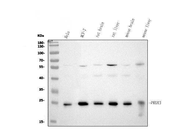
Western blot analysis of PRDX3 using anti-PRDX3 antibody (PA1836).
Electrophoresis was performed on a 5-20% SDS-PAGE gel at 70V (Stacking gel) / 90V (Resolving gel) for 2-3 hours. The sample well of each lane was loaded with 30 ug of sample under reducing conditions.
Lane 1: human Hela whole cell lysates,
Lane 2: human MCF-7 whole cell lysates,
Lane 3: rat brain tissue lysates,
Lane 4: rat liver tissue lysates,
Lane 5: mouse brain tissue lysates,
Lane 6: mouse liver tissue lysates.
After electrophoresis, proteins were transferred to a nitrocellulose membrane at 150 mA for 50-90 minutes. Blocked the membrane with 5% non-fat milk/TBS for 1.5 hour at RT. The membrane was incubated with rabbit anti-PRDX3 antigen affinity purified polyclonal antibody (Catalog # PA1836) at 0.5 μg/mL overnight at 4°C, then washed with TBS-0.1%Tween 3 times with 5 minutes each and probed with a goat anti-rabbit IgG-HRP secondary antibody at a dilution of 1:5000 for 1.5 hour at RT. The signal is developed using an Enhanced Chemiluminescent detection (ECL) kit (Catalog # EK1002) with Tanon 5200 system. A specific band was detected for PRDX3 at approximately 26 kDa. The expected band size for PRDX3 is at 28 kDa.

Click image to see more details
IHC analysis of PRDX3 using anti-PRDX3 antibody (PA1836).
PRDX3 was detected in a paraffin-embedded section of Rat Intestine tissue. Heat mediated antigen retrieval was performed in EDTA buffer (pH 8.0, epitope retrieval solution). The tissue section was blocked with 10% goat serum. The tissue section was then incubated with 1 μg/ml rabbit anti-PRDX3 Antibody (PA1836) overnight at 4°C. Peroxidase Conjugated Goat Anti-rabbit IgG was used as secondary antibody and incubated for 30 minutes at 37°C. The tissue section was developed using HRP Conjugated Rabbit IgG Super Vision Assay Kit (Catalog # SV0002) with DAB as the chromogen.

Click image to see more details
IHC analysis of PRDX3 using anti-PRDX3 antibody (PA1836).
PRDX3 was detected in a paraffin-embedded section of Human Mammary Cance tissue. Heat mediated antigen retrieval was performed in EDTA buffer (pH 8.0, epitope retrieval solution). The tissue section was blocked with 10% goat serum. The tissue section was then incubated with 1 μg/ml rabbit anti-PRDX3 Antibody (PA1836) overnight at 4°C. Peroxidase Conjugated Goat Anti-rabbit IgG was used as secondary antibody and incubated for 30 minutes at 37°C. The tissue section was developed using HRP Conjugated Rabbit IgG Super Vision Assay Kit (Catalog # SV0002) with DAB as the chromogen.

Click image to see more details
IF analysis of PRDX3 using anti-PRDX3 antibody (PA1836).
PRDX3 was detected in an immunocytochemical section of A549 cells. Enzyme antigen retrieval was performed using IHC enzyme antigen retrieval reagent (AR0022) for 15 mins. The cells were blocked with 10% goat serum. And then incubated with 5 μg/mL rabbit anti-PRDX3 Antibody (PA1836) overnight at 4°C. DyLight®488 Conjugated Goat Anti-Rabbit IgG (BA1127) was used as secondary antibody at 1:500 dilution and incubated for 30 minutes at 37°C. The section was counterstained with DAPI. Visualize using a fluorescence microscope and filter sets appropriate for the label used.

Click image to see more details
Flow Cytometry analysis of Hela cells using anti-PRDX3 antibody (PA1836).
Overlay histogram showing Hela cells stained with PA1836 (Blue line). To facilitate intracellular staining, cells were fixed with 4% paraformaldehyde and permeabilized with permeabilization buffer. The cells were blocked with 10% normal goat serum. And then incubated with rabbit anti-PRDX3 Antibody (PA1836, 1 μg/1x106 cells) for 30 min at 20°C. DyLight®488 conjugated goat anti-rabbit IgG (BA1127, 5-10 μg/1x106 cells) was used as secondary antibody for 30 minutes at 20°C. Isotype control antibody (Green line) was rabbit IgG (1 μg/1x106) used under the same conditions. Unlabelled sample (Red line) was also used as a control.
Specific Publications For Anti-Peroxiredoxin 3/PRDX3 Antibody Picoband® (PA1836)
Loading publications
Recommended Resources
Here are featured tools and databases that you might find useful.
- Boster's Pathways Library
- Protein Databases
- Bioscience Research Protocol Resources
- Data Processing & Analysis Software
- Photo Editing Software
- Scientific Literature Resources
- Research Paper Management Tools
- Molecular Biology Software
- Primer Design Tools
- Bioinformatics Tools
- Phylogenetic Tree Analysis
Customer Reviews
Have you used Anti-Peroxiredoxin 3/PRDX3 Antibody Picoband®?
Share your experimental results or join a short interview to earn up to $1,000 in product credits or other rewards.
0 Reviews For Anti-Peroxiredoxin 3/PRDX3 Antibody Picoband®
Customer Q&As
Have a question?
Find answers in Q&As, reviews.
Can't find your answer?
Submit your question
3 Customer Q&As for Anti-Peroxiredoxin 3/PRDX3 Antibody Picoband®
Question
We want to test anti-Peroxiredoxin 3/PRDX3 antibody PA1836 on mouse brain for research purposes, then I may be interested in using anti-Peroxiredoxin 3/PRDX3 antibody PA1836 for diagnostic purposes as well. Is the antibody suitable for diagnostic purposes?
Verified Customer
Verified customer
Asked: 2018-05-31
Answer
The products we sell, including anti-Peroxiredoxin 3/PRDX3 antibody PA1836, are only intended for research use. They would not be suitable for use in diagnostic work. If you have the means to develop a product into diagnostic use, and are interested in collaborating with us and develop our product into an IVD product, please contact us for more discussions.
Boster Scientific Support
Answered: 2018-05-31
Question
Is there a BSA free version of anti-Peroxiredoxin 3/PRDX3 antibody PA1836 available?
D. Wu
Verified customer
Asked: 2018-02-01
Answer
Thank you for your recent telephone inquiry. I can confirm that some lots of this anti-Peroxiredoxin 3/PRDX3 antibody PA1836 are BSA free. For now, these lots are available and we can make a BSA free formula for you free of charge. It will take 3 extra days to prepare. If you require this antibody BSA free again in future, please do not hesitate to contact me and I will be pleased to check which lots we have in stock that are BSA free.
Boster Scientific Support
Answered: 2018-02-01
Question
We appreciate helping with my inquiry over the phone. Here are the WB image, lot number and protocol we used for brain using anti-Peroxiredoxin 3/PRDX3 antibody PA1836. Let me know if you need anything else.
A. Jones
Verified customer
Asked: 2016-04-25
Answer
I appreciate the data. You have provided everything we needed. Our lab team are working to resolve your inquiry as quickly as possible, and we appreciate your patience and understanding! Please let me know if there is anything you need in the meantime.
Boster Scientific Support
Answered: 2016-04-25
