Click image to see more details
-

-
-
-
-
+5
Product Info Summary
| SKU: | A03625-1 |
|---|---|
| Size: | 0.1 mg |
| Reactive Species: | Human, Mouse, Rat |
| Host: | Rabbit |
| Application: | ELISA, IF, ICC, WB |
Customers Who Bought This Also Bought

Product info
Product Name
Anti-PHAP I ANP32A Antibody
SKU/Catalog Number
A03625-1
Size
0.1 mg
Form
Liquid
Description
Boster Bio Anti-PHAP I ANP32A Antibody (Catalog # A03625-1). Tested in ELISA, WB, ICC, IF applications. This antibody reacts with Human, Mouse, Rat.
Storage & Handling
PHAP I antibody can be stored at 4°C for three months and -20°C, stable for up to one year. Avoid repeated freeze-thaw cycles. Antibodies should not be exposed to prolonged high temperatures.
Cite This Product
Anti-PHAP I ANP32A Antibody (Boster Biological Technology, Pleasanton CA, USA, Catalog # A03625-1)
Host
Rabbit
Contents
PHAP I Antibody is supplied in PBS containing 0.02% sodium azide.
Clonality
Polyclonal
Isotype
IgG
Immunogen
Anti-PHAP I antibody was raised against a peptide corresponding to 15 amino acids near the carboxy terminus of human PHAP I. The immunogen is located within the last 50 amino acids of PHAP I.
Cross-reactivity
This polyclonal antibody has no cross-reaction to PHAP I2a and PHAP III.
Reactive Species
A03625-1 is reactive to ANP32A in Human, Mouse, Rat
Observed Molecular Weight
68 kDa
Calculated molecular weight
28.6 kDa
Background of ANP32A
Apoptosis is related to many diseases and development. Caspase-9 plays a central role in cell death induced by a variety of apoptosis activators. Cytochrome c, after released from mitochondria, binds to Apaf-1, which forms an apoptosome that in turn binds to and activate procaspase-9. Activated caspase-9 cleaves and activates the effector caspases (caspase-3, -6 and -7), which are responsible for the proteolytic cleavage of many key proteins in apoptosis. The tumor suppressor putative HLA-DR-associated proteins (PHAPs) were recently identified as important regulators of mitochondrion apoptosis. PHAP appears to facilitate apoptosome-medicated caspase-9 activation and to stimulate the mitochondrial apoptotic pathway. PHAP was also shown to oppose both Ras- and Myc-medicated cell transformation.
Antibody Validation
Boster validates all antibodies on WB, IHC, ICC, Immunofluorescence, and ELISA with known positive control and negative samples to ensure specificity and high affinity, including thorough antibody incubations.
Application & Images
Applications
A03625-1 is guaranteed for ELISA, IF, ICC, WB Boster Guarantee
Recommend Dilution
| Application | Dilution | Species |
|---|---|---|
| Antibody validated: Western Blot in human | mouse and rat samples; Immunocytochemistry in human samples; Immunofluorescence in human samples. All other applications and species not yet tested. Optimal dilutions for each application should be determined by the researcher. |
Validation Images & Assay Conditions
Click image to see more details
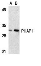
Western Blot Validation in Human Raji Cell Lysate
Loading: 15 μg of lysates per lane.
Antibodies: PHAP I A03625-1 (A: 2 μg/mL, B: 4 μg/mL), 1h incubation at RT in 5% NFDM/TBST.
Secondary: Goat anti-rabbit IgG HRP conjugate at 1:10000 dilution.

Click image to see more details
Independent Antibody Validation (IAV) via Protein Expression Profile in Cell Lines
Loading: 15 μg of lysates per lane.
Antibodies: PHAP I A03625-1 (2 μg/mL), PHAP I 3151 (1 μg/mL), and beta-actin (1 μg/mL), 1h incubation at RT in 5% NFDM/TBST.
Secondary: Goat anti-rabbit IgG HRP conjugate at 1:10000 dilution.

Click image to see more details
Western Blot Validation in Human Cell Lines
Loading: 15 μg of lysates per lane.
Antibodies: PHAP I A03625-1 (2 μg/mL), 1h incubation at RT in 5% NFDM/TBST.
Secondary: Goat anti-rabbit IgG HRP conjugate at 1:10000 dilution.

Click image to see more details
Immunofluorescence Validation of PHAP I in Raji Cells
Immunofluorescent analysis of 4% paraformaldehyde-fixed Raji Cells labeling PHAP I with A03625-1 at 10 μg/mL, followed by goat anti-rabbit IgG secondary antibody at 1/500 dilution (red).

Click image to see more details
Immunocytochemistry Validation of PHAP I in Raji Cells
Immunocytochemical analysis of Raji cells using anti-PHAP I antibody (A03625-1) at 2 μg/ml. Cells was fixed with formaldehyde and blocked with 10% serum for 1 h at RT; antigen retrieval was by heat mediation with a citrate buffer (pH6). Samples were incubated with primary antibody overnight at 4˚C. A goat anti-rabbit IgG H&L (HRP) at 1/250 was used as secondary. Counter stained with Hematoxylin.

Click image to see more details
KD Validation of PHAPI in Human Breast Cancer Cells (Schafer et al., 2006)
Human Breast Cancer Cells (T47D cells) were transfected with control or PHAPI siRNA duplex. PHAPI was detected via Western Blot analysis by using the anti-PHAPI antibody. PHAPI expression was reduced after PHAPI siRNA knockdown.

Click image to see more details
Increased Expression Validation of PHAPI in Patient Samples of Breast
Tumor Tissue (Schafer et al., 2006)
PHAPI was overexpressed in all breast tumor samples of patients and human breast cancer cells (MDA-MB-453), but not in the normal breast tissue or human primary mammary epithelial cells (HMEC).

Click image to see more details
Induced Expression Validation of PHAPI/Anp32a in Atxn1 KO Mice (Sa′nchez et al., 2013)
Western blot analysis of PHAPI/Anp32a from the cerebellum of WT and Atxn1 KO mice. PHAPI expression was significantly increased (2 folds) in Atxn1 KO mice as compared to WT mice. The same effect was observed in PHAPI mRNA levels.

Click image to see more details
Overexpression of PHAPI in Breast Cancer Cells (Schafer et al., 2006)
Western blot analysis with anti-PHAPI antibodies was performed for PHAPI in human cell lines from breast, prostate and lung. PHAPI was overexpressed in breast cancer cells when compared with normal cells (HMEC) whereas there were no significant differences in PHAPI expression in normal and cancer cells of either prostate or lung origin.
Specific Publications For Anti-PHAP I ANP32A Antibody (A03625-1)
Loading publications
Recommended Resources
Here are featured tools and databases that you might find useful.
- Boster's Pathways Library
- Protein Databases
- Bioscience Research Protocol Resources
- Data Processing & Analysis Software
- Photo Editing Software
- Scientific Literature Resources
- Research Paper Management Tools
- Molecular Biology Software
- Primer Design Tools
- Bioinformatics Tools
- Phylogenetic Tree Analysis
Customer Reviews
Have you used Anti-PHAP I ANP32A Antibody?
Share your experimental results or join a short interview to earn up to $1,000 in product credits or other rewards.
0 Reviews For Anti-PHAP I ANP32A Antibody
Customer Q&As
Have a question?
Find answers in Q&As, reviews.
Can't find your answer?
Submit your question
