Click image to see more details
Product Info Summary
| SKU: | P03794 |
|---|---|
| Size: | 100 μl |
| Reactive Species: | Human, Mouse, Rat |
| Host: | Rabbit |
| Application: | IHC, WB |
Customers Who Bought This Also Bought

Product info
Product Name
Anti-Phospho-Synapsin I (S9) Rabbit Monoclonal Antibody
SKU/Catalog Number
P03794
BM4501 is an alternative SKU for this antibody, used in previous lots.
Size
100 μl
Form
Liquid
Description
Boster Bio Anti-Phospho-Synapsin I (S9) Rabbit Monoclonal Antibody catalog # P03794. Tested in WB, IHC applications. This antibody reacts with Human, Mouse, Rat.
Storage & Handling
Store at -20°C for one year. For short term storage and frequent use, store at 4°C for up to one month. Avoid repeated freeze-thaw cycles.
Cite This Product
Anti-Phospho-Synapsin I (S9) Rabbit Monoclonal Antibody (Boster Biological Technology, Pleasanton CA, USA, Catalog # P03794)
Host
Rabbit
Contents
Rabbit IgG in stabilizing components, phosphate buffered saline, pH 7.4, 150mM NaCl, 0.02% sodium azide and 50% glycerol.
*This antibody is supplied in a stabilized formulation.
Compatibility with conjugation reactions depends on the chemistry of the conjugation method used.
For conjugation methods that are not compatible with the stabilizing components present in this formulation, a carrier-free antibody format is required.
Clonality
Monoclonal
Clone Number
FCE-19
Isotype
Rabbit IgG
Immunogen
A synthesized peptide derived from human Phospho-Synapsin I (S9)
Reactive Species
P03794 is reactive to SYN1 in Human, Mouse, Rat
Observed Molecular Weight
77 kDa
Calculated molecular weight
74.1 kDa
Antibody Validation
Boster validates all antibodies on WB, IHC, ICC, Immunofluorescence, and ELISA with known positive control and negative samples to ensure specificity and high affinity, including thorough antibody incubations.
Application & Images
Applications
P03794 is guaranteed for IHC, WB Boster Guarantee
Recommend Dilution
WB 1:500-2000
IHC 1:50-200
Tested application
Suggested blocking solution with 5% non-fat milk or BSA; (*)Recommended protein loading: 20-40 µg per lane
Validation Images & Assay Conditions
Click image to see more details
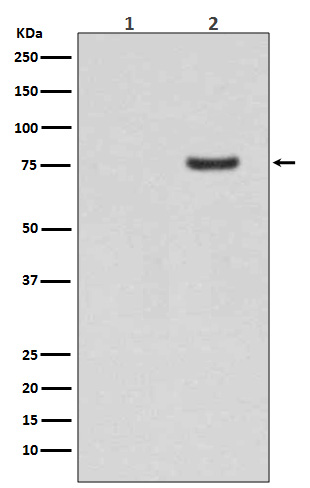
Western blot analysis of Phospho-Synapsin I (S9) expression in (1) Human brain lysate; (2) Human brain lysate treated with AP.
Specific Publications For Anti-Phospho-Synapsin I (S9) Rabbit Monoclonal Antibody (P03794)
Loading publications
Recommended Resources
Here are featured tools and databases that you might find useful.
- Boster's Pathways Library
- Protein Databases
- Bioscience Research Protocol Resources
- Data Processing & Analysis Software
- Photo Editing Software
- Scientific Literature Resources
- Research Paper Management Tools
- Molecular Biology Software
- Primer Design Tools
- Bioinformatics Tools
- Phylogenetic Tree Analysis
Customer Reviews
Have you used Anti-Phospho-Synapsin I (S9) Rabbit Monoclonal Antibody?
Share your experimental results or join a short interview to earn up to $1,000 in product credits or other rewards.
0 Reviews For Anti-Phospho-Synapsin I (S9) Rabbit Monoclonal Antibody
Customer Q&As
Have a question?
Find answers in Q&As, reviews.
Can't find your answer?
Submit your question
3 Customer Q&As for Anti-Phospho-Synapsin I (S9) Rabbit Monoclonal Antibody
Question
We are currently using anti-Phospho-Synapsin I (S9) Rabbit Monoclonal antibody P03794 for mouse tissue, and we are satisfied with the WB results. The species of reactivity given in the datasheet says human, mouse, rat. Is it possible that the antibody can work on feline tissues as well?
Verified Customer
Verified customer
Asked: 2019-08-09
Answer
The anti-Phospho-Synapsin I (S9) Rabbit Monoclonal antibody (P03794) has not been tested for cross reactivity specifically with feline tissues, though there is a good chance of cross reactivity. We have an innovator award program that if you test this antibody and show it works in feline you can get your next antibody for free. Please contact me if I can help you with anything.
Boster Scientific Support
Answered: 2019-08-09
Question
We have observed staining in mouse brain. Are there any suggestions? Is anti-Phospho-Synapsin I (S9) Rabbit Monoclonal antibody supposed to stain brain positively?
Verified Customer
Verified customer
Asked: 2019-07-12
Answer
Based on literature brain does express SYN1. Based on Uniprot.org, SYN1 is expressed in anterior cingulate cortex, brain, brain cortex, among other tissues. Regarding which tissues have SYN1 expression, here are a few articles citing expression in various tissues:
Brain, Pubmed ID: 2110562, 15772651
Brain cortex, Pubmed ID: 15822905
Boster Scientific Support
Answered: 2019-07-12
Question
We were happy with the WB result of your anti-Phospho-Synapsin I (S9) Rabbit Monoclonal antibody. However we have been able to see positive staining in brain cortex cell junction using this antibody. Is that expected? Could you tell me where is SYN1 supposed to be expressed?
Verified Customer
Verified customer
Asked: 2019-06-18
Answer
From what I have seen in literature, brain cortex does express SYN1. Generally SYN1 expresses in cell junction, synapse. golgi apparatus. Regarding which tissues have SYN1 expression, here are a few articles citing expression in various tissues:
Brain, Pubmed ID: 2110562, 15772651
Brain cortex, Pubmed ID: 15822905
Boster Scientific Support
Answered: 2019-06-18
