Click image to see more details
-

-
-
-
-
+8
Product Info Summary
| SKU: | M01091-1 |
|---|---|
| Size: | 100 μl/vial |
| Reactive Species: | Human, Mouse, Rat |
| Host: | Rabbit |
| Application: | IF, IHC, ICC, WB |
Customers Who Bought This Also Bought

Product info
Product Name
Anti-PI3 Kinase p110 beta Rabbit Monoclonal Antibody
SKU/Catalog Number
M01091-1
Size
100 μl/vial
Form
Liquid
Description
Boster Bio Anti-PI3 Kinase p110 beta Rabbit Monoclonal Antibody catalog # M01091-1. Tested in WB, IHC, ICC/IF applications. This antibody reacts with Human, Mouse, Rat.
Storage & Handling
Store at -20°C for one year. For short term storage and frequent use, store at 4°C for up to one month. Avoid repeated freeze-thaw cycles.
Cite This Product
Anti-PI3 Kinase p110 beta Rabbit Monoclonal Antibody (Boster Biological Technology, Pleasanton CA, USA, Catalog # M01091-1)
Host
Rabbit
Contents
Rabbit IgG in stabilizing components, phosphate buffered saline, pH 7.4, 150mM NaCl, 0.02% sodium azide and 50% glycerol.
*This antibody is supplied in a stabilized formulation.
Compatibility with conjugation reactions depends on the chemistry of the conjugation method used.
For conjugation methods that are not compatible with the stabilizing components present in this formulation, a carrier-free antibody format is required.
Clonality
Monoclonal
Clone Number
30P53
Isotype
IgG
Immunogen
A synthesized peptide derived from human PI3 Kinase p110 beta
Reactive Species
M01091-1 is reactive to PIK3CB in Human, Mouse, Rat
Observed Molecular Weight
123 kDa
Calculated molecular weight
122.8 kDa
Antibody Validation
Boster validates all antibodies on WB, IHC, ICC, Immunofluorescence, and ELISA with known positive control and negative samples to ensure specificity and high affinity, including thorough antibody incubations.
Application & Images
Applications
M01091-1 is guaranteed for IF, IHC, ICC, WB Boster Guarantee
Recommend Dilution
WB 1:500-2000
IHC 1:50-200
ICC/IF 1:50-200
Tested application
Use TE buffer pH 9.0 for antigen retrieval; (*) citrate buffer pH 6.0 is an alternative.
Validation Images & Assay Conditions
Click image to see more details
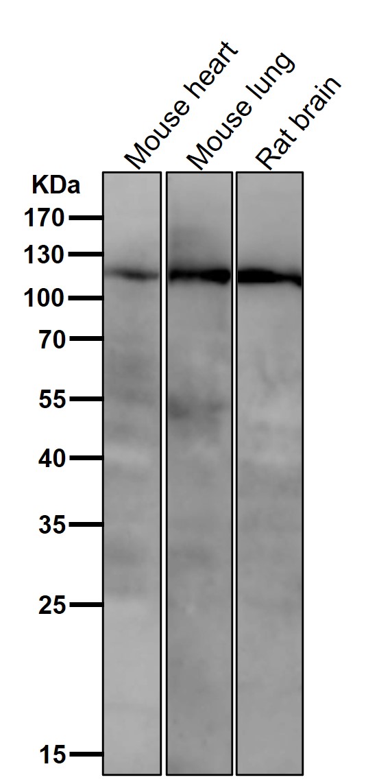
All lanes use the Antibody at 1:3K dilution for 1 hour at room temperature.

Click image to see more details
BECCs regulate PI3K, PDPK1, and mTOR protein levels in HAPH rats. (A–C) Primitive bands of p-PI3K, PI3K, p-PDPK1, PDPK1, p-mTOR, and mTOR by Western blots in lung tissues. (D–F) Quantitative evaluation of p-PI3K, PI3K, p-PDPK1, PDPK1, p-mTOR, and mTOR in lung tissues. n = 5. All data are represented as the mean ± SD. * p < 0.05 vs. control group and # p < 0.05 vs. hypoxia group.
Index in PubMed under a CC BY license. PMID: 40385484

Click image to see more details
Flavonoids inhibit the proliferation of PASMCs under hypoxic conditions by inhibiting the PI3K/AKT axis. (A, B) Primitive bands and quantitative densities of p-AKT1 Ser473 and AKT1 with or without Sc79 (20 μM) by Western blots in PASMCs under 3% O 2 . (C, D) Primitive bands and quantitative densities of p-PI3K, PI3K, p-AKT1 Ser473, and AKT1 with or without 740Y-P (10 μM) by Western blots in PASMCs under 3% O 2 . (E, F) Primitive bands and quantitative densities of p-PDPK1, PDPK, p-AKT1 Ser473, and AKT1 with or without MYH1485 (2 μM) in PASMCs by Western blots under 3% O 2 . n = 3. All data are represented as the mean ± SD. * p < 0.05 vs. control group, # p < 0.05 vs. 3% O 2 group, and p < 0.05 vs. 3% O 2 + Fla-50 μg/ml group.
Index in PubMed under a CC BY license. PMID: 40385484

Click image to see more details
Eriocitrin and quercetin are responsible for anti-proliferation by targeting the PI3K protein in PASMCs under hypoxic conditions. ERI, eriocitrin; QCT, quercetin. (A, B) Primitive bands and quantitative evaluation of p-mTOR, mTOR, p-AKT1 (Ser473), and AKT1 with or without PS210 (2 μM) by Western blotting in PASMCs under 3% O 2 . n = 3. All data are represented as the mean ± SD. * p < 0.05 vs. control group, # p < 0.05 vs. 3% O 2 group, and p < 0.05 vs. 3% O 2 + FLA-50 μg/ml group. (C–G) Primitive bands and quantitative densities of p-PI3K and PI3K by Western blots. n = 3. All data are represented as the mean ± SD. * p < 0.05 vs. control group and # p < 0.05 vs. 3% O 2 group. (H–N) BECC, ERI, and QCT treatment increased the stability of PI3K in PASMC protease lysates by the DARTS experiment. (H–K) Primitive Western blots of PI3K. (L–N) Quantitative evaluation of PI3K levels. n = 3. All data are represented as the mean ± SD. * p < 0.05 vs. DMSO group.
Index in PubMed under a CC BY license. PMID: 40385484

Click image to see more details
Treatment with eriocitrin in PASMC proliferation and antioxidation under hypoxic conditions. (A, B) Eriocitrin in cell proliferation were assessed using Ki67 immunofluorescence and quantitative evaluation in hypoxia-induced PASMCs (n = 3, scale bar = 100 μm). (C–H) Primitive Western blots and quantitative densities of PCNA, p-PI3K, PI3K, p-AKT1 (Ser473), AKT1 with or without 740Y-P (10 μM), LY294002 (10 μM), or eriocitrin (11 μM) in PASMCs under 3% O 2 for 24 h (I–L) Quantitative evaluation of SOD and GSH-Px activities and GSH and MDA contents in 3% O 2 -induced PASMCs. n = 3. All data are represented as the mean ± SD. * p < 0.05 vs. control group, # p < 0.05 vs. 3% O 2 group, and p < 0.05 vs. 3% O 2 + ERI-11 μM.
Index in PubMed under a CC BY license. PMID: 40385484

Click image to see more details
Treatment with quercetin in PASMC proliferation and antioxidation under hypoxia. (A, B) Quercetin in cell proliferation were assessed using Ki67 immunofluorescence and quantitative evaluation in hypoxia-induced PASMCs (n = 3, scale bar = 100 μm). (C–H) Primitive Western blots and quantitative densities of PCNA, p-PI3K, PI3K, p-AKT1 Ser473, AKT1 with or without 740Y-P(10 μM), LY294002(10 μM), or quercetin (18 μM) in PASMCs under 3% O 2 for 24 h (I–L) Quantitative evaluation of SOD and GSH-Px activities and GSH and MDA contents in 3% O 2 -induced PASMCs. n = 3. All data represent mean ± SD. * p < 0.05 vs. control group, # p < 0.05 vs. 3% O 2 group, and p < 0.05 vs. 3% O 2 + QCT-18 μM.
Index in PubMed under a CC BY license. PMID: 40385484

Click image to see more details
All lanes use the Antibody at 1:3K dilution for 1 hour at room temperature.

Click image to see more details
Western blot analysis of PI3 Kinase p110 beta expression in (1) 293 cell lysate; (2) Mouse Brain lysate.

Click image to see more details
IHC analysis of PI3 Kinase p110 Beta/PIK3CB using anti-PI3 Kinase p110 Beta/PIK3CB antibody (M01091-1).
PI3 Kinase p110 Beta/PIK3CB was detected in a paraffin-embedded section of human colon tissue. Heat mediated antigen retrieval was performed in EDTA buffer (pH 8.0, epitope retrieval solution). The tissue section was blocked with 10% goat serum. The tissue section was then incubated with 1: 50 rabbit anti-PI3 Kinase p110 Beta/PIK3CB Antibody (M01091-1) overnight at 4°C. Peroxidase Conjugated Goat Anti-rabbit IgG was used as secondary antibody and incubated for 30 minutes at 37°C. The tissue section was developed using HRP Conjugated Rabbit IgG Super Vision Assay Kit (Catalog # SV0002) with DAB as the chromogen.

Click image to see more details
IHC analysis of PI3 Kinase p110 Beta/PIK3CB using anti-PI3 Kinase p110 Beta/PIK3CB antibody (M01091-1).
PI3 Kinase p110 Beta/PIK3CB was detected in a paraffin-embedded section of mouse brain tissue. Heat mediated antigen retrieval was performed in EDTA buffer (pH 8.0, epitope retrieval solution). The tissue section was blocked with 10% goat serum. The tissue section was then incubated with 1: 50 rabbit anti-PI3 Kinase p110 Beta/PIK3CB Antibody (M01091-1) overnight at 4°C. Peroxidase Conjugated Goat Anti-rabbit IgG was used as secondary antibody and incubated for 30 minutes at 37°C. The tissue section was developed using HRP Conjugated Rabbit IgG Super Vision Assay Kit (Catalog # SV0002) with DAB as the chromogen.

Click image to see more details
IHC analysis of PI3 Kinase p110 Beta/PIK3CB using anti-PI3 Kinase p110 Beta/PIK3CB antibody (M01091-1).
PI3 Kinase p110 Beta/PIK3CB was detected in a paraffin-embedded section of rat brain tissue. Heat mediated antigen retrieval was performed in EDTA buffer (pH 8.0, epitope retrieval solution). The tissue section was blocked with 10% goat serum. The tissue section was then incubated with 1: 50 rabbit anti-PI3 Kinase p110 Beta/PIK3CB Antibody (M01091-1) overnight at 4°C. Peroxidase Conjugated Goat Anti-rabbit IgG was used as secondary antibody and incubated for 30 minutes at 37°C. The tissue section was developed using HRP Conjugated Rabbit IgG Super Vision Assay Kit (Catalog # SV0002) with DAB as the chromogen.

Click image to see more details
IHC analysis of PI3 Kinase p110 Beta/PIK3CB using anti-PI3 Kinase p110 Beta/PIK3CB antibody (M01091-1).
PI3 Kinase p110 Beta/PIK3CB was detected in a paraffin-embedded section of human colon cancer tissue. Heat mediated antigen retrieval was performed in EDTA buffer (pH 8.0, epitope retrieval solution). The tissue section was blocked with 10% goat serum. The tissue section was then incubated with 1: 50 rabbit anti-PI3 Kinase p110 Beta/PIK3CB Antibody (M01091-1) overnight at 4°C. Peroxidase Conjugated Goat Anti-rabbit IgG was used as secondary antibody and incubated for 30 minutes at 37°C. The tissue section was developed using HRP Conjugated Rabbit IgG Super Vision Assay Kit (Catalog # SV0002) with DAB as the chromogen.
Specific Publications For Anti-PI3 Kinase p110 beta Rabbit Monoclonal Antibody (M01091-1)
Loading publications
Recommended Resources
Here are featured tools and databases that you might find useful.
- Boster's Pathways Library
- Protein Databases
- Bioscience Research Protocol Resources
- Data Processing & Analysis Software
- Photo Editing Software
- Scientific Literature Resources
- Research Paper Management Tools
- Molecular Biology Software
- Primer Design Tools
- Bioinformatics Tools
- Phylogenetic Tree Analysis
Customer Reviews
Have you used Anti-PI3 Kinase p110 beta Rabbit Monoclonal Antibody?
Share your experimental results or join a short interview to earn up to $1,000 in product credits or other rewards.
0 Reviews For Anti-PI3 Kinase p110 beta Rabbit Monoclonal Antibody
Customer Q&As
Have a question?
Find answers in Q&As, reviews.
Can't find your answer?
Submit your question
