Click image to see more details
-

-
-
-
-
+1
Product Info Summary
| SKU: | A00449-3 |
|---|---|
| Size: | 100 μg/vial |
| Reactive Species: | Human |
| Host: | Rabbit |
| Application: | ELISA, Flow Cytometry, IF, ICC, WB |
Customers Who Bought This Also Bought

Product info
Product Name
Anti-PPAR gamma/PPARG Antibody Picoband®
SKU/Catalog Number
A00449-3
Size
100 μg/vial
Form
Lyophilized
Description
Boster Bio Anti-PPAR gamma/PPARG Antibody Picoband® catalog # A00449-3. Tested in ELISA, IF, ICC, Flow Cytometry, WB applications. This antibody reacts with Human. The brand Picoband indicates this is a premium antibody that guarantees superior quality, high affinity, and strong signals with minimal background in Western blot applications. Only our best-performing antibodies are designated as Picoband, ensuring unmatched performance.
Storage & Handling
At -20°C for one year from date of receipt. After reconstitution, at 4°C for one month. It can also be aliquotted and stored frozen at -20°C for six months. Avoid repeated freezing and thawing.
Cite This Product
Anti-PPAR gamma/PPARG Antibody Picoband® (Boster Biological Technology, Pleasanton CA, USA, Catalog # A00449-3)
Host
Rabbit
Contents
Each vial contains 4 mg Trehalose, 0.9 mg NaCl, 0.2 mg Na2HPO4.
Clonality
Polyclonal
Isotype
Rabbit IgG
Immunogen
E.coli-derived human PPAR gamma/PPARG recombinant protein (Position: H58-K329).
Cross-reactivity
No cross-reactivity with other proteins.
Reactive Species
A00449-3 is reactive to PPARG in Human
Observed Molecular Weight
65 kDa
Calculated molecular weight
57.6 kDa
Background of PPARG
Peroxisome proliferator- activated receptor gamma (PPAR-γ or PPARG), also known as the glitazone reverse insulin resistance receptor, or NR1C3 (nuclear receptor subfamily 1, group C, member 3) is a type II nuclear receptor (protein regulating genes) that in humans is encoded by the PPARG gene. This gene encodes a member of the peroxisome proliferator-activated receptor (PPAR) subfamily of nuclear receptors. PPARs form heterodimers with retinoid X receptors (RXRs) and these heterodimers regulate transcription of various genes. Three subtypes of PPARs are known: PPAR-alpha, PPAR-delta, and PPAR-gamma. The protein encoded by this gene is PPAR-gamma and is a regulator of adipocyte differentiation. Additionally, PPAR-gamma has been implicated in the pathology of numerous diseases including obesity, diabetes, atherosclerosis and cancer. Alternatively spliced transcript variants that encode different isoforms have been described.
Antibody Validation
Boster validates all antibodies on WB, IHC, ICC, Immunofluorescence, and ELISA with known positive control and negative samples to ensure specificity and high affinity, including thorough antibody incubations.
Application & Images
Applications
A00449-3 is guaranteed for ELISA, Flow Cytometry, IF, ICC, WB Boster Guarantee
Recommend Dilution
| Application | Dilution | Species |
|---|---|---|
| Western blot | 0.25-0.5 μg/ml | Human |
| Immunocytochemistry/Immunofluorescence | 5 μg/ml | Human |
| Flow Cytometry (Fixed) | 1-3 μg/1x106 cells | Human |
| ELISA | 0.1-0.5 μg/ml | - |
Tested application
Suggested blocking solution with 5% non-fat milk or BSA; (*)Recommended protein loading: 20-40 µg per lane
Validation Images & Assay Conditions
Click image to see more details
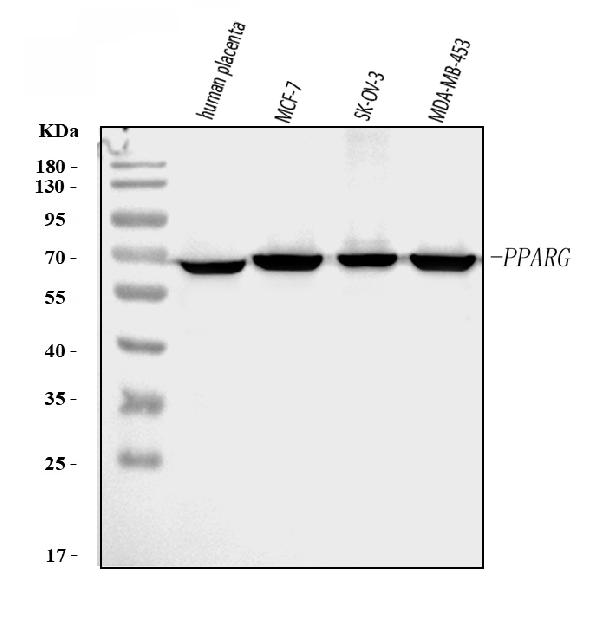
Western blot analysis of PPAR Gamma/PPARG using anti-PPAR Gamma/PPARG antibody (A00449-3).
Electrophoresis was performed on a 5-20% SDS-PAGE gel at 70V (Stacking gel) / 90V (Resolving gel) for 2-3 hours. The sample well of each lane was loaded with 30 ug of sample under reducing conditions.
Lane 1: human placenta tissue lysates,
Lane 2: human MCF-7 whole cell lysates,
Lane 3: human SK-OV-3 whole cell lysates,
Lane 4: human MDA-MB-453 whole cell lysates.
After electrophoresis, proteins were transferred to a nitrocellulose membrane at 150 mA for 50-90 minutes. Blocked the membrane with 5% non-fat milk/TBS for 1.5 hour at RT. The membrane was incubated with rabbit anti-PPAR Gamma/PPARG antigen affinity purified polyclonal antibody (Catalog # A00449-3) at 0.5 μg/mL overnight at 4°C, then washed with TBS-0.1%Tween 3 times with 5 minutes each and probed with a goat anti-rabbit IgG-HRP secondary antibody at a dilution of 1:5000 for 1.5 hour at RT. The signal is developed using an Enhanced Chemiluminescent detection (ECL) kit (Catalog # EK1002) with Tanon 5200 system. A specific band was detected for PPAR Gamma/PPARG at approximately 65 kDa. The expected band size for PPAR Gamma/PPARG is at 65 kDa.

Click image to see more details
ANXA1 mediated PPARγ-CEBPα pathway to regulate osteoclast differentiation (A) The mRNA level of PPARγ in RAW264.7 cultured with siANXA1-EVs. (B) The mRNA level of CEBPα in RAW264.7 cultured with siANXA1-EVs. (C) The protein level of PPARγ and CEBPα in RAW264.7 cultured with siANXA1-EVs. (D) Quantitative analysis of PPARγ protein expression. (E) Quantitative analysis of CEBPα protein expression. (F) Schematic illustration of PPARγ inhibited RAW264.7 and DFSC-EVs co-culture system. (G) Representative images of TRAP staining. Scale bar = 200 μm. (H) Quantitative analysis of TRAP-positive area. (I) PPARγ inhibited RAW264.7 construction. (J) The mRNA level of CEBPα in PPARγ inhibited RAW264.7. (K) The protein level of PPARγ and CEBPα in PPARγ inhibited RAW264.7. (L) Quantitative analysis of PPARγ protein expression. (M) Quantitative analysis of CEBPα protein expression. (N) The mRNA level of ACP5 , CTSK and CFOS in PPARγ inhibited RAW264.7. (O) The protein level of ACP5, CTSK and CFOS in PPARγ inhibited RAW264.7. (P) Western blotting quantification. * p < 0.05, ** p < 0.01, *** p < 0.001, **** p < 0.0001. n = 3.
Index in PubMed under a CC BY license. PMID: 39834384

Click image to see more details
DFSCs-EVs/ANXA1 regulating tooth eruption by affecting osteoclast differentiation. (A) Representative micro-CT images of detecting tooth eruption distance. (B) Analysis of tooth eruption distance based on micro-CT. (C) Representative H&E staining images of the first mandibular molar area. (D) Analysis of tooth eruption distance based on H&E staining. (E) Representative images of TRAP staining. (F) Quantitative analysis of TRAP-positive area. (G) Representative immunohistochemistry staining images of PPARγ expression in the first mandibular molar area. (H) Quantitative analysis of PPARγ expression in the first mandibular molar area. (I) Representative immunohistochemistry staining images of CEBPα expression in the first mandibular molar area. (J) Quantitative analysis of CEBPα expression in the first mandibular molar area. ns, not significant. Scale bar = 1 mm ** p < 0. 01. n = 3.
Index in PubMed under a CC BY license. PMID: 39834384

Click image to see more details
IF analysis of PPAR gamma/PPARG using anti-PPAR gamma/PPARG antibody (A00449-3).
PPAR gamma/PPARGNOX4 was detected in an immunocytochemical section of A431 cells. Enzyme antigen retrieval was performed using IHC enzyme antigen retrieval reagent (AR0022) for 15 mins. The cells were blocked with 10% goat serum. And then incubated with 5 μg/mL rabbit anti-PPAR gamma/PPARG Antibody (A00449-3) overnight at 4°C. DyLight488 Conjugated Goat Anti-Rabbit IgG (BA1127) was used as secondary antibody at 1:500 dilution and incubated for 30 minutes at 37°C. The section was counterstained with DAPI. Visualize using a fluorescence microscope and filter sets appropriate for the label used.

Click image to see more details
Flow Cytometry analysis of A431 cells using anti-PPAR Gamma/PPARG antibody (A00449-3).
Overlay histogram showing A431 cells stained with A00449-3 (Blue line). To facilitate intracellular staining, cells were fixed with 4% paraformaldehyde and permeabilized with permeabilization buffer. The cells were blocked with 10% normal goat serum. And then incubated with rabbit anti-PPAR Gamma/PPARG Antibody (A00449-3, 1 μg/1x106 cells) for 30 min at 20°C. DyLight®488 conjugated goat anti-rabbit IgG (BA1127, 5-10 μg/1x106 cells) was used as secondary antibody for 30 minutes at 20°C. Isotype control antibody (Green line) was rabbit IgG (1 μg/1x106) used under the same conditions. Unlabelled sample without incubation with primary antibody and secondary antibody (Red line) was used as a blank control.
Specific Publications For Anti-PPAR gamma/PPARG Antibody Picoband® (A00449-3)
Loading publications
Recommended Resources
Here are featured tools and databases that you might find useful.
- Boster's Pathways Library
- Protein Databases
- Bioscience Research Protocol Resources
- Data Processing & Analysis Software
- Photo Editing Software
- Scientific Literature Resources
- Research Paper Management Tools
- Molecular Biology Software
- Primer Design Tools
- Bioinformatics Tools
- Phylogenetic Tree Analysis
Customer Reviews
Have you used Anti-PPAR gamma/PPARG Antibody Picoband®?
Share your experimental results or join a short interview to earn up to $1,000 in product credits or other rewards.
0 Reviews For Anti-PPAR gamma/PPARG Antibody Picoband®
Customer Q&As
Have a question?
Find answers in Q&As, reviews.
Can't find your answer?
Submit your question
