Click image to see more details
-

-
-
-
-
+2
Product Info Summary
| SKU: | A09399 |
|---|---|
| Size: | 0.1 mg |
| Reactive Species: | Human, Mouse, Rat |
| Host: | Rabbit |
| Application: | ELISA, IHC-P, ICC, WB |
Customers Who Bought This Also Bought

Product info
Product Name
Anti-Protein orai-3 ORAI3 Antibody
SKU/Catalog Number
A09399
Size
0.1 mg
Form
Liquid
Description
Boster Bio Anti-Protein orai-3 ORAI3 Antibody (Catalog # A09399). Tested in ELISA, WB, ICC, IHC-P applications. This antibody reacts with Human, Mouse, Rat.
Storage & Handling
ORAI3 antibody can be stored at 4°C for three months and -20°C, stable for up to one year. Avoid repeated freeze-thaw cycles. Antibodies should not be exposed to prolonged high temperatures.
Cite This Product
Anti-Protein orai-3 ORAI3 Antibody (Boster Biological Technology, Pleasanton CA, USA, Catalog # A09399)
Host
Rabbit
Contents
ORAI3 Antibody is supplied in PBS containing 0.02% sodium azide.
Clonality
Polyclonal
Isotype
IgG
Immunogen
ORAI3 antibody was raised against a 15 amino acid synthetic peptide from near the amino terminus of human ORAI3. The immunogen is located within the first 50 amino acids of ORAI3.
Cross-reactivity
This antibody is predicted to have no cross-reactivity to ORAI1 or ORAI2.
Reactive Species
A09399 is reactive to ORAI3 in Human, Mouse, Rat
Observed Molecular Weight
68 kDa
Calculated molecular weight
31.5 kDa
Background of ORAI3
Antigen stimulation of immune cells triggers Ca++ entry through Ca++ release-activated Ca++ (CRAC) channels. ORAI3 is one of two mammalian homologs to ORAI1, a recently identified four-transmembrane spanning protein that is an essential component of CRAC. All three homologs have been shown to function as Ca++ plasma membrane channels gated through interactions with STIM1, the store-activated endoplasmic reticulum Ca++ sensor. However, ORAI3 channels failed to produce detectable Ca++ selective currents in cells co-transfected with ORAI3 and STIM1, indicating that ORAI3 channels undergo a lesser degree of depotentiation than ORAI1 or ORAI2. Na+ currents through ORAI1, 2 and 3 channels were equally inhibited by extracellular Ca++, indicating that each have similar affinities for Ca++ within the selectivity filter.
Antibody Validation
Boster validates all antibodies on WB, IHC, ICC, Immunofluorescence, and ELISA with known positive control and negative samples to ensure specificity and high affinity, including thorough antibody incubations.
Application & Images
Applications
A09399 is guaranteed for ELISA, IHC-P, ICC, WB Boster Guarantee
Recommend Dilution
ORAI3 antibody can be used for detection of ORAI3 by Western blot at 1 - 4 μg/mL. Antibody can also be used for immunocytochemistry starting at 10 μg/mL and Immunohistochemistry starting at 2 μg/mL
Antibody validated: Western Blot in mouse samples; Immunohistochemistry in mouse samples and Immunocytochemistry in mouse samples. All other applications and species not yet tested. Optimal dilutions for each application should be determined by the researcher.
Validation Images & Assay Conditions
Click image to see more details
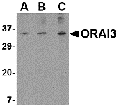
Western blot analysis of ORAI3 in A20 cell lysate with ORAI3 antibody at (A) 1, (B) 2 and (C) 4 μg/mL.

Click image to see more details
Immunocytochemistry of ORAI3 in A20 cells with ORAI3 antibody at 10 μg/mL.

Click image to see more details
Immunohistochemistry of ORAI3 in mouse spleen tissue with ORAI3 antibody at 2 μg/mL.

Click image to see more details
Orai3 and Stim1 silencing blocks TGF-β induced SNAI1 transcription. NMuMG ( A – D ) or MDA-MB-231 ( E – H ) cells were transfected with indicated siRNAs for 96 h using a final concentration of 100 picomoles of siRNA. The cells were treated with TGF-β for 2 h prior to RNA and protein isolation. RNA was converted to cDNA and RT-PCR performed to analyze both the gene knockdown efficiency for each gene, and Snai1 transcript levels. Western blots were performed against each protein to test efficiency of knockdown and normalized using GAPDH. All data are representative of at least 3 biological replicates (See for quantitation). Statistical analyses were performed with Graphpad Prism software. * = p -value ≤ 0.05, ** = p -value ≤ 0.01; *** = p -value ≤ 0.001.
Index in PubMed under a CC BY license. PMID: 30034631

Click image to see more details
Orai3 silencing blocks 2APB dependent increase in SOCE. Calcium imaging was performed in control ( A ) and Orai3 knockdown ( B ) NMuMG cells. Analog plots of the fluorescence ratio (340/380) from an average of 40–60 cells are shown. ( C ) Quantification (mean ± SD) of fluorescence ratio (340/380). All data are representative of at least 3 biological replicates. Statistical analyses were performed with Graphpad Prism software. * = p -value ≤ 0.05.
Index in PubMed under a CC BY license. PMID: 30034631

Click image to see more details
Orai3 silencing inhibits both cell migration and Snai1 transcription in response to TGF-β. ( A ) NMuMG cells were treated as indicated with TGF-β, TGF-β+2APB or DMSO, in the presence or absence of siOrai3 and RNA isolated. RNA was converted to cDNA and analyzed by real-time PCR using primers specific to mouse Snai1, Snai2 or Twist1, and normalized to Gapdh. Data represent the average of 3 individual biological replicates. ( B ) Proteins isolated from the same cells as in (A) were evaluated for SNAI1 expression by immunoblotting. Antibody to ACTIN was used a loading control, and the blots are representative of at least 3 independent biological replicates. Blots were quantitated using the LiCOR imaging software and are represented as SNAI1/ACTB signal, after normalizing to DMSO control. Error bars represent SEM and statistical analyses were performed Graphpad PRISM. * = p -value ≤ 0.05, ** = p -value ≤ 0.01 relative to control. ( C ) Confluent NMuMG cells in a 6-well plate were serum starved for 4 h prior to treatment, and TGF-β (for 8 h) and/or 2APB (for 24 h) were added to the wells prior to wounding using a sterile 200 ul tip. Three representative fields were marked and imaged immediately at time of (0 h) and a time period after (8 h) wounding as described in materials and methods. The images were captured using an Olympus IX71 microscope camera. All data are representative of at least 3 biological replicates. Statistical analyses were performed with Graphpad Prism software. * = p -value ≤ 0.05, ** = p -value ≤ 0.01; *** = p -value ≤ 0.001; **** = p -value ≤ 0.0001. ( D ) Model for ORAI3-mediated Snai1 upregulation. AKT (green oval) pathway can be activated by both calcium (black circles) and by TGF-β signaling. 2APB prevents SOCE via ORAI1 and ORAI2, while increasing calcium influx through ORAI3. Activation of AKT triggers increased binding of p65 at the Snai1 promoter, leading to increased recruitment of Pol II and hence transcription of Snai1 .
Index in PubMed under a CC BY license. PMID: 30034631
Specific Publications For Anti-Protein orai-3 ORAI3 Antibody (A09399)
Loading publications
Recommended Resources
Here are featured tools and databases that you might find useful.
- Boster's Pathways Library
- Protein Databases
- Bioscience Research Protocol Resources
- Data Processing & Analysis Software
- Photo Editing Software
- Scientific Literature Resources
- Research Paper Management Tools
- Molecular Biology Software
- Primer Design Tools
- Bioinformatics Tools
- Phylogenetic Tree Analysis
Customer Reviews
Have you used Anti-Protein orai-3 ORAI3 Antibody?
Share your experimental results or join a short interview to earn up to $1,000 in product credits or other rewards.
0 Reviews For Anti-Protein orai-3 ORAI3 Antibody
Customer Q&As
Have a question?
Find answers in Q&As, reviews.
Can't find your answer?
Submit your question
