Click image to see more details
-

-
-
-
-
+7
Product Info Summary
| SKU: | M00006-1 |
|---|---|
| Size: | 100 μl |
| Reactive Species: | Human, Mouse, Rat |
| Host: | Rabbit |
| Application: | Flow Cytometry, IF, IHC, ICC, WB |
Customers Who Bought This Also Bought

Product info
Product Name
Anti-PTEN Rabbit Monoclonal Antibody
SKU/Catalog Number
M00006-1
BM4114 is an alternative SKU for this antibody, used in previous lots.
Size
100 μl
Form
Liquid
Description
Boster Bio Anti-PTEN Rabbit Monoclonal Antibody catalog # M00006-1. Tested in WB, IHC, ICC/IF, Flow Cytometry applications. This antibody reacts with Human, Mouse, Rat.
Storage & Handling
Store at -20°C for one year. For short term storage and frequent use, store at 4°C for up to one month. Avoid repeated freeze-thaw cycles.
Cite This Product
Anti-PTEN Rabbit Monoclonal Antibody (Boster Biological Technology, Pleasanton CA, USA, Catalog # M00006-1)
Host
Rabbit
Contents
Rabbit IgG in stabilizing components, phosphate buffered saline, pH 7.4, 150mM NaCl, 0.02% sodium azide and 50% glycerol.
*This antibody is supplied in a stabilized formulation.
Compatibility with conjugation reactions depends on the chemistry of the conjugation method used.
For conjugation methods that are not compatible with the stabilizing components present in this formulation, a carrier-free antibody format is required.
Clonality
Monoclonal
Clone Number
BEI-16
Isotype
Rabbit IgG
Immunogen
A synthesized peptide derived from human PTEN
Reactive Species
M00006-1 is reactive to PTEN in Human, Mouse, Rat
Observed Molecular Weight
54 kDa
Calculated molecular weight
47.2 kDa
Antibody Validation
Boster validates all antibodies on WB, IHC, ICC, Immunofluorescence, and ELISA with known positive control and negative samples to ensure specificity and high affinity, including thorough antibody incubations.
Application & Images
Applications
M00006-1 is guaranteed for Flow Cytometry, IF, IHC, ICC, WB Boster Guarantee
Recommend Dilution
WB 1:500-2000
IHC 1:50-200
ICC/IF 1:50-200
FC 1:20
Tested application
Suggested blocking solution with 5% non-fat milk or BSA; (*)Recommended protein loading: 20-40 µg per lane
Use TE buffer pH 9.0 for antigen retrieval; (*) citrate buffer pH 6.0 is an alternative.
Validation Images & Assay Conditions
Click image to see more details
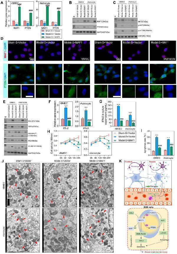
Overexpression of MAF1 promoted PTEN expression, increased cellular activity, decreased cell apoptosis, and normalized autophagy. A , B qPCR and western blot analyses of MAF1 and PTEN expression in the sham-S + vector, model-S + vector, and model-S + MAF1 groups. C Western blot analyses of AKT/mTOR signaling pathway proteins. D MAF1 and PTEM expression was measured by IF. E Western blot analyses of RIG-I and IRF3, the apoptosis-related protein caspase-3, and autophagy-related proteins p62 and LC3B. F , G qPCR and flow cytometry were used to detect IFNβ expression levels. H CCK-8 analyses for detection of cellular activity in rBMECs and rAstrocytes after transfection for 0, 6, 12, 18, and 24 h. I Flow cytometry was used to detect the apoptosis of transfected cells. J Transmission electron microscopy was used to detect autophagy in rBMECs and rAstrocytes. K Schematic diagram of the mechanism by which MAF1 expression level affects BBB function in SAE. *** P < 0.001 model-S + vector versus sham-S + vector. # P < 0.05, ### P < 0.001 model-S + MAF1 versus sham-S + vector
Index in PubMed under a CC BY license. PMID: 39833662

Click image to see more details
Interference with FOXO1 inhibited cellular activity and enhanced cell apoptosis. The rBMECs and rAstrocytes were treated with 10% serum harvested from the sham (sham-S) or model rats (model-S). A , B qPCR and western blot analyses of MAF1 , PTEN , and FOXO1 expression in the model-S + vector + siCtrl, model-S + PTEN + siCtrl, and model-S + PTEN + siFOXO1 groups. C MAF1 expression was measured by IF. D Western blot analyses evaluating the protein expression levels of RIG-I and IRF3 and apoptosis-related protein caspase-3. E qPCR and flow cytometry were used to detect IFNβ expression levels. F CCK-8 analyses for detection of cellular activity in rBMECs and rAstrocytes after transfection for 0, 6, 12, 18, and 24 h. G Flow cytometry was used to detect the apoptosis of transfected cells. *** P < 0.001 model-S + PTEN + siCtrl versus model-S + vector + siCtrl. # P < 0.05, ### P < 0.001 model-S + PTEN + siFOXO1 versus model-S + PTEN + siCtrl
Index in PubMed under a CC BY license. PMID: 39833662

Click image to see more details
The AKT/mTOR signaling pathway inhibited cellular activity and promoted cell apoptosis. A Levels of MAF1 expression were evaluated in the control, AKT1/2-IN, and AKT1/2-IN + CHX groups. B , C Western blot analyses were performed to assess the levels of MAF1 and FOXO1 protein expression in the control, AKT1/2-IN, and AKT1/2-IN + CHX groups. D , E The levels of cell activity and cell apoptosis in the control, AKT1/2-IN, and AKT1/2-IN + CHX groups. F An EMSA confirming the binding relationship between MAF1 and the PTEN promoter. *** P < 0.001 AKT1/2-IN versus control. # P < 0.05, ### P < 0.001 AKT1/2-IN + CHX versus AKT1/2-IN
Index in PubMed under a CC BY license. PMID: 39833662

Click image to see more details
Immunohistochemical analysis of paraffin-embedded human kidney, using PTEN Antibody .

Click image to see more details
Overexpression of PTEN attenuated the RIG-I/IRF3 signal pathway and apoptosis and recovered normal autophagy. The rBMECs and rAstrocytes were treated with 10% serum harvested from the sham (sham-S) or model rats (model-S). A , B qPCR and western blot analyses of MAF1 and PTEN expression in the sham-S + vector, model-S + vector, and model-S + PTEN groups. C Western blot analyses of AKT/mTOR signaling pathway. D MAF1 and CUL2 expression was measured by IF. E Western blot analyses of proteins RIG-I and IRF3, caspase-3, p62, and LC3B. F , G qPCR and flow cytometry were used to detect IFNβ expression levels. H CCK-8 analysis of rBMEC and rAstrocyte activity at 0, 6, 12, 18, and 24 h after transfection. I Flow cytometry analysis of apoptosis in transfected cells. J Transmission electron microscopy was used to detect autophagy in rBMECs and rAstrocytes. *** P < 0.001 model-S + vector versus sham-S + vector. # P < 0.05, ### P < 0.001 model-S + PTEN versus sham-S + vector
Index in PubMed under a CC BY license. PMID: 39833662

Click image to see more details
Immunofluorescent analysis of HeLa cells, using PTEN Antibody .

Click image to see more details
Knockdown of CUL2 increased MAF1 and PTEN expression and relieved cellular damage. A EBD extravasation and absorbance at 620 nm in brain tissues from the sham + Lv-shCtrl, model + Lv-shCtrl, and model + Lv-shCUL2 groups. B The levels of MAF1 and PTEN expression were measured by qPCR. C A Pearson correlation analysis was performed on the expression levels of MAF1 and PTEN . D , E Western blot analyses evaluating the levels of CUL2, MAF1, PTEN, AKT, p-AKT, and p-mTOR protein expression. F H&E staining showing the brain tissue morphology. G – I IHC analyses of CUL2, MAF1, and PTEN expression in brain tissues. *** P < 0.001 model + Lv-shCtrl versus sham + Lv-shCtrl. # P < 0.05, ### P < 0.001, model + Lv-shCUL2 versus model + Lv-shCtrl
Index in PubMed under a CC BY license. PMID: 39833662

Click image to see more details
Immunofluorescent analysis using the Antibody at 1:50 dilution.

Click image to see more details
Western blot analysis of PTEN using anti-PTEN antibody (M00006-1).
Electrophoresis was performed on a 5-20% SDS-PAGE gel at 70V (Stacking gel) / 90V (Resolving gel) for 2-3 hours. The sample well of each lane was loaded with 30 ug of sample under reducing conditions.
Lane 1: human Hela whole cell lysates,
Lane 2: human 293T whole cell lysates,
Lane 3: human MCF-7 whole cell lysates,
Lane 4: rat liver tissue lysates,
Lane 5: mouse heart tissue lysates,
Lane 6: mouse liver tissue lysates.
After electrophoresis, proteins were transferred to a nitrocellulose membrane at 150 mA for 50-90 minutes. Blocked the membrane with 5% non-fat milk/TBS for 1.5 hour at RT. The membrane was incubated with rabbit anti-PTEN antigen affinity purified monoclonal antibody (Catalog # M00006-1) at 1:500 overnight at 4°C, then washed with TBS-0.1%Tween 3 times with 5 minutes each and probed with a goat anti-rabbit IgG-HRP secondary antibody at a dilution of 1:500 for 1.5 hour at RT. The signal is developed using an Enhanced Chemiluminescent detection (ECL) kit (Catalog # EK1002) with Tanon 5200 system. A specific band was detected for PTEN at approximately 54 kDa. The expected band size for PTEN is at 47 kDa.

Click image to see more details
Immunofluorescent analysis using the Antibody at 1:150 dilution.

Click image to see more details
Western blot analysis of PTEN using anti-PTEN antibody (M00006-1).
Electrophoresis was performed on a 5-20% SDS-PAGE gel at 80V (Stacking gel) / 120V (Resolving gel) for 2 hours. The sample well of each lane was loaded with 30 ug of sample under reducing conditions.
Lane 1: control group-human mouse hippocampus tissue lysates,
Lane 2: model group-human mouse hippocampus tissue lysates,
Lane 3:Drug treatment (100mg/kg) – Mouse hippocampus tissue lysates,
Lane 4:Drug treatment (500mg/kg) – Mouse hippocampus tissue lysates.
After electrophoresis, proteins were transferred to a nitrocellulose membrane at 150 mA for 50-90 minutes. Blocked the membrane with 5% non-fat milk/TBS for 1 hour at RT. The membrane was incubated with rabbit anti-PTEN antigen affinity purified monoclonal antibody (A04887-1) overnight at 4°C, then washed with TBS-0.1%Tween 3 times with 5 minutes each and probed with a goat anti-rabbit IgG-HRP secondary antibody at a dilution of 1:5000 for 1.5 hour at RT. The signal is developed using an ECL Plus Western Blotting Substrate (Catalog # AR1196-200) with ChemiDoc MP system. A specific band was detected for PTEN at approximately 52 kDa. The expected band size for PTEN is at 54 kDa.
Specific Publications For Anti-PTEN Rabbit Monoclonal Antibody (M00006-1)
Loading publications
Recommended Resources
Here are featured tools and databases that you might find useful.
- Boster's Pathways Library
- Protein Databases
- Bioscience Research Protocol Resources
- Data Processing & Analysis Software
- Photo Editing Software
- Scientific Literature Resources
- Research Paper Management Tools
- Molecular Biology Software
- Primer Design Tools
- Bioinformatics Tools
- Phylogenetic Tree Analysis
Customer Reviews
Have you used Anti-PTEN Rabbit Monoclonal Antibody?
Share your experimental results or join a short interview to earn up to $1,000 in product credits or other rewards.
1 Reviews For Anti-PTEN Rabbit Monoclonal Antibody
This antibody is suitable for detecting PTEN protein in mouse hippocampus by Western blot, showing clear, distinct, and highly specific bands.
Excellent

| SKU | M00006-1 |
|---|---|
| Application | Western Blot |
| Sample | Mouse hippocampus tissue |
| Sample Processing Description | The mouse hippocampus was lysed with RIPA buffer containing a protease inhibitor cocktail. After protein quantification, samples were mixed with 5× protein loading buffer and heated for 10 minutes to denature. Load 5 μL of protein per lane and apply to SDS-PAGE. |
| Other Reagents | 5% Non-fat milk |
| Primary Antibody | Anti-PTEN Rabbit Monoclonal Antibody |
| Primary Incubation | overnight at 4 ℃ |
| Secondary Antibody | HRP-conjugated Anti-Rabbit IgG Secondary Antibody |
| Secondary Incubation | 1 hour in room temperature |
| Detection | Substrate: Ultra-sensitive ECL luminescent reagent, Imaging system:ChemiDoc MP |
| Results Summary | This antibody is suitable for detecting PTEN protein in mouse hippocampus by Western blot, showing clear, distinct, and highly specific bands. |
Changbin Yuan, LNUTCM
Verified customer
Submitted 2025-11-10
Customer Q&As
Have a question?
Find answers in Q&As, reviews.
Can't find your answer?
Submit your question
16 Customer Q&As for Anti-PTEN Rabbit Monoclonal Antibody
Question
We are currently using anti-PTEN Rabbit Monoclonal antibody M00006-1 for rat tissue, and we are happy with the IF results. The species of reactivity given in the datasheet says human, mouse, rat. Is it true that the antibody can work on dog tissues as well?
Verified Customer
Verified customer
Asked: 2020-03-26
Answer
The anti-PTEN Rabbit Monoclonal antibody (M00006-1) has not been tested for cross reactivity specifically with dog tissues, but there is a good chance of cross reactivity. We have an innovator award program that if you test this antibody and show it works in dog you can get your next antibody for free. Please contact me if I can help you with anything.
Boster Scientific Support
Answered: 2020-03-26
Question
Would M00006-1 anti-PTEN Rabbit Monoclonal antibody work on parafin embedded sections? If so, which fixation method do you recommend we use (PFA, paraformaldehyde, other)?
Verified Customer
Verified customer
Asked: 2020-02-20
Answer
It shows on the product datasheet, M00006-1 anti-PTEN Rabbit Monoclonal antibody as been validated on IF. It is best to use PFA for fixation because it has better tissue penetration ability. PFA needs to be prepared fresh before use. Long term stored PFA turns into formalin, as the PFA molecules congregate and become formalin.
Boster Scientific Support
Answered: 2020-02-20
Question
Is a blocking peptide available for product anti-PTEN Rabbit Monoclonal antibody (M00006-1)?
Verified Customer
Verified customer
Asked: 2019-12-19
Answer
We do provide the blocking peptide for product anti-PTEN Rabbit Monoclonal antibody (M00006-1). If you would like to place an order for it please contact support@bosterbio.com and make a special request.
Boster Scientific Support
Answered: 2019-12-19
Question
Thanks for helping with my inquiry over the phone. Here are the WB image, lot number and protocol we used for epithelium using anti-PTEN Rabbit Monoclonal antibody M00006-1. Let me know if you need anything else.
Verified Customer
Verified customer
Asked: 2019-11-29
Answer
Thank you for the data. You have provided everything we needed. Our lab team are working to resolve your inquiry as quickly as possible, and we appreciate your patience and understanding! Please let me know if there is anything you need in the meantime.
Boster Scientific Support
Answered: 2019-11-29
Question
Our lab want to know about to test anti-PTEN Rabbit Monoclonal antibody M00006-1 on mouse epithelium for research purposes, then I may be interested in using anti-PTEN Rabbit Monoclonal antibody M00006-1 for diagnostic purposes as well. Is the antibody suitable for diagnostic purposes?
Verified Customer
Verified customer
Asked: 2019-09-02
Answer
The products we sell, including anti-PTEN Rabbit Monoclonal antibody M00006-1, are only intended for research use. They would not be suitable for use in diagnostic work. If you have the means to develop a product into diagnostic use, and are interested in collaborating with us and develop our product into an IVD product, please contact us for more discussions.
Boster Scientific Support
Answered: 2019-09-02
Question
I was wanting to use your anti-PTEN Rabbit Monoclonal antibody for IF for mouse epithelium on frozen tissues, but I want to know if it has been tested for this particular application. Has this antibody been tested and is this antibody a good choice for mouse epithelium identification?
Verified Customer
Verified customer
Asked: 2019-08-15
Answer
As indicated on the product datasheet, M00006-1 anti-PTEN Rabbit Monoclonal antibody has been tested for IP, IF, WB on human, mouse, rat tissues. We have an innovator award program that if you test this antibody and show it works in mouse epithelium in IHC-frozen, you can get your next antibody for free.
Boster Scientific Support
Answered: 2019-08-15
Question
Would anti-PTEN Rabbit Monoclonal antibody M00006-1 work on goat IF with spleen?
Verified Customer
Verified customer
Asked: 2019-05-24
Answer
Our lab technicians have not validated anti-PTEN Rabbit Monoclonal antibody M00006-1 on goat. You can run a BLAST between goat and the immunogen sequence of anti-PTEN Rabbit Monoclonal antibody M00006-1 to see if they may cross-react. If the sequence homology is close, then you can perform a pilot test. Keep in mind that since we have not validated goat samples, this use of the antibody is not covered by our guarantee. However we have an innovator award program that if you test this antibody and show it works in goat spleen in IF, you can get your next antibody for free.
Boster Scientific Support
Answered: 2019-05-24
Question
Please see the WB image, lot number and protocol we used for epithelium using anti-PTEN Rabbit Monoclonal antibody M00006-1. Please let me know if you require anything else.
C. Johnson
Verified customer
Asked: 2018-12-14
Answer
Thank you very much for the data. Our lab team are working to resolve this as quickly as possible, and we appreciate your patience and understanding! You have provided everything we needed. Please let me know if there is anything you need in the meantime.
Boster Scientific Support
Answered: 2018-12-14
Question
We have been able to see staining in mouse intestine. What should we do? Is anti-PTEN Rabbit Monoclonal antibody supposed to stain intestine positively?
W. Krishna
Verified customer
Asked: 2018-03-19
Answer
According to literature intestine does express PTEN. According to Uniprot.org, PTEN is expressed in intestine, epithelium, spleen, lung, liver, among other tissues. Regarding which tissues have PTEN expression, here are a few articles citing expression in various tissues:
Epithelium, Pubmed ID: 9187108, 9090379
Liver, Pubmed ID: 24275569
Lung, Pubmed ID: 15489334
Spleen, Pubmed ID: 14702039
Boster Scientific Support
Answered: 2018-03-19
Question
I see that the anti-PTEN Rabbit Monoclonal antibody M00006-1 works with IF, what is the protocol used to produce the result images on the product page?
K. Anderson
Verified customer
Asked: 2018-02-08
Answer
You can find protocols for IF on the "support/technical resources" section of our navigation menu. If you have any further questions, please send an email to support@bosterbio.com
Boster Scientific Support
Answered: 2018-02-08
Question
We ordered your anti-PTEN Rabbit Monoclonal antibody for IP on spleen last year. I am using rat, and I plan to use the antibody for WB next. Our lab want to know about examining spleen as well as epithelium in our next experiment. Could you please give me some suggestion on which antibody would work the best for WB?
C. Huang
Verified customer
Asked: 2018-02-05
Answer
I took a look at the website and datasheets of our anti-PTEN Rabbit Monoclonal antibody and it seems that M00006-1 has been validated on rat in both IP and WB. Thus M00006-1 should work for your application. Our Boster satisfaction guarantee will cover this product for WB in rat even if the specific tissue type has not been validated. We do have a comprehensive range of products for WB detection and you can check out our website bosterbio.com to find out more information about them.
Boster Scientific Support
Answered: 2018-02-05
Question
Do you have a BSA free version of anti-PTEN Rabbit Monoclonal antibody M00006-1 available?
Verified Customer
Verified customer
Asked: 2018-02-01
Answer
Thank you for your recent telephone inquiry. I can confirm that some lots of this anti-PTEN Rabbit Monoclonal antibody M00006-1 are BSA free. For now, these lots are available and we can make a BSA free formula for you free of charge. It will take 3 extra days to prepare. If you require this antibody BSA free again in future, please do not hesitate to contact me and I will be pleased to check which lots we have in stock that are BSA free.
Boster Scientific Support
Answered: 2018-02-01
Question
Is this M00006-1 anti-PTEN Rabbit Monoclonal antibody reactive to the isotypes of PTEN?
Verified Customer
Verified customer
Asked: 2018-01-09
Answer
The immunogen of M00006-1 anti-PTEN Rabbit Monoclonal antibody is A synthesized peptide derived from human PTEN . Could you tell me which isotype you are interested in so I can help see if the immunogen is part of this isotype?
Boster Scientific Support
Answered: 2018-01-09
Question
We were content with the WB result of your anti-PTEN Rabbit Monoclonal antibody. However we have been able to see positive staining in epithelium cytoplasm using this antibody. Is that expected? Could you tell me where is PTEN supposed to be expressed?
K. Rodriguez
Verified customer
Asked: 2015-04-23
Answer
From literature, epithelium does express PTEN. Generally PTEN expresses in cytoplasm. Regarding which tissues have PTEN expression, here are a few articles citing expression in various tissues:
Epithelium, Pubmed ID: 9187108, 9090379
Liver, Pubmed ID: 24275569
Lung, Pubmed ID: 15489334
Spleen, Pubmed ID: 14702039
Boster Scientific Support
Answered: 2015-04-23
Question
My question regarding product M00006-1, anti-PTEN Rabbit Monoclonal antibody. I was wondering if it would be possible to conjugate this antibody with biotin. I would need it to be without BSA or sodium azide. I am planning on using a buffer exchange of sodium azide with PBS only. Would there be problems for me to conjugate the antibody and store it in -20 degrees in small aliquots?
B. Edwards
Verified customer
Asked: 2014-05-28
Answer
We do not recommend storing this antibody with PBS buffer only in -20 degrees. If you want to store it in -20 degrees it is best to add some cryoprotectant like glycerol. If you want carrier free M00006-1 anti-PTEN Rabbit Monoclonal antibody, we can provide it to you in a special formula with trehalose and/or glycerol. These molecules will not interfere with conjugation chemistry and provide a good level of protection for the antibody from degradation. Please be sure to specify this in your purchase order.
Boster Scientific Support
Answered: 2014-05-28
Question
Will anti-PTEN Rabbit Monoclonal antibody M00006-1 work for IF with epithelium?
T. Krishna
Verified customer
Asked: 2013-05-24
Answer
According to the expression profile of epithelium, PTEN is highly expressed in epithelium. So, it is likely that anti-PTEN Rabbit Monoclonal antibody M00006-1 will work for IF with epithelium.
Boster Scientific Support
Answered: 2013-05-24
