Click image to see more details
Product Info Summary
| SKU: | A03775 |
|---|---|
| Size: | 100 μg/vial |
| Reactive Species: | Human, Mouse, Rat |
| Host: | Rabbit |
| Application: | ELISA, WB |
Customers Who Bought This Also Bought

Product info
Product Name
Anti-Retinal S antigen/SAG Antibody Picoband®
SKU/Catalog Number
A03775
Size
100 μg/vial
Form
Lyophilized
Description
Boster Bio Anti-Retinal S antigen/SAG Antibody Picoband® catalog # A03775. Tested in ELISA, WB applications. This antibody reacts with Human, Mouse, Rat. The brand Picoband indicates this is a premium antibody that guarantees superior quality, high affinity, and strong signals with minimal background in Western blot applications. Only our best-performing antibodies are designated as Picoband, ensuring unmatched performance.
Storage & Handling
Store at -20˚C for one year from date of receipt. After reconstitution, at 4˚C for one month. It can also be aliquotted and stored frozen at -20˚C for six months. Avoid repeated freeze-thaw cycles.
Cite This Product
Anti-Retinal S antigen/SAG Antibody Picoband® (Boster Biological Technology, Pleasanton CA, USA, Catalog # A03775)
Host
Rabbit
Contents
Each vial contains 4mg Trehalose, 0.9mg NaCl, 0.2mg Na2HPO4, 0.05mg NaN3.
Clonality
Polyclonal
Isotype
Rabbit IgG
Immunogen
E. coli-derived human Retinal S antigen recombinant protein (Position: R193-E405).
Cross-reactivity
No cross-reactivity with other proteins.
Reactive Species
A03775 is reactive to SAG in Human, Mouse, Rat
Observed Molecular Weight
45-55 kDa
Calculated molecular weight
45.1 kDa
Background of SAG
S-arrestin is a protein that in humans is encoded by the SAG gene. Members of arrestin/beta-arrestin protein family are thought to participate in agonist-mediated desensitization of G-protein-coupled receptors and cause specific dampening of cellular responses to stimuli such as hormones, neurotransmitters, or sensory signals. S-arrestin, also known as S-antigen, is a major soluble photoreceptor protein that is involved in desensitization of the photoactivated transduction cascade. It is expressed in the retina and the pineal gland and inhibits coupling of rhodopsin to transducin in vitro. Additionally, S-arrestin is highly antigenic, and is capable of inducing experimental autoimmune uveoretinitis. Mutations in this gene have been associated with Oguchi disease, a rare autosomal recessive form of night blindness.
Antibody Validation
Boster validates all antibodies on WB, IHC, ICC, Immunofluorescence, and ELISA with known positive control and negative samples to ensure specificity and high affinity, including thorough antibody incubations.
Application & Images
Applications
A03775 is guaranteed for ELISA, WB Boster Guarantee
Recommend Dilution
| Application | Dilution | Species |
|---|---|---|
| Western blot | 0.1-0.5μg/ml | |
| ELISA | 0.1-0.5μg/ml |
Tested application
Suggested blocking solution with 5% non-fat milk or BSA; (*)Recommended protein loading: 20-40 µg per lane
Validation Images & Assay Conditions
Click image to see more details
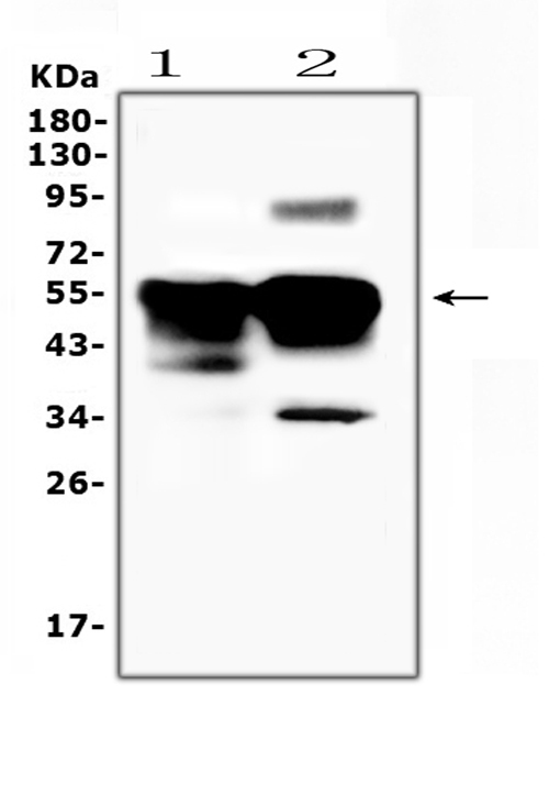
Western blot analysis of Retinal S antigen using anti-Retinal S antigen antibody (A03775).
Electrophoresis was performed on a 5-20% SDS-PAGE gel at 70V (Stacking gel) / 90V (Resolving gel) for 2-3 hours. The sample well of each lane was loaded with 50ug of sample under reducing conditions.
Lane 1: rat eye ball tissue lysates,
Lane 2: mouse eye ball tissue lysates.
After Electrophoresis, proteins were transferred to a Nitrocellulose membrane at 150mA for 50-90 minutes. Blocked the membrane with 5% Non-fat Milk/ TBS for 1.5 hour at RT. The membrane was incubated with rabbit anti-Retinal S antigen antigen affinity purified polyclonal antibody (Catalog # A03775) at 0.5 ug/mL overnight at 4 then washed with TBS-0.1%Tween 3 times with 5 minutes each and probed with a goat anti-rabbit IgG-HRP secondary antibody at a dilution of 1:10000 for 1.5 hour at RT. The signal is developed using an Enhanced Chemiluminescent detection (ECL) kit (Catalog # EK1002) with Tanon 5200 system. A specific band was detected for Retinal S antigen at approximately 45-55KD. The expected band size for Retinal S antigen is at 45KD.
Specific Publications For Anti-Retinal S antigen/SAG Antibody Picoband® (A03775)
Loading publications
Recommended Resources
Here are featured tools and databases that you might find useful.
- Boster's Pathways Library
- Protein Databases
- Bioscience Research Protocol Resources
- Data Processing & Analysis Software
- Photo Editing Software
- Scientific Literature Resources
- Research Paper Management Tools
- Molecular Biology Software
- Primer Design Tools
- Bioinformatics Tools
- Phylogenetic Tree Analysis
Customer Reviews
Have you used Anti-Retinal S antigen/SAG Antibody Picoband®?
Share your experimental results or join a short interview to earn up to $1,000 in product credits or other rewards.
0 Reviews For Anti-Retinal S antigen/SAG Antibody Picoband®
Customer Q&As
Have a question?
Find answers in Q&As, reviews.
Can't find your answer?
Submit your question
1 Customer Q&As for Anti-Retinal S antigen/SAG Antibody Picoband®
Question
We are currently using anti-Retinal S antigen/SAG antibody A03775 for mouse tissue, and we are happy with the WB results. The species of reactivity given in the datasheet says human, mouse, rat. Is it possible that the antibody can work on primate tissues as well?
Verified Customer
Verified customer
Asked: 2019-03-28
Answer
The anti-Retinal S antigen/SAG antibody (A03775) has not been tested for cross reactivity specifically with primate tissues, but there is a good chance of cross reactivity. We have an innovator award program that if you test this antibody and show it works in primate you can get your next antibody for free. Please contact me if I can help you with anything.
Boster Scientific Support
Answered: 2019-03-28
